In this video, we demonstrate the use of spectral-domain optical coherence tomography (SD-OCT) to image retinal layers and assess structural changes in a genetically modified mouse model with optic neuropathy.
A subscription to JoVE is required to view this content. Sign in or start your free trial.
Method Article
In Vivo Imaging of the Mouse Retina Using Optical Coherence Tomography
1.4K views
⸱
May 29th, 2025
In This Article
Abstract
Protocol
All procedures involving animal models have been reviewed by the local institutional animal care committee and the JoVE veterinary review board.
Access restricted. Please log in or start a trial to view this content.
Results
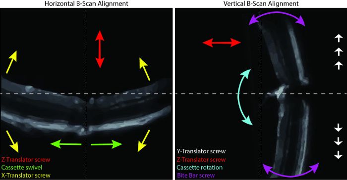
Access restricted. Please log in or start a trial to view this content.
Materials
| Name | Company | Catalog Number | Comments |
|---|---|---|---|
| Mice | |||
| Opa1delTTAG mouse | |||
| Equipment | |||
| EnVisu R2200 SD-OCT Imaging System | |||
| EnVisu R2200 SD-OCT Imaging System Software | |||
| ImageJ 1.48v | |||
| Self-regulating heating plate | |||
| Supplies | |||
| Gauze pads 3"x3" | |||
| Dual Ended Cotton tip applicator | |||
| Cotton Twists | |||
| Reagents and Drugs | |||
| Néosynéphrine Faure 10% | |||
| Mydriaticum 0.5% | |||
| Cebesine 0.4% | |||
| Imalgene 1000 | |||
| Rompun | |||
| NaCl 0,9% | |||
| Systene Ultra | |||
| GenTeal' | |||
| Aniospray Surf 29 |
