This video demonstrates the use of micro-CT imaging to visualize cerebral cavernous malformation (CCM) lesions in a mouse brain. It details the steps for sample preparation, imaging, and 3D image reconstruction to evaluate the density and location of CCM lesions in brain tissue.
A subscription to JoVE is required to view this content. Sign in or start your free trial.
Method Article
Contrast-Enhanced Micro-CT Imaging of Cerebral Cavernous Malformation Lesions in a Mouse Brain
999 views
⸱
May 29th, 2025
In This Article
Abstract
Protocol
All procedures involving animal models have been reviewed by the local institutional animal care committee and the JoVE veterinary review board.
1. Sample Preparation for Micro-Computed Tomography (CT) Scans
- Euthanize neonatal pups at P8 via carbon dioxide asphyxiation.
- Perform intra-cardiac perfusion by inserting a 29-gauge needle with 3 mL of 2% paraformaldehyde in phosphate buffered saline (PBS) in a 10 mL syringe into the ventricle of the mouse heart.
CAUTION: Paraformaldehyde is toxic; wear appropriate protection. - Detach the head from the body using scissors. Remove all the skin off the head using a scissors and peel off the skull using forceps to dissect the whole brain.
- Take images of the brain with a stereomicroscope at 7.82x magnification, 1x gain, 0.6 gamma, and 20 ms exposure time.
- Fix the dissected brains with 4% paraformaldehyde in PBS solution overnight.
CAUTION: Paraformaldehyde is toxic; wear appropriate protection. - On the following day, detach hindbrains using forceps and wash with PBS solution.
- Incubate the hindbrains in Lugol's iodine solution for 48 h.
- Following the incubation, briefly air dry the hindbrains to remove excess Lugol's iodine solution.
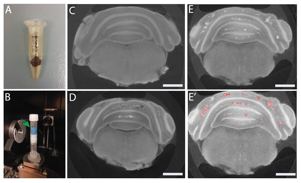
- Pack the Lugol's iodine-stained hindbrains in 0.65 mL microcentrifuge tubes and seal completely with plastic paraffin film to avoid tissue shrinkage (Figure 1A).
- Place microcentrifuge tubes in 5 mL plastic tubes with sponges to prevent them from moving during scan (Figure 1B).
2. Micro-CT Scan of cerebral cavernous malformations (CCMs) in the Mouse Brain
- Vertically mount the hindbrain packed in a tube on an aluminum holder in the micro-CT system.
- Set the scanning parameters to 540 projections and 2 s exposure time with source conditions of 50 kV and 10 W to acquire the tomographic datasets (image resolution 9.5 μm/pixel).
- Radiographs from the scan are reconstructed automatically by hardware-based projection reconstruction software supplied by the micro-CT system, producing an image series of 16-bit axial slices ("TXM" file).
3. Analysis of Micro-CT Datasets
- Open the reconstructed image file ("TXM" file) in the 3-D analysis software and visualize the brain using the "volume rendering" function.
- Transform the hindbrain to the desired orientation using the "bounding box" and "clipping plane" functions.
- Resample the transformed image in extended mode and preserve the voxel size.
- Create "edit new label field" on the transformed image (3.3) and highlight only the hindbrain using greyscale intensity.
- Add the highlighted areas and run "label analysis" to obtain measurements of the whole hindbrain (i.e., total volume and area). Export measurements in an Excel file.
- Visualize the hindbrain using the "isosurface rendering" function.
- Create another "edit new label field" on the transformed image and highlight areas with lesions using greyscale intensity.
- Add the highlighted areas and run "label" analysis to obtain measurements of the lesions within the hindbrain (i.e., total volume and area). Export measurements in an Excel file.
- Visualize the lesions using the "generate surface" and "surface view" functions.
- Snapshot the images of the hindbrain at desired orientations or generate a movie for 3-D visualization of the lesions.
Access restricted. Please log in or start a trial to view this content.
Results
Access restricted. Please log in or start a trial to view this content.
Materials
| Name | Company | Catalog Number | Comments |
|---|---|---|---|
| 4-hydroxy tamoxifen | |||
| Corn oil | |||
| Stereomicroscope | |||
| Lugol's Iodine solution | |||
| Plastic paraffin film | |||
| Micro-CT | |||
| 3D rendering software |
