This video demonstrates a live-imaging technique using differential interference contrast (DIC) microscopy to observe the beating of ependymal cilia in sagittal mouse brain sections. Ependymal cilia propel cerebrospinal fluid (CSF), and their activity is recorded in brain sections maintained in a nutrient-rich medium and imaged in a climate-controlled chamber. High-contrast videos enable the identification of functional cilia and quantify their beating frequency.
A subscription to JoVE is required to view this content. Sign in or start your free trial.
Method Article
Live Imaging of Ependymal Cell Ciliary Activity in a Mouse Brain Section
1.7K views
⸱
May 29th, 2025
In This Article
Abstract
Protocol
All procedures involving animal samples have been reviewed and approved by the appropriate animal ethical review committee.
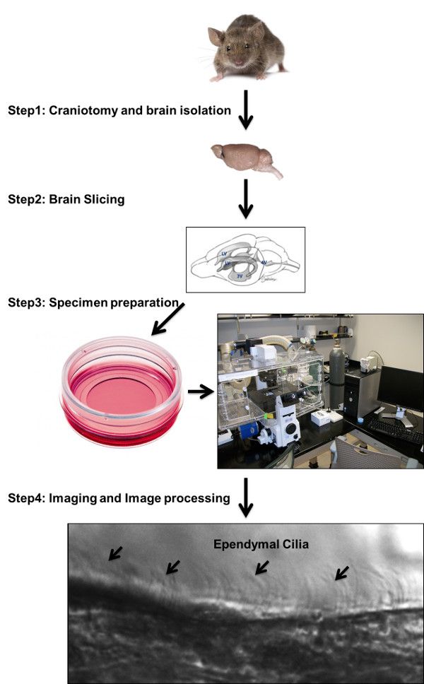
1. Brain Extraction, Sectioning and Tissue Preparation
- Sacrifice wild-type mouse strain C57BL/6 by deeply euthanizing with CO2 asphyxiation for 5 min. Assure death by cervical dislocation.
- Clean the mouse head with 70% ethanol.
- Perform craniectomy using sterile scissors and forceps by first pulling the skin off, starting with the top of the head to expose the skull.
- Then, when the skull is exposed, remove the skull by peeling the bone piece-by-piece, starting from the posterior side and moving toward the anterior side. Be cautious not to destroy the brain ventricles.
- Collect the whole brain.
- Place the brain in a 100 mm Petri dish containing Dulbecco’s Modified Eagle’s Medium (DMEM)/High-Glucose supplemented with 10% fetal bovine serum (FBS) and 1% penicillin/streptomycin solution containing 10,000 units/ml of penicillin and 10,000 µg/ml of streptomycin and pre-warmed to 37 °C.
- Slice the brain on the median sagittal plane by hand with a sharp blade, and use a vibratome to obtain the first 100-200 mm section from each half.
- Rinse the brain tissue with pre-warmed 37 °C phosphate-buffered saline (1x PBS) solution.
- Immediately place the brain section in DMEM/High-Glucose media pre-warmed to 37 °C.
2. Live Imaging Configuration and Setup
- Place the brain tissue sections in 30 mm glass-bottom culture dishes containing 1 ml of DMEM/High-Glucose media. Adjust the microscope’s enclosed chamber environment to 37 °C, 95%/5% O2/CO2 content (Figure 1).
- Using a 60X objective oil immersion lens, collect the ependymal cells/cilia images by placing an oil drop on the 60X objective lens and focusing on the cells with regular differential interference contrast (DIC) transmitted light.
- Then, follow the direction of the DMEM bubble movement as a guide to the location of the motile ependymal cilia as ciliary beatings create a kind of bubble movement in the area. Choose an area containing healthy cells with motile cilia in the brain’s lateral ventricle using the DIC filter. Once the ependymal cilia are found, adjust the light and focus to obtain a satisfactory image.
- Using Metamorph imaging software, set the live imaging parameters according to a specific purpose. In the present demonstration, acquire twenty-four-bit images with the camera binning set to 1 x 1 combined with a 60X objective and a 5-10 msec exposure time.
- Collect the DIC images by opening the microscope aperture to an optimal level for a minimum exposure time. Observe live images streamed to the camera to provide fast and immediate image acquisition without delay. Calculate the speed of cilia beating based on the requirement of the minimal exposure times to obtain sufficient image contrast.
Access restricted. Please log in or start a trial to view this content.
Results
Access restricted. Please log in or start a trial to view this content.
Materials
| Name | Company | Catalog Number | Comments |
|---|---|---|---|
| DMEM/HIGH GLUCOSE | |||
| Fetal bovine serum (FBS) | |||
| Penicillin/Streptomycin | |||
| Phosphate buffered saline | |||
| Vibratome | |||
| Cell Culture plate | |||
| Inverted Fluorescence Microscope |
