In this video, a rheometer was used to apply controlled oscillatory rotational motion to brain tissue to generate shear strain and evaluate the tissue's viscous and elastic properties. The rheometer, as a functional tool, it provides insights into brain mechanics during injury or neurodegenerative diseases.
A subscription to JoVE is required to view this content. Sign in or start your free trial.
Method Article
Measuring the Viscoelastic Properties of Mouse Brain Tissue Using Rheometry
928 views
⸱
June 17th, 2025
In This Article
Abstract
Protocol
All procedures involving animal samples have been reviewed and approved by the appropriate animal ethical review committee.
- Rheology
- Set up and calibrate the rheometer as per the manufacturer's instructions.
- Initialize the rheometer by opening the device/control panel. On the control panel tab, click "initialize."
- Mount the 25 mm-diameter measurement plate (PP25) and the thermal system.
- (Optional) To reduce slip between the rheometer plates and the tissue, cut out adhesive sandpaper slices that match the shape of the top rheometer plate and adhere the sandpaper to the top and bottom plate.
- Make contact between the top and bottom plates by clicking "set zero gap" on the control panel.
- Zero the normal force transducer by clicking "reset normal force."
- Conduct an inertia test by opening the service tab on the control panel, clicking "measurement system," and then clicking "inertia test". Record the old and new inertia. Verify that the inertia is within the allowable limit for the probe, as listed by the manufacturer.
- Set up and calibrate the rheometer as per the manufacturer's instructions.
- Load the sample into the rheometer.
- After harvesting the tissue and slicing a coronal segment of the pig brain to ~5 mm thickness, store it on ice in a CO2-independent medium.
- Place the brain between the two plates. Remove large water droplets from the top and bottom surface of the sample to prevent slippage, but do not dry out the sample.
- Slowly lower the measurement plate until the plate is in full contact with the top surface of the tissue and the measured normal force is consistent at 0.01 mN after a 5-10 min relaxation period.
- In the control panel, enter successively lower heights in the measurement position box and click "measurement position" to slowly lower the measurement plate.
- When within a millimeter of contact with the tissue, lower the measurement plate in 0.1 mm increments until the plate is fully in contact with the top surface of the tissue. Ensure that the measured-normal force is consistently at 0.01 mN after a 5-10 min relaxation period.
- Record the initial measured normal force. Repeated measurements should be taken at the same compressive stresses/strains.
- If the sample exceeds the diameter of the plate, trim it with a plastic blade. Pipette a small volume (~1-2 ml) of media on the edges of the sample to hydrate the tissue.
- (Optional) Lower the thermal hood. On the control panel, set the temperature to 37 °C and click "set".
- Perform an amplitude sweep to establish the linear viscoelastic range of the material (i.e., the shear strains at which G' and G'' are constant) at frequencies of interest (e.g., 1 rad/sec).
- Select "file/new". Under the gel tab, select "Amplitude sweep: LVE-range." Select window and click "Measurement 1: Amplitude sweep." Double click on the oscillation box. Enter the initial and final strain (e.g., 0.01 to 105), the frequency (e.g., 1 rad/sec), and the number of points per decade (e.g., 6 points/dec). Select "ok" and click “Start."
- Repeat this procedure for several slices with repeated trials to ensure consistency of the linear elastic range. The axial compression of the sample should remain constant between samples.
- Conduct a frequency sweep of the tissue at a strain in the linear viscoelastic range of the tissue (e.g., 1% strain), and at a frequency range of interest (e.g., 0.1-100 rad/sec).
- Click "file/new" and under the gel tab select "Frequency sweep." Click Window/Measurement 1: Frequency sweep. Double-click on the oscillation box. Enter the frequency range (e.g., 0.1 to 100 rad/sec), the strain (e.g., 1% strain), and the number of points per decade (e.g., 6 points/dec). Select "ok" and click "Start" to initiate the frequency sweep.
- Repeat frequency sweep (step 1.4) in duplicates or triplicates.
Review the data that are automatically calculated and exported by the rheometer: G' and G" as a function of frequency (frequency sweep) or shear strain (amplitude sweep). NOTE: G' and G'' are calculated from the sample's (maximum) reactional torque amplitude T'0, and rotational displacement angle (or deflection angle) φ°, and phase lag ϕ, of the sample's response to the applied oscillatory strain (Figure 1):
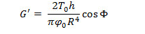
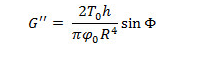
where R and h are the radius and height of the sample.
Access restricted. Please log in or start a trial to view this content.
Results
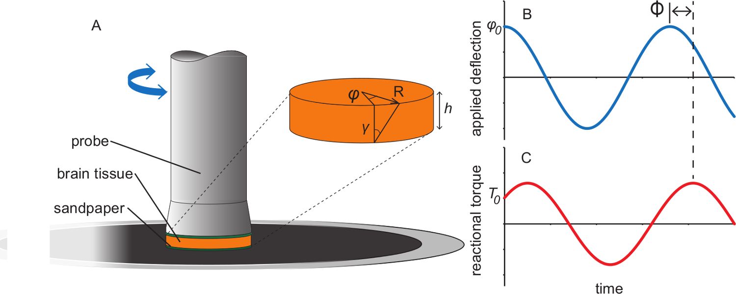
Figure 1. Illustration of parallel plate rheometer experiments. (A) Schematic of parallel plate r...
Access restricted. Please log in or start a trial to view this content.
Materials
| Name | Company | Catalog Number | Comments |
|---|---|---|---|
| Xylaxine | Lloyd Laboratoried | perscription drug | |
| Ketamine | AnaSed Injections | perscription drug | |
| Hibernate-A Medium | Gibco | A1247501 | CO2-independent neural medium for adult tissue |
| Parallel Plate Rheometer MCR501 | Anton-Parr | - | |
| PP25 | Anton-Parr | - | 25 mm diameter flat measurement plate |
| Adhesive Sandpaper | McMaster-Carr | 4184A48 | alternate suppliers can be used |
| Loctite 4013 Instant Adhesive | Henkel | 20268 | alternate suppliers can be used |
