This video demonstrates the use of confocal microscopy to visualize dendritic dynamics in live mouse retinal explants. It outlines the steps for stabilizing the retina under a confocal microscope, performing epifluorescence imaging to visualize starburst amacrine interneurons, and capturing time-lapse 3D images to monitor changes in amacrine dendritic morphology.
A subscription to JoVE is required to view this content. Sign in or start your free trial.
Method Article
Time-Lapse Confocal Imaging of Neuronal Dendritic Dynamics in Mouse Retinal Explants
831 views
⸱
June 17th, 2025
In This Article
Abstract
Protocol
All procedures involving animal samples have been reviewed and approved by the appropriate animal ethical review committee.
1. Time-lapse confocal imaging of live whole-mount retina preparations
- Assemble the live-imaging incubation chamber for an upright confocal microscope, as seen in Figure 1.
NOTE: For inverted confocal systems, flat-mounts are placed retinal ganglion cells (RGC) side down directly onto the glass bottom coverslip of the incubation chamber. Once the retinas make contact with the coverslip, they cannot be moved. - Fill the chamber with oxygenated artificial cerebrospinal fluid (aCSF) and turn on the pump and temperature controller (temperature 32-34 °C, flow rate 1 mL/min). Do not allow the temperature to rise above 34 °C.
- To transfer the retinal flat-mount to the perfusion chamber, stop the pump and remove the aCSF that is in the chamber. Place the mixed cellulose ester (MCE) disc with the retinal flat-mount into the (empty) incubation chamber.
- Place a sample weight onto the flat-mount; pre-wet the weight to break the surface tension. Refill the chamber with the warmed aCSF, and circulate aCSF at ~1 mL/min.
- Position the nosepiece with the 25x water dipping objective (numerical aperture 0.95) into the imaging chamber. Screen for the labeled cells of interest using epifluorescent light (Figure 1C).
- Adjust the imaging volume to capture dendritic features of interest.
NOTE: This study captured the complete dendritic arbor at 1024 x 1024 pixels per frame, z-step 1 µm, and a frame rate of 2 min between each z-stack. Final image sizes are ~100 µm x 100 µm x 20 µm. - To adjust the laser power to an optimal setting, use a look-up table that identifies both oversaturated and undersaturated pixels. While scanning, adjust the laser power such that no pixels are oversaturated (i.e., at an intensity of 255 or above). Continue imaging as long as required or until there is a significant and detectable decline in fluorescent signal and an increase in noise (typically 2-4 h).
NOTE: Reduced laser power is recommended as deconvolution algorithms work optimally when pixels are distributed over the full dynamic range. Pixel intensity should not exceed 254; empirical analyses of neurites revealed that pixel values below 170 are ideal for deconvolution. Fast scan speeds (400-600 Hz) with line averaging (2-3) are preferable to single, slower scans of the same total pixel dwell time. The area of prolonged imaging often photobleaches, but other explant sections remain viable. Multiple regions in a flat-mount can be imaged, each for 2-4 h, with a total incubation time of 6 hours. Imaging sessions beyond 6 h have not been systematically tested. Neurite degradation and blebbing are signs that the explant viability is declining.
Access restricted. Please log in or start a trial to view this content.
Results
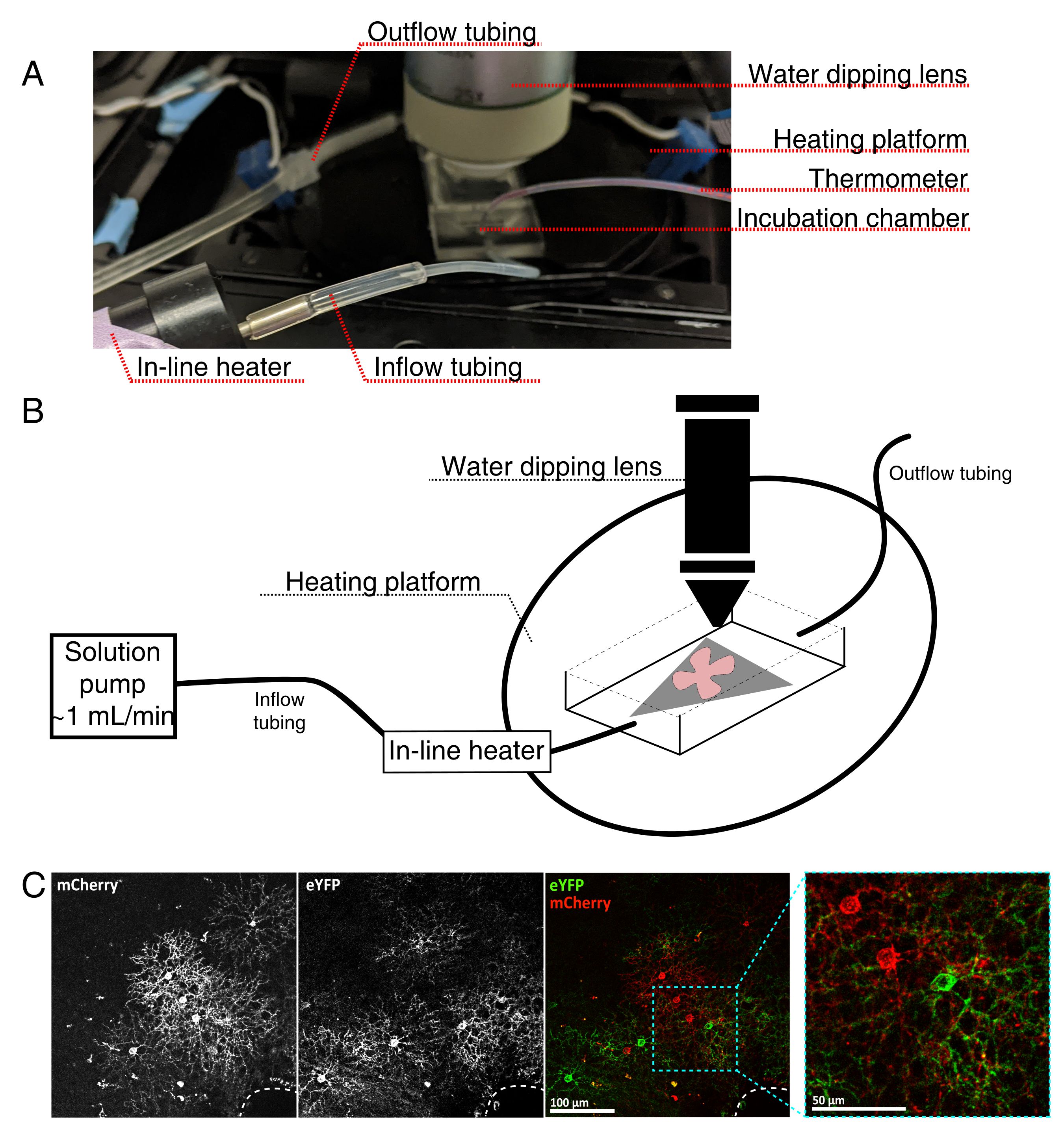
Figure 1: Live-cell imaging incubation chamber setup. (A) Live-imaging incubation apparatus sh...
Access restricted. Please log in or start a trial to view this content.
Materials
| Name | Company | Catalog Number | Comments |
|---|---|---|---|
| Mixed cellulose ester membrane (MCE) filter papers, hydrophilic, 0.45 µm pore size | Millipore | HABG01 300 | |
| Polyethylene disposable transfer pipette | VWR | 470225-034 | |
| Chamber polyethylene tubing, PE-160 10' | Warner Instruments | 64-0755 | |
| Dual channel heater controller, Model TC-344C | Warner Instruments | 64-2401 | |
| HC FLUOTAR L 25x/0.95 W VISIR dipping objective | Leica | 15506374 | |
| Heater controller cable | Warner Instruments | CC-28 | |
| Large bath incubation chamber with slice support | Warner Instruments | RC-27L | |
| MPII Mini-Peristaltic Pump | Harvard Apparatus | 70-2027 | |
| PM-6D Magnetic Heated Platform (incubation chamber heater) | Warner Instruments | PM-6D | |
| Pump Head Tubing Pieces For MPII Mini-Peristaltic Pump | Harvard Apparatus | 55-4148 | |
| Sample anchor (Harps) | Warner Instruments | 64-0260 | Sample anchor must be compatible with incubation chamber |
| Sloflo In-line Solution Heater | Warner Instruments | SF-28 | |
| Calcium chloride dihydrate | Sigma-Aldrich | C7902 | |
| Carbogen (5% CO₂, 95% O₂) | AirGas | X02OX95C2003102 | Supplier may vary depending on region |
| D-(+)-Glucose | Sigma-Aldrich | G7021 | |
| HEPES, Free Acid | Bio Basic | HB0264 | |
| Hydrochloric acid solution, 1 N | Sigma-Aldrich | H9892 | |
| Magnesium chloride hexahydrate | Sigma-Aldrich | M2670 | |
| pH-Test strips (6.0-7.7) | VWR | BDH35317.604 | |
| Potassium chloride (KCl) | Sigma-Aldrich | P9541 | |
| Sodium chloride (NaCl) | Bio Basic | DB0483 | |
| Sodium phosphate monobasic | Sigma-Aldrich | RDD007 | |
| ImageJ | National Institutes of Health (NIH) | Open source |
