In this video, cortical slice cultures from transgenic mouse embryos are used to monitor gene expression through Three-dimensional imaging. Bioluminescence, fluorescence, and bright-field images are used to visualize real-time gene expression during neuronal differentiation within the native tissue.
A subscription to JoVE is required to view this content. Sign in or start your free trial.
Method Article
Real-Time Gene Expression Monitoring in Brain Slices via Three-Dimensional Imaging
568 views
⸱
June 17th, 2025
In This Article
Abstract
Protocol
All procedures involving animal samples have been reviewed and approved by the appropriate animal ethical review committee.
- Visualization of luciferase reporter expression in slice cultures
- Start the luminescence live-imaging system before starting the dissection. Use the following conditions for cortical slice cultures: 40% O2, 5% CO2 and 37 °C.
- Put the immersion oil on the 40x objective lens. Put the sample dish on the microscope stage. Acquire the fluorescent test image and set the position and focus plane to the region of interest under the illumination of excitation light.
- Run the time-lapse acquisition by 3-dimensional (luminescence, fluorescence, and bright field) acquisitions for 24 h (Figure 1F-H). For luminescence image acquisition, use the following camera settings: low-transfer rate (50 kHz), 2x2/4x4 binning, 10 min/5 min exposure time. For the fluorescence and bright field image acquisition, use the following settings: intermediate-transfer rate (1 MHz), 1x1 binning, and 100 ms exposure time.
Access restricted. Please log in or start a trial to view this content.
Results
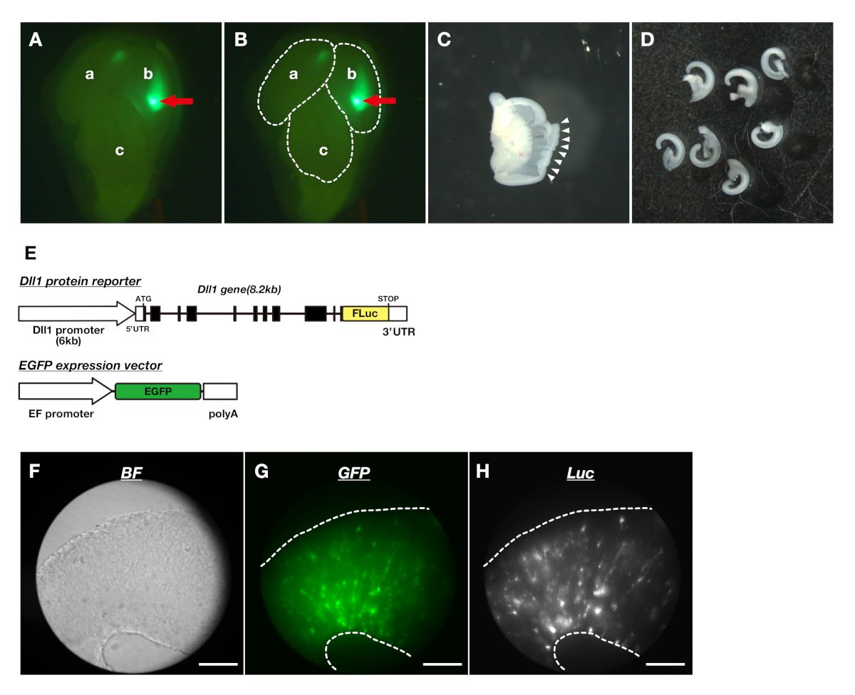
Figure 1: Making the cortical slices of the embryonic telencephalon and visualizing Dll1 protein dynami...
Access restricted. Please log in or start a trial to view this content.
Materials
| Name | Company | Catalog Number | Comments |
|---|---|---|---|
| Chilled water circulator (chiller) | Julabo | Model: F12-ED | |
| Cooled CCD camera | Andor Technology | Model: iKon-M 934 | |
| Incubator system | TOKAI HIT | Model: INU-ONICS | |
| Inverted microscope | Olympus | Model: IX81 | |
| Inverted microscope | Olympus | Model: IX83 | |
| LED illumination device | CoolLED | Model: pE1 | |
| MetaMorph | MOLECULAR DEVICES | Model: 40000 | |
| Mix gas controller | Tokken | Model: TK-MIGM OLO2 | |
| Objective lens | Olympus | Model: UPLFLN 40X O |
