$$\rightleftharpoonup{xx}$$
$$\longleftharp{xx}$$,
$$\longrightharp{xx}$$,
All procedures involving animal models have been reviewed by the local institutional animal care committee and the JoVE veterinary review board.
1. Blood Oxygen Level-Dependent Functional Magnetic Resonance Imaging (BOLD fMRI) Acquisition
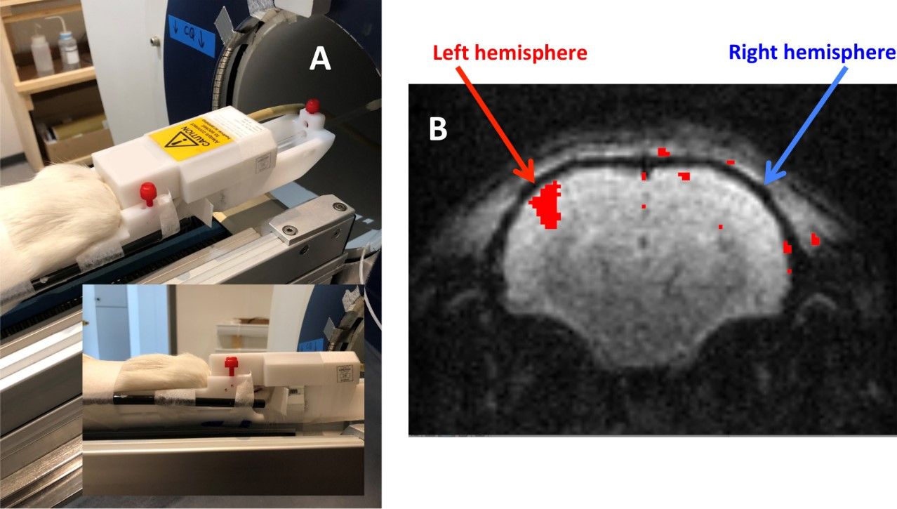
- Place the rat brain so that it is in an upright position and use the ear bars to maintain it. Place the volume array coil above the rat’s head (Figure 1A) and fix it using tape. Check that the sail is moving correctly (anteroposterior movement, no rotation, and no friction of sail) when the air-puff system is turned On; then, switch it Off.
- Insert the bed and the coil in the center of the magnet. Check that the sail is still moving correctly once the bed is inside the magnet when the air-puff system is On; then, switch it Off. Switch completely from isoflurane to medetomidine (perfusion rate: 20 µL/min).
- Check that the rat is well located using a localization sequence (echo time, TE = 2.5 ms; repetition time, TR = 100 ms; average = 1; repetition = 1; slice = 1 mm; image size = 256 x 256; field of view (FOV) = 50 x 50 mm; scan time = 12 s 800 ms). Drag the Localizer sequence tab into the Instruction name and click on Continue.
- Drag the T2_Star_FID_EPI (T2*-weighted free induction decay echo planar imaging) sequence tab into the Instruction name, center the FOV on the middle of the brain, and click on the Adjustment platform tab to open the edited scan instruction. Record a B0 map and proceed to a scan shim.
NOTE: For a magnetic field homogeneity (B0) map, use the following parameters: first echo time = 1.65 ms; TR = 20 ms, average = 1; flip angle = 30°; echo spacing = 3.805 ms; slice = 58 mm; image size = 64 x 64 x 64; FOV = 58 x 58 x 58 mm; scan time = 1 m 24 s 920 ms. For scan shim, use the following parameters: voxel selective excitation = stimulated echo acquisition mode (STEAM) Gaussian pulse; TE = 5 ms; mixing time = 10 ms; acquisition duration = 204.8 ms; bandwidth = 10,000 Hz; dwell time = 50 μs). - Start the T2 Star_FID_EPI sequence (TE = 16.096 ms; TR = 500 ms; average = 1; repetition = 600; slice = 1 mm; four consecutive slices; image size = 128 x 128; FOV = 20 x 20 mm; bandwidth = 333,333.3 Hz; scan time = 5 min 00 s).
NOTE: Due to the Transistor-transistor logic (TTL) port, an external trigger signal will start the air-puff system at the same time. The paradigm = [20 s activation + 10 s rest] x 10, for the total duration of the 600 scans, 500 ms per scan. The slices are centered in the middle of the barrel field area. - Acquire another localization sequence (same as the one described in step 5.3) to compare with the first one and check whether the rat has moved during the T2_Star_FID_EPI sequence.
- Bring the bed to its initial position, remove the volume array coil, and connect the surface coil.
2. BOLD Processing
- Open the T2 Star_FID_EPI file and read the T2 Star_FID_EPI image in Image Display. Open the start-up window of the functional controller, called FunController.
- In this Processing tab, select the functional imaging window and define the stimulation protocol (duration and alternation of On/Off periods corresponding to the paradigm used).
- Select the protocol window (dataset with 600 frames) and insert the value of 40 in the On Period tab and 20 in the Off Period tab. Click on the Invert Attribution tab and drag the Stimulation States slider to the left to select the value 1.
- In the preprocessing window, click on the Median filter in the plane for preprocessing and the Median filter (2D, 3D) for postprocessing.
- Click on the Execute tab and drag the cursors to adjust the overlay lookup table. Visualize the activated brain area (Figure 1B).
3. Proton Magnetic Resonance Spectroscopy (1H-MRS) Acquisitions
- To correctly position the surface coil, modify the position of the rat head. Rotate the head (approximately 30° clockwise) so that the surface coil (Figure 1A) can be placed just above the left barrel cortex while being horizontal and located at the magnet center when inside the magnet.
- Place the surface coil, fix it on the rat brain using tape (Figure 2B), and check that the sail is moving correctly (anteroposterior movement, no rotation, and no friction of the sail) when the air-puff system is turned On; then, switch it Off at the main switch.
- Check that the sail is moving correctly once the bed is inside the magnet when the air-puff system is On. Then, switch it Off.
- Check that the rat is positioned correctly using a localization sequence. Set parameters as follows: TE = 2.5 ms; TR = 100 ms; average = 1; repetition = 1; slice = 1 mm; image size = 256 x 256; FOV = 50 x 50 mm; scan time = 12 s 800 ms.
- Drag the Localizer sequence tab into the Instruction name window and click on the Continue tab to execute the scan program.
- When the brain localization is correct, drag the T2-weighted turbo rapid acquisition with relaxation enhancement (T2_TurboRARE) sequence tab in the Instruction name window and click on Continue to execute the scan program. These anatomical images, together with the previous BOLD fMRI acquisition, will allow the correct localization of the voxel in the primary somatosensory barrel field or S1BF, for MRS.
NOTE: The T2_TurboRARE parameters are 14 slices, 2 mm per slice, FOV = 2.5 x 2.5 cm, TE = 100 ms, TR = 5,000 ms, matrix = 128 x 128, sequence time = 2 min 40 s. - Drag the localization by adiabatic selective refocusing (LASER) sequence tab into the Instruction name window, and place the voxel (2 mm high, 2.5 mm long, 3 mm deep) at the center of the S1BF area.
- Use a rat brain atlas and the BOLD fMRI enhancement to localize the zone on the T2 images (Figure 3). Click on the Adjustment platform tab to open the edited scan instructions. Click on the Wobble tab and change the impedance (electronic loading) of the receive coil slightly to tune it. Click on the Apply tab when the tuning is finished to close the instruction editor and apply the changes in the edited instruction.
- Record a B0 map and proceed to scan the shim and then perform a local shim.
NOTE: For the B0 map, use the following parameters: first echo time = 1.65 ms; TR = 20 ms, average = 1; flip angle = 30°; echo spacing = 3.805 ms; slice = 58 mm; image size = 64 x 64 x 64; FOV = 58 x 58 x 58 mm; scan time = 1 m 24 s 920 ms. For the scan shim, use the following parameters: voxel selective excitation, STEAM Gaussian pulse; TE = 5 ms; mixing time = 10 ms; acquisition duration = 204.8 ms; bandwidth = 10,000 Hz; dwell time = 50 μs. For the local shim, use the following parameters: water suppression, variable power, and optimized relaxation (VAPOR) acquisition duration = 1,363.15 ms; points = 4,096; bandwidth in Hz = 3,004.81 Hz; bandwidth in ppm (parts per million) = 10 ppm; dwell time = 166.40 μs; spectral resolution = 0.37 Hz/points. The LASER parameters are: echo time = 19.26 ms; TR = 2,500 ms; averages = 128 or 32; scan time = 5 min 20 s or 1 min 20 s; acquisition points = 4,096. - Perform 1H-MRS.
- Start the 1H-MRS acquisition first during a resting period (4 x 32 LASER scans + 128 LASER scans; 2,500 ms per scan).
- Acquire another localization sequence (same as the one described in step 5.3) to compare with the first one recorded and ensure that the rat has not moved during the LASER acquisition.
- Perform 1H-MRS during whisker activation using the LASER sequence (4 x 32 LASER scans + 128 LASER scans; 2,500 ms per scan) with the air-puff system On (paradigm = 20 s of activation and 10 s of rest).
- Once again, perform a localization sequence to check whether the rat has moved.
NOTE: The number of scans and resting/activated periods can be adapted and modified, but always ensure that the rat is not moving by regularly performing a localization sequence.