Protein complexes catalyze key cellular functions. Detailed functional and structural characterization of many essential complexes requires recombinant production. MultiBac is a baculovirus/insect cell system particularly tailored for expressing eukaryotic proteins and their complexes. MultiBac was implemented as an open-access platform, and standard operating procedures developed to maximize its utility.
Method Article
The MultiBac Protein Complex Production Platform at the EMBL
In This Article
Summary
Abstract
Proteomics research revealed the impressive complexity of eukaryotic proteomes in unprecedented detail. It is now a commonly accepted notion that proteins in cells mostly exist not as isolated entities but exert their biological activity in association with many other proteins, in humans ten or more, forming assembly lines in the cell for most if not all vital functions.1,2 Knowledge of the function and architecture of these multiprotein assemblies requires their provision in superior quality and sufficient quantity for detailed analysis. The paucity of many protein complexes in cells, in particular in eukaryotes, prohibits their extraction from native sources, and necessitates recombinant production. The baculovirus expression vector system (BEVS) has proven to be particularly useful for producing eukaryotic proteins, the activity of which often relies on post-translational processing that other commonly used expression systems often cannot support.3 BEVS use a recombinant baculovirus into which the gene of interest was inserted to infect insect cell cultures which in turn produce the protein of choice. MultiBac is a BEVS that has been particularly tailored for the production of eukaryotic protein complexes that contain many subunits.4 A vital prerequisite for efficient production of proteins and their complexes are robust protocols for all steps involved in an expression experiment that ideally can be implemented as standard operating procedures (SOPs) and followed also by non-specialist users with comparative ease. The MultiBac platform at the European Molecular Biology Laboratory (EMBL) uses SOPs for all steps involved in a multiprotein complex expression experiment, starting from insertion of the genes into an engineered baculoviral genome optimized for heterologous protein production properties to small-scale analysis of the protein specimens produced.5-8 The platform is installed in an open-access mode at EMBL Grenoble and has supported many scientists from academia and industry to accelerate protein complex research projects.
Introduction
Biological activity is controlled by assemblies of proteins and other biomolecules that act in concert to catalyze cellular functions. Notable examples include the machinery that transcribes the hereditary information contained in DNA into messenger RNA. In humans, more than 100 proteins come together in a defined and regulated process to transcribe genes, forming large multiprotein complexes with 10 and more subunits including RNA polymerase II and the general transcription factors such as TFIID, TFIIH and others.9 Other examples are the ribosome, consisting of many proteins and RNA molecules, that catalyzes protein synthesis, or the nuclear pore complex which is responsible for shuttling biomolecules through the nuclear envelope in eukaryotes. A detailed architectural and biochemical dissection of essentially all multicomponent machines in the cell is vital to understand their function. The structure elucidation of prokaryotic and eukaryotic ribosomes, for instance, constituted hallmark events yielding unprecedented insight into how these macromolecular machines carry out their designated functions in the cell.10,11
Ribosomes can be obtained in sufficient quality and quantity for detailed study by purifying the endogenous material from cultured cells, due to the fact that up to 30% of the cellular mass consists of ribosomes. RNA polymerase II is already less abundant by orders of magnitude, and many thousand liters of yeast culture had to be processed to obtain a detailed atomic view of this essential complex central to transcription.12 The overwhelming majority of the other essential complexes are however present in much lower amounts in native cells, and thus cannot be purified adequately from native source material. To render such complexes accessible to detailed structural and functional analysis requires heterologous production by using recombinant techniques.
Recombinant protein production had a major impact on life science research. Many proteins were produced recombinantly, and their structure and function dissected at high resolution. Structural genomics programs have taken advantage of the elucidation of the genomes of many organisms to address the gene product repertoire of entire organisms in high-throughput (HT) mode. Thousands of protein structures have thus been determined. To date, the most prolifically used system for recombinant protein production has been E. coli, and many expression systems have been developed and refined over the years for heterologous production in this host. The plasmids harboring a plethora of functionalities to enable protein production in E. coli fill entire catalogues of commercial providers.
However, E. coli has certain limitations which make it unsuitable to produce many eukaryotic proteins and in particular protein complexes with many subunits. Therefore, protein production in eukaryotic hosts has become increasingly the method of choice in recent years. A particularly well-suited system to produce eukaryotic proteins is the baculovirus expression vector system (BEVS) that relies on a recombinant baculovirus carrying the heterologous genes to infect insect cell cultures cultivated in the laboratory. The MultiBac system is a more recently developed BEVS which is particularly tailored for the production of eukaryotic protein complexes with many subunits (Figure 1). MultiBac was first introduced in 2004.13 Since its introduction, MultiBac has been continuously refined and stream-lined to simplify handling, improve target protein quality and generally making the system accessible to non-specialist users by designing efficient standard operating procedures (SOPs).4 MultiBac has been implemented in many laboratories world-wide, in academia and industry. At the EMBL in Grenoble, transnational access programs were put in place by the European Commission to provide expert training at the MultiBac platform for scientists who wished to use this production system for advancing their research. The structure and function of many protein complexes that were hitherto not accessible was elucidated by using samples produced with MultiBac.4 In the following, the essential steps of MultiBac production are summarized in protocols as they are in operation at the MultiBac facility at EMBL Grenoble.
Protocol
1. Tandem Recombineering (TR) for Creating Multigene Expression Constructs
- Planning the co-expression strategy. Design approach for inserting your genes of interest into Donors and Acceptors. Potential physiological submodules of your complex should be grouped together on specific Acceptors and Donors. Use Multiplication Module consisting of Homing endonuclease (HE) - BstXI pairs to combine expression cassettes on individual Donor and Acceptor plasmids.7,8 Create all relevant constructs in silico and validate strategy thoroughly before proceeding to experimental work. For example, genes of interest should be checked not to contain HE or other restriction sites and the presence of correct open reading frames (ORFs) should be validated. Consider ordering synthetic genes optimized for insect cell codon usage and mRNA secondary structure to improve protein production levels as well as removal of any existing HE sites from the genes of interest. Consider placing purification tags based on data from the literature about flexible or exposed N- or C-termini of your proteins of choice. Consider applying polyprotein strategies that aim at producing several protein subunits in your complex if the relative amounts of individual proteins need to be controlled due to stoichiometry issues in the complex.4 Prepare detailed "How-To" document (electronic lab book is recommended) containing all projected experimental steps of the project leading up to the complete multigene construct(s). Create electronic files of the Cre-LoxP fused plasmids for example by using the Cre-ACEMBLER software which can be downloaded from the Berger group home page (www.embl.fr/multibac/ multiexpression_technologies/cre-acembler).
- Insert your genes of interest into selected Donors and Acceptors by using restriction enzymes and ligase, or, alternatively, by using ligation independent methods following published protocols.5,6,14 If you have access to a liquid handling work-station and if you plan a large number of constructs to be generated (for example for combinatorial approaches) consider using robotics scripts developed and implemented by the Berger group (Figure 2).14,15 If a liquid handling work-station is not available, manual operation using microtitre plates allows gene insertion in a HT like fashion.
- Constraints imposed by the need to control the stoichiometry of the expressed subunits may materialize. In the case of stoichiometrically imbalanced expression levels of individual subunits of a protein complex, consider applying polyprotein strategies to conjoin several subunits of your complex and a specific protease (for example tobacco etch virus NIa protease) in single large ORFs spaced by specific proteolytic sites.4,8 Consider co-expressing one or several polyproteins with single expression cassettes if you have a very large complex with many subunits and widely ranging molecular weights of individual subunits. Consider co-expressing several genes encoding for the same protein in a polyprotein or as several identical expression cassettes if that protein is characterized by low production yield.4,13
- Validate all Donor and Acceptor constructs cloned by restriction mapping (optionally in high-throughput) and sequencing. Proceed to fuse Donor-Acceptor combinations by Cre-LoxP recombination to generate the multigene expression constructs of choice. Validate purified Acceptor-Donor fusion plasmids by restriction mapping, use electronic sequences created by Cre-ACEMBLER or similar programs as a reference.
- Store purified and validated Donors, Acceptors and Donor-Acceptor fusions at -20 °C or -80 °C. Archive plasmids and their sequences (Microsoft Excel, Filemaker, others) carefully for later usage.
2. Composite Multigene Baculovirus Generation, Amplification and Storage
- Integrate multigene transfer vectors the MultiBac baculoviral genome by transforming into DH10 cells harboring the viral genome and the functionalities required for Tn7 transposition. Note that the Multibac baculoviral genome can be preloaded with particular genes of interest (YFP marker, chaperones, etc.) in its own LoxP site (engineered into the genome distal to the Tn7 attachment site) by an in vivo Cre reaction preceding Tn7 integration.13 After Tn7 transposition, cells with composite baculovirus containing the genes of interest are selected by blue/white screening (successful Tn7 transposition results in loss of α-complementation of the β-galactosidase; therefore, colonies with correct Tn7 transposition remain white on selective agar plates containing X-gal) and the genome is prepared by alkaline lysis and ethanol/isopropanol precipitation.5,6
- Transfection and initial virus production. Place 6-well tissue culture plate into sterile hood. From log-phase Sf21 insect cell culture, seed out aliquots of cells in the wells and transfect by adding the purified baculoviral genome and a transfection reagent mixed in culture media as described.6 Harvest initial virus after 48-60 hr by removing the media (high quality, low titer virus V0, typically 3 ml per well). Supplement fresh media, and test for protein production (and, if a YFP marker is present, for fluorescence) after an additional 2-3 days.6,7
- Amplification of virus, low MOI regimen. Use V0 virus to infect 25-50 ml of cells in log phase (cell density <1x10⁶ cells per ml) in small (100-250 ml) Erlenmeyer shaker flasks agitated on orbital platform shakers (Figure 3). Count cells and split every 24 hr until cells stop doubling (proliferation arrest). Follow a low MOI (multiplicity of infection i.e. number of virus particles per cell) regimen: Cells must double (at least) once (MOI < 1), otherwise repeat experiment with a smaller volume of V0 added. Normally, 3 ml of V0 are used to infect 25 ml of Sf21 insect cells at a density of 0.5x10⁶ cells / ml. This is essential to prevent detrimental over-amplification and auto-deletion of virus that can result in loss of the heterologous genes of interest. Harvest V1 virus (25-50 ml) after 48-60 hr by pelleting cells and removing the media containing the virus. Supplement with fresh media and test for YFP and protein production by removing 1x10⁶ cells every 12 or 24 hr, pelleting and validating protein production and marker protein (YFP) signal.6, Amplify virus (to V2) further if larger expression volumes are aimed at by infecting up to 400 ml cells in 2 L shaker flasks with V1 virus respecting the above low MOI regimen (cells must double at least once after infection with V1). Stringently test protein production and marker protein signal during amplification to avoid accumulation of defective viruses no longer containing your genes of interest.5-7 Use cell pellets accumulating at each amplifications step already for establishing purification protocols for the expressed protein complex of interest.
- BIIC storage of production virus. We strongly recommend to store V2 virus as the production virus by using the BIIC (Baculovirus-infected insect cells) method, to prevent modifications (e.g. loss of the gene of interest) of the recombinant virus and to preserve high expression levels.16 Pellet infected cells 24 hr after proliferation arrest is observed - at this stage cells contain complete viral particles just before they would be released (by budding) into the media. Remove media and freeze aliquots of the cell pellet in liquid nitrogen and store indefinitely.7,16
3. Protein Production and Downstream Processing
- Infecting large(r) cultures and monitoring YFP. Use V1, V2 or frozen BIIC aliquots to infect larger cell cultures for production runs (typically 400 ml in 2 L flasks). Adhere to low MOI regimen (adjust virus volume used for infection such that infected culture doubles at least once). Enlarge infected culture volumes if needed by multiplying number of flasks. If YFP marker protein is present, withdraw at defined intervals 1x10⁶ cells, pellet and lyse cells and monitor evolution of the YFP signal until a plateau is reached indicating maximum recombinant protein production. YFP levels can be measured in a standard 96 well plate reader capable of recording fluorescence signals (e.g. Tecan SPECTRAFluor). Harvest cells at this stage. Store cell pellets at -20 °C (short term) or -80 °C (long term).
- Cell lysis and fractionation. Lyse cells by your favorite method of choice, tailored to the requirements of your protein (freeze-thaw, sonication, French press, others).5-7 Fractionate cytosol and nuclei and test for the presence of your proteins of interest. Develop purification protocols based on the results to simplify protein purification. Consider applying soaking procedures to extract your protein from the nuclear fraction under high KCl conditions if your proteins reside in the nucleus.7,18
- Protein purification (micro-scale, large scale). Note that often small volumes (10 or 25 ml) of cell culture are sufficient for obtaining cell pellets for purifying substantial amounts of your proteins of interest due to the typically high or very high production levels of heterologous proteins in the baculovirus/insect cell systems (often 10-100 mg of protein per L culture and more). In conjunction with micro-purification (multiwell plates, microtip methods, GE Healthcare ÄKTAmicro system, others) it is possible to obtain biochemical and activity data and often also sufficient amount of the desired proteins and complexes for nanoliter-scale high-throughput crystallization (HTX). Consider using metal affinity purification (Clonetech Takara TALON, Qiagen NiNTA metal chelator resins) and an oligo-histidine (6-10 residues) tag on exposed subunits of your protein complex to facilitate purification, in conjunction with ion exchange and size exclusion chromatography in small volumes using for example the ÄKTAmicro or a similar small volume purification machine (Figure 4). Other affinity purification steps and ion exchange (IEX) steps in addition to the affinity purification with oligo-histidine tags or other tags (choose from GST, MBP, CBP, others) can and should be considered, according to the biochemical properties of the proteins of interest and individual preferences. Consider tagging more than one subunit with affinity tags to enhance purification efficiency. Concentrate your purified protein complexes and establish storage protocols such as freezing with or without glycerol. Develop quality control criteria that can be applied standardly (activity assays, biochemical and biophysical tests) to assess batch-to-batch variation of your purified proteins.
Results
Strong co-expression of heterologous proteins achieved by the MultiBac system is shown in Figure 1d (probes taken 48 hr after infecting a suspension cell culture). The overexpressed protein bands are clearly discernible in the whole cell extract (SNP) and the cleared lysate (SN). The quality and quantity of the protein material produced is often sufficient to enable structure determination of protein complexes, such as the mitotic checkpoint complex MCC shown in Figure 1e.17
Figure 2 displays the work-flow of a gene assembly experiment by robot-assisted tandem recombineering (TR). Robust DNA assembly protocols were scripted into robotics routines for parallelized assembly of multigene expression constructs. Individual robotic steps are shown in snap-shots (Figure 2c, I - IV). The DNA components to be assembled are generated by PCR and quality controlled by e-gels (Figure 2d, left); the assembled multigene constructs are likewise validated by PCR with specifically designed sets of primers (Figure 2d, right).14,15
Recombinant baculovirus generation and amplification follows standard operating procedures (schematically displayed in Figure 3a). Snapshots of cells cultured in a monolayer are shown (Figure 3b, I. & II.) following infection with a MultiBac virus.
Downstream processing of the recombinant protein complexes can be miniaturized by using multi-well-plate or microtip-based chromatography for affinity purification followed by size exclusion chromatography (SEC) of the complexes by integrating into the work-flow small-scale systems such as the ÄKTAmicro (Figure 4a). A representative SEC profile of a ~700 kDa human transcription factor complex is shown. Sample purified by using the ÄKTAmicro in small-scale is typically sufficient for characterization by biochemical and biophysical means including electron microscopy (Figure 4b).
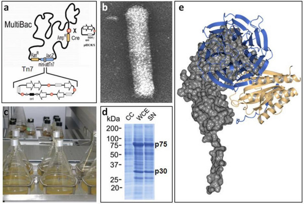
Figure 1. MultiBac platform technology for multiprotein complex production. (a) Genes of interest are integrated into the Multibac baculoviral genome by using Tn7 transposition in conjunction with blue/white screening. A LoxP site on the virus backbone allows addition of further functionalities such as a fluorescent marker protein to monitor virus performance and heterologous protein production. (b) The baculovirus adopts an elongated stick shape and is characterized by a flexible envelope that can augment to accommodate the large (>130 kb) circular double-stranded DNA genome. Large heterologous gene insertions in the genome are tolerated by elongating the envelope. (c) Standard Erlenmeyer flasks on orbital shaker platforms can be used for growing large scale insect cell cultures for heterologues protein production. (d) The MultiBac system efficiently produces recombinant proteins which are often clearly visible already in the whole-cell extract (SNP). (e) The structure of the mitotic checkpoint complex was elucidated by X-ray diffraction from crystals grown from sample produced with the MultiBac system.17 Click here to view larger figure.
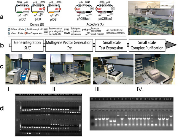
Figure 2. (Automated) Tandem Recombineering (TR). (a) Tandem recombineering utilizes small arrays of synthetic plasmid DNA molecules called Donors and Acceptors for assembling multigene expression constructs, optionally in robot-assisted mode using a liquid handling work-station (right). (b) The TR workflow is shown. SLIC stands for sequence and ligation independent cloning, Cre stands for the Cre-LoxP fusion concatenating Donors and Acceptors into which genes of interest have been inserted by SLIC. Multigene expression constructs generated by this recombineering procedure using SLIC and Cre in tandem are then integrated into the MultiBac baculovirus genome and used for small and large scale expression in insect cell cultures infected by the recombinant virus. (c) Snapshots of the robot-assisted TR process are shown including provision of template DNA and primers (I), preparation of PCR reactions in multiwell plates (II), PCR amplification of DNAs (III) and preparation of multigene constructs grown in bacterial culture by alkaline lysis in multi-well plates (IV). (d) PCR products used for TR are visualized by using the e-gel system (left). Completed multigene constructs are validated by analytical PCR reactions loaded on an e-gel (right). Click here to view larger figure.
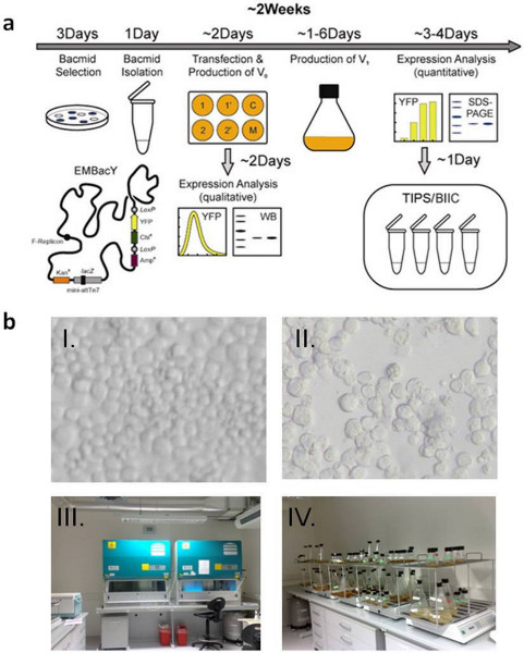
Figure 3. MultiBac virus generation, amplification, storage. (a) The standard operating procedure (SOP) of the MultiBac platform at the EMBL Grenoble is shown in a schematic view. Recombinant MultiBac virus is identified by blue-white screening and prepared from bacterial cultures. Initial transfection takes place on 6-well plates seeded with monolayers of insect cells (Sf21, Sf9, Hi5, others). Virus is amplified and target protein produced in Erlenmeyer shakers. Virus is stored by freezing aliquots of infected insect cells (BIIC). Florescence is recorded as an analytical tool if YFP (or another fluorescent protein) has been integrated as a marker protein for example into the loxP site present on the MultiBac baculoviral genome. (b) Snapshots of insect cells infected with MultiBac baculovirus are shown. The cells stop proliferating, increase in size (I.). Cell fusions are observed (II). Virus budded off the infected cells into the media is collected and used to infect larger cell cultures for protein complex production (III, IV). Click here to view larger figure.
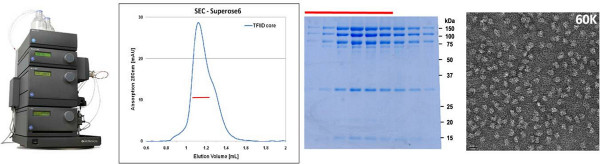
Figure 4. Protein complex production and down-stream processing. Protein complex sample can be already conveniently purified from small-scale initial cell cultures by utilizing miniaturized purification methods such as multiwall or microtip-based purification, optionally in conjunction with small-volume chromatography systems (left). Often the yield of already these "analytical" purification runs (middle) is sufficient for analyzing the structure and function of the purified complex by a variety of means including electron microscopy (right). Click here to view larger figure.
Discussion
Video snap-shots in Figures 2 and 3 illustrate the entire process from robot-assisted generation from cDNA of multigene expression constructs all the way to infection of insect cell cultures for protein production. New reagents (plasmids and virus) and robust protocols have been developed to enable a pipeline relying on SOPs. The entire pipeline has been implemented as a platform technology at the EMBL in Grenoble. The MultiBac platform has been accessed by many scientists from academia and industry who are engaged in multiprotein research. The training access is supported by dedicated access programs funded by the European Commission (P-CUBE, BioSTRUCT-X).
The availability of SOPs to carry out protein complex expression by using the MultiBac system has rendered this technology easily amenable also to non-specialist users. Robot-assisted operation accelerates multiprotein complex production in particular when a sufficiently large number of complexes, for example variants and mutants of a specimen of interest, need to be generated in parallel. However, manual operation at low- to medium throughput also greatly benefits from the availability of SOPs. In our experience, processes that could be successfully scripted into robotics routines needed to be refined first with significant effort until sufficiently robust protocols were obtained that are compatible with using a robot. Such protocols form the basis of our SOPs.5,6,14,15 Indeed, the implementation of these robust protocols for the robot lead to a very considerable efficiency gain also of manual operations in our laboratory.
Many proteins and protein complexes have been and are being produced by using the MultiBac system that we developed, and close to 500 laboratories world-wide have obtained the reagents. MultiBac has catalyzed research not only in structural biology but also in many other areas of life sciences that investigate or exploit the interactions between proteins in large assemblies. MultiBac has also been used to produce protein targets of considerable pharmacological interest including virus-like particles, which may become useful vaccine candidates.4 More recently, MultiBac has also been used to deliver genes into mammalian cells and cell cultures, or even entire organisms by gene therapy.4 We anticipate that approaches such as those illustrated in this contribution will prove to be useful for many areas of research involving multiprotein assemblies and complex interplay of biological macromolecules that form the basis of cellular processes in health and disease.
Disclosures
IB is inventor on patents and patent applications detailing parts of the technology here described.
Acknowledgements
We thank Christoph Bieniossek, Simon Trowitzsch, Daniel Fitzgerald, Yuichiro Takagi, Christiane Schaffitzel, Yvonne Hunziker, Timothy Richmond and all past and present members of the Berger laboratory for help and advice. The MultiBac platform and its development have been and are generously supported by funding agencies including the Swiss National Science Foundation (SNSF), the Agence National de Recherche (ANR) and the Centre National de Recherche Scientifique (CNRS) and the European Commission (EC) in Framework programs (FP) 6 and 7. Support for transnational access is provided by the EC FP7 projects P-CUBE (www.p-cube.eu) and BioStruct-X (www.biostruct-x.eu). The French Ministry of Science is particularly acknowledged for supporting the MultiBac platform at the EMBL through the Investissement d'Avenir project FRISBI.
Materials
| Name | Company | Catalog Number | Comments |
|---|---|---|---|
| Bluo-Gal | Invitrogen | 15519-028 (1 g) | |
| Tetracycline | Euromedex | UT2965-B (25 g) | 1,000X at 10 mg/ml |
| Kanamycine | Euromedex | EU0420 (25 g) | 1,000X at 50 mg/ml |
| Gentamycine | SIGMA | G3632 (5 g) | 1,000X at 10 mg/ml |
| IPTG | Euromedex | EU0008-B (5 g) | 1,000X at 1M |
| Cre-recombinase | New England BioLabs | M0298 | |
| X-Treme GENE HP transfection reagent | Roche | 06 366 236 001 | |
| Hyclone SFM4 Insect | Thermo Scientific | SH 30913.02 | |
| 6-well plate Falcon | Dominique Dutscher | 353046 | |
| 2 ml pipette Falcon | Dominique Dutscher | 357507 | |
| 5 ml pipette Falcon | Dominique Dutscher | 357543 | |
| 10 ml pipette Falcon | Dominique Dutscher | 357551 | |
| 25 ml pipette Falcon | Dominique Dutscher | 357535 | |
| 50 ml pipette Falcon | Dominique Dutscher | 357550 | |
| 50 ml tube Falcon | Dominique Dutscher | 352070 | |
| 15 ml tube Falcon | Dominique Dutscher | 352096 | |
| 1.8 ml cryotube Nunc | Dominique Dutscher | 55005 | |
| 100 ml shaker flasks Pyrex | Dominique Dutscher | 211917 | |
| 250 ml shaker flasks Pyrex | Dominique Dutscher | 211918 | |
| 500 ml shaker flasks Pyrex | Dominique Dutscher | 211919 | |
| 2 L shaker flasks Pyrex | Dominique Dutscher | 211921 | |
| Certomat Orbital Shaker + plateau | Sartorius | 4445110, 4445233 | |
| Liquid nitrogen tank dewar 35 L | Fisher Scientific | M76801 | |
| Biological Safety Cabinet Faster | Sodipro | FASV20000606 | |
| Optical Microscope | Zeiss | 451207 | |
| Sf21 Insect cells | |||
| Hi5 Insect cells | Invitrogen | B855-02 | |
| Tecan freedom EVO running Evoware plus | TECAN | ||
| 10 μl conductive tips (black), | TECAN | 10 612 516 | |
| 200 μl conductive tips (black) | TECAN | 10 612 510 | |
| disposable trough for reagents, 100 ml | TECAN | 10 613 049 | |
| twin.tec PCR plate 96, skirted | Eppendorf | 0030 128.648 | |
| 96 well V bottom, non sterile | BD falcon | 353263 | |
| 96 deepwell plate color natural, PP) | Fisher | M3752M | |
| PS microplate, 96 well flat bottom | Greiner | 655101 | |
| 96 deepwell plate | Thermo scientific | AB-0932 | |
| 24 well blocks RB | Qiagen | 19583 | |
| DpnI restriction enzyme | NEB | R0176L | 20 U/uL |
| NEBuffer 4 10X | NEB | B7004S | |
| 2X phusion mastermix HF | Finnzyme | ref F-531L | |
| 2X phusion mastermix GC | Finnzyme | ref F-532L | |
| DGLB 1.5X | homemade | 7.5% glycerol, 0.031% Bromophenol blue, 0.031% Xylen cyanol FF | |
| High DNA Mass Ladder for e-gel | Life Technologies | 10496-016 | |
| Low DNA Mass Ladder for e-gel | Life Technologies | 10068-013 | |
| E-gel 48 1% agarose GP | Life Technologies | G8008-01 | |
| Nucleo Spin- robot-96 plasmid kit | Macherey Nagel | 740 708.24 | |
| PCR clean-up kit, Nucleospin Robot-96 Extract | Macherey Nagel | 740 707.2 | |
| Gotaq green master mix | Promega | M7113 | |
| T4 DNA polymerase, LIC-qualified | Novagen | 70099-3 | |
| DTT 100 mM | homemade | ||
| Urea 2 M | homemade | ||
| EDTA 500 mM pH 8.0 | Homemade | ||
| LB broth (Miller) 500 g | Athena ES | 103 |
References
- Nie, Y., Viola, C., Bieniossek, C., Trowitzsch, S., Vijay-Achandran, L. S., Chaillet, M., Garzoni, F., Berger, I. Getting a Grip on Complexes. Curr. Genomics. 10 (8), 558-572 (2009).
- Robinson, C. V., Sali, A., Baumeister, W. The molecular sociology of the cell. Nature. 450 (7172), 973-982 (2007).
- Kost, T. A., Condreay, J. P., Jarvis, D. L. Baculovirus as versatile vectors for protein expression in insect and mammalian cells. Nat. Biotechnol. 23 (5), 567-575 (2005).
- Bieniossek, C., Imasaki, T., Takagi, Y., Berger, I. MultiBac: expanding the research toolbox for multiprotein complexes. Trends Biochem. Sci. 37 (2), 49-57 (2012).
- Fitzgerald, D. J., Berger, P., Schaffitzel, C., Yamada, K., Richmond, T. J., Berger, I. Protein complex expression by using multigene baculoviral vectors. Nat. Methods. 3 (12), 1021-1032 (2006).
- Bieniossek, C., Richmond, T. J., Berger, I. MultiBac: multigene baculovirus-based eukaryotic protein complex production. Curr. Protoc. Protein Sci. Chapter 5, Unit 5.20(2008).
- Trowitzsch, S., Bieniossek, C., Nie, Y., Garzoni, F., Berger, I. New baculovirus expression tools for recombinant protein complex production. J. Struct. Biol. 172 (1), 45-54 (2010).
- Vijayachandran, L. S., Viola, C., Garzoni, F., Trowitzsch, S., Bieniossek, C., Chaillet, M., Schaffitzel, C., Busso, D., Romier, C., Poterszman, A., Richmond, T. J., Berger, I. Robots, pipelines, polyproteins: enabling multiprotein expression in prokaryotic and eukaryotic cells. J. Struct. Biol. 175 (2), 198-208 (2011).
- Thomas, M. C., Chiang, C. M. The general transcription machinery and general cofactors. Crit. Rev. Biochem. Mol. Biol. 41 (3), 105-178 (2006).
- Klinge, S., Voigts-Hoffmann, F., Leibundgut, M., Ban, N. Atomic structures of the eukaryotic ribosome. Trends Biochem. Sci. 37 (5), 189-198 (2012).
- Melnikov, S., Ben-Shem, A., Garreau de Loubresse, N., Jenner, L., Yusupova, G., Yusupov, M. One core, two shells: bacterial and eukaryotic ribosomes. Nat. Struct. Mol. Biol. 19 (6), 560-567 (2012).
- Cramer, P., Bushnell, D. A., Fu, J., Gnatt, A. L., Maier-Davis, B., Thompson, N. E., Burgess, R. R., Edwards, A. M., David, P. R., Kornberg, R. D. Architecture of RNA polymerase II and implications for the transcription mechanism. Science. 288 (5466), 640-649 (2000).
- Berger, I., Fitzgerald, D. J., Richmond, T. J. Baculovirus expression system for heterologous multiprotein complexes. Nat. Biotechnol. 22 (12), 1583-1587 (2004).
- Bieniossek, C., Nie, Y., Frey, D., Olieric, N., Schaffitzel, C., Collinson, I., Romier, C., Berger, P., Richmond, T. J., Steinmetz, M. O., Berger, I. Automated unrestricted multigene recombineering for multiprotein complex production. Nat. Methods. 6 (6), 447-450 (2009).
- Nie, Y., Bieniossek, C., Frey, D., Olieric, N., Schaffitzel, C., Steinmetz, M. O., Berger, I. ACEMBLing multigene expression constructs by recombineering. Nat. Protocols. , (2009).
- Wasilko, D. J., Lee, S. E., Stutzman-Engwall, K. J., Reitz, B. A., Emmons, T. L., Mathis, K. J., Bienkowski, M. J., Tomasselli, A. G., Fischer, H.D. titerless infected-cells preservation and scale-up (TIPS) method for large-scale production of NO-sensitive human soluble guanylate cyclase (sGC) from insect cells infected with recombinant baculovirus. Protein Expr. Purif. 65 (2), 122-132 (2009).
- Chao, W. C., Kulkarni, K., Zhang, Z., Kong, E. H., Barford, Structure of the mitotic checkpoint complex. Nature. 484 (7393), 208-213 (2012).
- Yamada, K., Frouws, T. D., Angst, B., Fitzgerald, D. J., DeLuca, C., Schimmele, K., Sargent, D. F., Richmond, T. J. Structure and mechanism of the chromatin remodelling factor ISW1a. Nature. 472 (7344), 448-453 (2011).
Reprints and Permissions
