The giant ciliate Stentor coeruleus is a classical system for studying regeneration and wound healing in single cells. By imaging Stentor cells simultaneously at low and high magnification it is possible to measure cytoplasmic flows before, during, and after wounding.
Method Article
Visualizing Cytoplasmic Flow During Single-cell Wound Healing in Stentor coeruleus
In This Article
Summary
Abstract
Although wound-healing is often addressed at the level of whole tissues, in many cases individual cells are able to heal wounds within themselves, repairing broken cell membrane before the cellular contents leak out. The giant unicellular organism Stentor coeruleus, in which cells can be more than one millimeter in size, have been a classical model organism for studying wound healing in single cells. Stentor cells can be cut in half without loss of viability, and can even be cut and grafted together. But this high tolerance to cutting raises the question of why the cytoplasm does not simply flow out from the size of the cut. Here we present a method for cutting Stentor cells while simultaneously imaging the movement of cytoplasm in the vicinity of the cut at high spatial and temporal resolution. The key to our method is to use a "double decker" microscope configuration in which the surgery is performed under a dissecting microscope focused on a chamber that is simultaneously viewed from below at high resolution using an inverted microscope with a high NA lens. This setup allows a high level of control over the surgical procedure while still permitting high resolution tracking of cytoplasm.
Introduction
Regeneration and wound healing are important biological processes whose mechanisms remain areas of active investigation. Indeed, the ability to heal wounds and regenerate damaged parts is one of the features that sets living things apart from inanimate objects. Normally, regeneration is thought to be mediated by differentiation of stem cells into various cell types needed to rebuild a damaged or severed animal part, while wound healing is viewed in terms of new cells invading a wound to rebuild an intact epithelium. Thus both processes appear to be mediated by collectives of individual cells, and seem therefore to be quintessentially multicellular processes. It is therefore potentially surprising that individual cells are, in some cases, able to repair wounds and regenerate lost components1. Wound healing within single cells can involve multiple pathways including actin-myosin purse string contraction around the wound and membrane patching by rapid fusion of exocytotic vesicles1-3. Although many cell types are capable of some degree of wound healing4-6, perhaps the most dramatic example is the giant ciliate Stentor coeruelus7, shown in Figure 1. Individual Stentor cells are 1 mm long, and are covered with rows of cilia. Stentor cells are cone-shaped with an oral apparatus, consisting of thousands of cilia, at one end, and a narrow hold-fast structure at the other end. Stentor was a classical model organism for studying regeneration because if any part of the cell was cut off, it could regenerate7. For example, in a classic series of experiments, Thomas Hunt Morgan8 showed that a Stentor cell could be cut in half and each half would regenerate a normally proportioned cell in a matter of hours8. The molecular pathways responsible for regeneration in Stentor remain completely unknown.
The ability of Stentor cells to serve as a regeneration model system relies on the amazing ability of these cells to heal themselves after being cut. Why doesn't the cytoplasm of a Stentor cell leak out when the cell is cut? Wound healing is not due to immediate contraction of the cell cortex, because it is possible to take two Stentor cells, cut them, and then join the two cut cells together in various configurations7. During such grafting experiments, the cytoplasm of the two cells must remain exposed since it fuses when the cells are pushed together. Somehow, the cytoplasm is exposed enough to the surrounding media to allow grafting, but yet it does not flow out of the cell. Is the cytoplasm of Stentor sufficiently elastic that it simply cannot flow out even when a large hole is made in the plasma membrane? Do cells increase the viscosity of cytoplasm locally around a wound to prevent the cytoplasm from flowing out? Cytoplasm is a highly complex active material9-11, and the Stentor wound healing response represents a unique opportunity to study cytoplasmic flows and mechanics during single cell wound healing. However, the use of surgical methods to induce wounding in Stentor poses a technical challenge. In order to cut a 1 mm long cell using glass needles, the conventional surgical paradigm, it is necessary to operate at low magnification with long working distance. Under these imaging conditions, it is not possible to track the flow of cytoplasm, and all one can determine is whether the cytoplasm remains inside the cell or not. The real question we would like to ask is how the movement and viscosity of cytoplasm may change during cutting and healing. This requires high resolution imaging. Here we present an adaptation of Tartar's protocol for cutting Stentor7 that permits the cutting to be done under lower magnification optics to see the whole cell while simultaneously observing cytoplasm flow at high resolution, allowing this system to be used as a platform for exploring the mechanisms of single-cell wound healing.
Protocol
1. Cell Culture and Collection
- Obtain Stentor coeruleus cells either from natural ponds as described by Tartar7 or else from a commercial supplier (see Reagents).
- Maintain cultures by growing in the dark at 20 ºC in Modified Stentor Medium (MSM), which is adapted from recipes described by Tartar7 and De Terra12 containing 0.75 mM Na2CO3, 0.15 mM KHCO3, 0.15 mM NaNO3, 0.15 mM KH2PO4, 0.15 mM MgSO4, 0.5 mM CaCl2, and 1.47 mM NaCl.
- Supplement the MSM medium with a carbon source by adding living prey to the medium. Grow cultures of Chlamydomonas reinhardtii TAP medium using standard culture methods13. Prior to feeding, wash Chlamydomonas cells in MSM. Feed 300 ml Stentor cultures ~3 x 10⁷ Chlamydomonas cells 2-3x per week.
- Collect Stentor cells for surgery using a pipette. Stentor tend to anchor to the bottom and sides of the culture dish and so, in order to collect cells, agitate the culture by bubbling air through a pipette tip until cells begin to detach and swim freely.
- Capture swimming cells using a pipette equipped with a wide-bore tip to avoid shearing the cells.
- Wash collected cells in fresh MSM.
2. Preparation of Tools for Microsurgery
- Hold two glass stirring rods over a Bunsen burner or other flame until the ends begin to melt.
- Touch the ends of the two rods together so the molten glass fuses.
- After creating a small droplet of molten glass between the two rods, remove them from the flame, allow to cool for 1-2 sec, and then pull them apart to make a clean break. Ideally, the glass will pull out into thin strings and break smoothly as the glass cools resulting in a needle that is approximately 1-2 cm in length with fine point at the end (Figure 2).
- If the resulting needle is suboptimal the tips may be broken into glass waste and the rods may be reused.
3. Mounting Cells for Microsurgery and Observation (Normal Surgery)
- Prepare a square Vaseline walled chamber using a square rubber stamp coated in Vaseline and pressed onto a glass coverslip.
- Add MSM containing 2% Methyl Cellulose to the Vaseline square.
- Place the coverslip in a plastic adaptor, which allows the coverslip to be placed onto a microscope with a standard slide holder (Figure 3).
- Add washed cells from step 1.6 to the MSM plus methyl cellulose to impede the motility of the Stentor cells so that they stay in place during surgery.
- Arrange cells in the methyl cellulose so that they are evenly spaced and are pushed close enough to the surface of the coverslip to be in focus during imaging.
4. High Resolution Imaging of Wound Healing in Live Stentor
To simultaneously image a Stentor cell at high resolution while cutting it surgically under low magnification, construct a "double decker" microscope with a dissecting scope attached to the eyepiece head of the inverted scope so that it is focused on the top of the slide while the inverted scope is focused from beneath the slide.
- Put a slide on the inverted scope (use either a Zeiss Axiovert 200M or an Olympus IX80).
- Remove dissecting microscope body from its stand and determine approximate position above inverted microscope eyepiece for placing dissecting scope to allow the slide to be visualized at low magnification.
- Modify a cardboard box to have the same thickness as the desired spacing between inverted scope eyepiece head and dissecting scope, then tape into inverted scope using lab tape which can be easily removed when experiment is completed. Adjust the thickness of the box according to the exact configuration of the eyepiece head of the inverted microscope and on the size of the dissecting microscope so that the sample will be in the field of view without needing to move the dissecting scope after it is attached.
- Holding dissecting scope against box, move it until the slide is in focus, then tape onto the box again using lab tape. The final configuration is shown in Figure 4.
- Image cells through inverted scope using DIC optics and a 20X air objective lens. Use a long working distance DIC condenser to provide enough space to perform the microsurgery from above.
- Acquire images at a frame rate of 2 images/sec using a 512 x 512 CCD camera.
- Begin acquisition before starting to cut the cells so as to capture the initial cytoplasmic flow pattern and observe all stages of the wounding and wound healing process.
- Terminate acquisition when cell has resumed normal appearance and/or active motility, in order to visualize and measure cytoplasmic movements throughout the regeneration process.
5. Analyzing Cytoplasmic Flow During Wound Healing
Measure cytoplasmic flow using particle image velocimetry (PIV). This technique is routine in the study of fluid dynamics, and can generate flow-fields in the absence of individually trackable particles in the fluid. PIV has been used to measure flows of cytoplasm based on imaging subcellular vesicles and other features visible in the cytoplasm by transmitted light microscopy14.
- Download PIVLab, a MATLAB package for PIV, from the web site http://pivlab.blogspot.com/.
- Load the tiff image stack for each video sequence into the PIV program within MATLAB.
- Manually generate a mask for each image stack to mask out regions of the raw data outside the body of Stentor.
- Set PIVLab to use the fast-Fourier transform setting and a bin size of 64 x 64 pixels, which will bin the pixels in each frame into larger square pixels.
- Run PIVLab to compute cross-correlations between the pixels in each frame with nearby pixels in the next frame to yield a time-dependent flow vector centered at each binned pixel, which has the same time resolution as our raw data.
- Compute time averages of the resulting vector field output to generate streamlines for comparison between data taken with the Stentor before cutting and during wound healing, and calculate histograms of flow velocities (to study flow strength) and change of velocity at a given pixel over time (to study flow fluctuations).
- For image sequences with Stentor cleaved in half and cytoplasm visibly leaking into the surrounding buffer, calculate total flux of cytoplasm back into the cell body proper by multiplying the average flow velocity by the cross-sectional area of the base of the extracellular bubble of cytoplasm.
Results
Using the protocols described above, it was found that the cytoplasm of Stentor is remarkably dynamic, both before and after cutting. Video 1 shows the extensive flow of cytoplasm present in un-cut cells. Despite our original expectation that Stentor's ability to survive cutting would involve a stiffening or gelling of cytoplasm, in fact when Stentor cells are cut with enough violence to draw the cytoplasm out of the cell, the cytoplasm remains highly dynamic and manages to flow back into the cell in a highly directed fashion (Video 2).
In addition to demonstrating a previously unsuspected degree of cytoplasmic motion during wound healing, the high resolution of imaging in the assay can be used to quantify the flow rates using particle image velocimetry. The ability to make such measurements will allow the effect of chemical inhibitors to be rigorously tested.
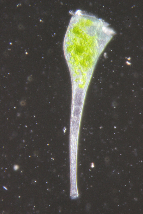
Figure 1. Morphology of a living Stentor cell. Darkfield image of a living Stentor cell that has been fed with Chlamydomonas as described in Procedure 1.3, visible as green material inside food vacuoles. The oral apparatus through which the cell eats is visible as a rim of cilia at the top of the image.
Click here to view larger image.

Figure 2. Preparation of glass needle for surgery. (A) Needle is drawn from the end of a solid glass rod. (B) High magnification view of needle tip indicating the length and sharpness necessary for surgery.
Click here to view larger image.
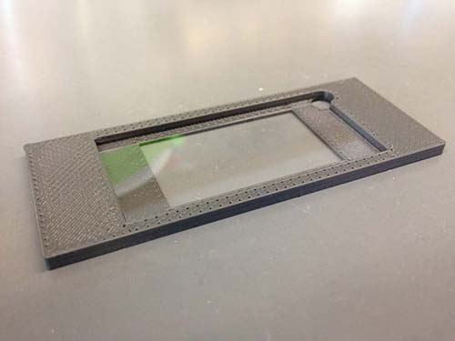
Figure 3. Custom coverslip holder for microsurgery. In order to allow open access for surgery while allowing optimal imaging conditions, cells are mounted on a coverslip rather than a thick class slide. In order to allow the coverslip to fit on the microscope stage a plastic holder can be fabricated using a 3D printer. The holder has the same dimensions as a standard microscope slide while the depression in the middle matches the dimensions of the coverslip. A hole in one corner allows the coverslip to be popped out of the holder.
Click here to view larger image.
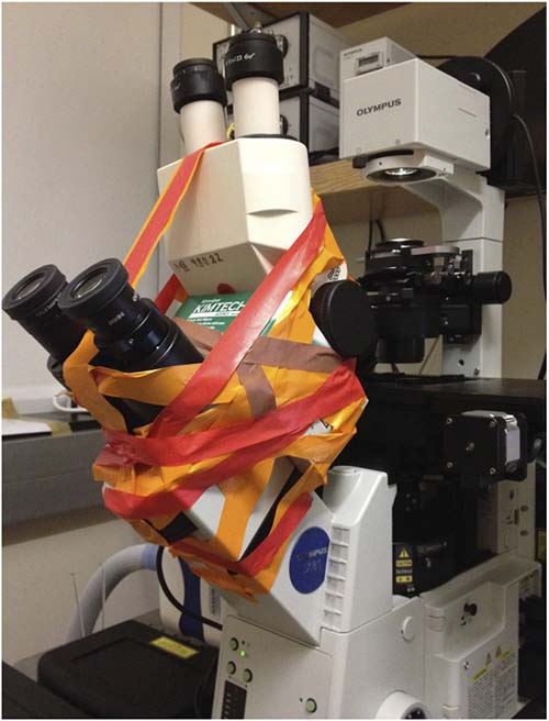
Figure 4. Double-decker microscope configuration. A dissecting microscope head is mounted on top of the eyepiece head of an inverted microscope. Lab tape allows the apparatus to be rapidly assembled and disassembled when the experiment is completed.
Click here to view larger image.
Video 1. Example of cytoplasmic flow observed in living Stentor cell. Cell imaged with 10X objective with darkfield optics at a rate of 2 frames/sec.
Video 2. Imaging of a Stentor cell as it is cut and disrupted with a glass needle. After cutting a substantial quantity of cytoplasm has been pulled out of the main cell body, but it rapidly flows back into the cell.
Discussion
The most important application of this method will be to use it to determine the molecular pathways that regulate cytoplasmic flow during wound healing in Stentor. A wide range of chemical inhibitors are available that target motor proteins as well as components of the cytoskeleton. Inhibitors of calcium signaling would also be likely candidates. It would also be of interest to remove or reduce the pigment granules (extrusomes) containing the blue pigment Stentorin, whose release from the cell is triggered under stress, and ask how the exocytotic release of Stentorin may affect flow of nearby cytoplasm. The pigment can be depleted using a simple bleaching treatment15 which could be done just prior to cutting. There does not appear to be any simple way to perform the wound healing assay in a parallel fashion since the cells need to be cut and imaged one at a time. Thus using a single microscope, it would be possible to operate on and measure cells at a rate of roughly 1 per 0.5 hr, so that during a single day of work it should be possible to obtain enough data points for a single compound to determine if it has a statistically significant effect on the process. While the method is thus not in any sense high throughput, it will be absolutely feasible to screen though a selected set of candidate compounds specific for a few carefully chosen target pathways.
A major limitation of the current approach is the relatively crude method for assembling the double-decker microscope. Assuming that the inverted microscope used for imaging is a general use instrument that other workers need to use for other purposes, an important consideration is the ease with which the apparatus can be disassembled and reassembled. While the present method using lab tape is indeed rapid and easily disassembled, it does require careful alignment by the user during assembly each time. An important modification would therefore be to construct a customized removable holder with slots for the eyepiece head of the inverted scope and the body of the dissecting scope. This should be easy to fabricate using modern 3D printer technology.
A second limitation of the approach is the reliance on DIC imaging of intracellular organelles as markers of flow. Because the identity of these organelles observed in the microscope is not currently known, it is not possible to know their size or potential interactions with other cellular components. It might therefore be advantageous to inject extrinsic tracers such as fluorescent beads and use them to track the flow. However this would require an extra step - microinjection - before the experiment could be done, thus further slowing the assay.
The final key limitation is the reliance on a single image plane for data acquisition. The primary importance of this assay is that it goes beyond simply asking whether a wound heals or not, and allows detailed analysis of the flow pattern to be obtained. In this regard, it will be extremely important to extend the imaging to three dimensional imaging. The current method images a single focal plane and thus when the PIV analysis is performed, the result is a slice through the full three dimensional flow field. This is informative but incomplete. By acquiring 3D images at each time step, it should be possible to obtain a fully three dimensional flow field. This will be particularly critical for observing exactly how the cytoplasm moves in the immediate vicinity of the wound site.
Disclosures
The authors declare that they have no competing financial interests.
Acknowledgements
This work was supported by NIH grant R01 GM090305 (WFM). The procedure described here was developed at the Physiology Course of the Marine Biological Laboratory during the summer of 2012, and we thank the students, instructors, and course directors for many helpful and stimulating discussions.
Materials
| Name | Company | Catalog Number | Comments |
|---|---|---|---|
| Stentor coeruleus live culture | Carolina Biological | 131598 | |
| Methyl Cellulose, 1,500 cP | Sigma | M0387 | |
EQUIPMENT: | |||
| Material Name | Company | Catalogue Number | |
| Soft glass stirring rod 5 in long | Fisher Scientific | 11-380A | |
| Culture Dish 4.5 in 250 ml glass | Carolina Biological | 741004 | |
| IX81 inverted microscope | Olympus | ||
| Axiovert 200M inverted microscope | Zeiss | ||
| Stemi 2000 Stereo Microscope | Zeiss | ||
| Evolve 512 x 512 EMCCD camera | Photometrics | ||
| Plan Apo 20X objective | Olympus | UPLSAPO |
References
- Sonnemann, K. J., Bement, W. M. Wound repair: toward understanding and integration of single-cell and multicellular wound responses. Annu. Rev. Cell Dev. Biol. 27, 237-264 (2011).
- Idone, V., Tam, C., Andrews, N. W. Two-way traffic on the road to plasma membrane repair. Trends Cell Biol. 18 (11), 552-559 (2008).
- Reddy, A., Caler, E. V., Andrews, N. W. Plasma membrane repair is mediated by Ca2+-regulated exocytosis of lysosomes. Cell. 106 (2), 157-169 (2001).
- Foissner, I., Wastenays, G. O. The characean internodal cell as a model system for studying wound healing. J. Microsc. 247 (1), 10-12 (2012).
- Bement, W. M., Mandato, C. A., Kirsch, M. N. Wound-induced assembly and closure of an actomyosin purse string in Xenopus oocytes. Curr. Biol. 9 (11), 579-587 (1999).
- Kim, H. G., Klotchkova, T. A., Kang, Y. M. Life without a cell membrane: regeneration of protoplasts from disintegrated cells of the marine green alga Bryopsis plumosa. J. Cell Sci. 114 (11), 2009-2014 (2001).
- Tartar, V. The Biology of Stentor. , Pergamon Press. 434 (1961).
- Morgan, T. H. Regeneration of proportionate structures in Stentor. Biol. Bull. 2 (6), 311-328 (1901).
- Heidemann, S. R., Wirtz, D. Towards a regional approach to cell mechanics. Trends Cell Biol. 14 (4), 160-166 (2004).
- Kasza, K. E., Rowat, A. C., Liu, J., Angelini, T. E., Brangwynne, C. P., Koenderinck, G. H., Weitz, D. A. The cell as a material. Curr. Opin. Cell Biol. 19 (1), 101-107 (2007).
- Mitchison, T. J., Charras, G. T., Mahadevan, L. Implications of a poroelastic cytoplasm for the dynamics of animal cell shape. Sem. Cell Dev. Biol. 19 (3), 215-223 (2008).
- De Terra, N. Culture of Stentor coeruleus on Colpidium campylum. J. Eukar. Microbiol. 13 (3), 491-492 (1966).
- Harris, E. H. The Chlamydomonas Sourcebok. 1, (2009).
- Vallotton, P., Ponti, A., Waterman-Storer, C. M., Salmon, E. D., Danuser, G. Recovery, visualization, and analysis of actin and tubulin polymer flow in live cells: a fluorescent speckle microscopy study. Biophys. J. 85 (2), 1289-1306 (2003).
- Miyake, A., Harumoto, T., Iio, H. Defence function of pigment granules in Stentor coeruleus. Eur. J. Protistol. 37 (1), 7-88 (2001).
Reprints and Permissions
