Unlike cell-free HIV-1 particles, infected CD4+ T cells are effectively sensed by plasmacytoid dendritic cells (pDCs). This manuscript describes a method where peripheral blood mononuclear cells (PBMCs) or isolated pDCs are co-cultured with HIV-1 infected T cells to evaluate innate sensing by pDCs as assessed by release of type-I IFN.
Method Article
Assessing the Innate Sensing of HIV-1 Infected CD4+ T Cells by Plasmacytoid Dendritic Cells Using an Ex vivo Co-culture System.
In This Article
Summary
Abstract
HIV-1 innate sensing requires direct contact of infected CD4+ T cells with plasmacytoid dendritic cells (pDCs). In order to study this process, the protocols described here use freshly isolated human peripheral blood mononuclear cells (PBMCs) or plasmacytoid dendritic cells (pDCs) to sense infections in either T cell line (MT4) or heterologous primary CD4+ T cells. In order to ensure proper sensing, it is essential that PBMC are isolated immediately after blood collection and that optimal percentage of infected T cells are used. Furthermore, multi-parametric flow cytometric staining can be used to confirm that PBMC samples contain the different cell lineages at physiological ratios. A number of controls can also be included to evaluate viability and functionality of pDCs. These include, the presence of specific surface markers, assessing cellular responses to known agonist of Toll-Like Receptors (TLR) pathways, and confirming a lack of spontaneous type-I interferon (IFN) production. In this system, freshly isolated PBMCs or pDCs are co-cultured with HIV-1 infected cells in 96 well plates for 18-22 hr. Supernatants from these co-cultures are then used to determine the levels of bioactive type-I IFNs by monitoring the activation of the ISGF3 pathway in HEK-Blue IFN-α/β cells. Prior and during co-culture conditions, target cells can be subjected to flow cytometric analysis to determine a number of parameters, including the percentage of infected cells, levels of specific surface markers, and differential killing of infected cells. Although, these protocols were initially developed to follow type-I IFN production, they could potentially be used to study other imuno-modulatory molecules released from pDCs and to gain further insight into the molecular mechanisms governing HIV-1 innate sensing.
Introduction
Type-I IFN-producing pDCs represent the first line of defense against viral infections, and as such serve as a vital link between innate and adaptive immunity 1. While cell-free HIV-1 particles are poorly detected by the innate immune system, HIV-1-infected CD4+ T cells are effectively sensed by pDCs 2. The sensing mechanism requires an initial contact between the viral envelope glycoprotein (gp120) at the surface of the infected T cell and CD4 molecules on the pDC, which is then followed by virus capture and internalization by the pDC. Subsequent recognition of transferred HIV-1 RNA by TLR7 triggers the activation of the Myd88/IRF7 pathway and ultimately leads to type-I IFN production 3. Importantly, the second sensing pathway present in pDCs, which is mediated by TLR9, is inhibited by the interaction of the viral glycoprotein, gp120, with the pDC-specific surface marker BDCA2 4.
The TLR7 and TLR9 sensing pathways of pDC can potentially be negatively modulated by the engagement of BST2 (also designated Tetherin or CD317) with another pDC-specific surface inhibitory receptor called ILT7 5. BST2 is expressed at high levels at the surface of pDCs and some cancer cells, but at relatively lower levels in other cell types, such as B cells, macrophages, and T cells. Importantly, BST2 can be induced by type-I IFN in a number of transformed cell lines as well as in primary cell cultures of human and murine origins 6. Apart from its immune-regulatory function, BST2 was recently found to inhibit the release of HIV-1 and other enveloped viral particles by cross-linking nascent virus particles onto the infected cell surface 7. In the case of HIV-1, the virus-encoded accessory protein U (Vpu) was shown to act as a BST2 antagonist. It is believed that Vpu downregulates BST2 from the cell surface of HIV-1-infected cells, the site of its tethering activity and as a result enhances viral release and spread 7, although this notion has been challenged 8,9. In the absence of Vpu, a large number of fully formed and mature progeny HIV-1 particles are retained at the cell surface. Whether these tethered particles could represent effective targets for immune sensing still remains an open question. Additionally, it is unclear whether Vpu could dysregulate type-I IFN production from pDCs by modulating surface BST2 levels.
Early studies designed to assess HIV-1 sensing were conducted using cell-free HIV-1 particles, which are generally considered poor inducers of type-I IFN 3,10-12. Given that recent studies suggest that PBMCs and pDCs efficiently sense HIV-infected CD4+ T lymphocytes 2,13,14, we describe here a simple method to measure innate responses triggered by pDCs upon recognition of HIV-1-infected cells in vitro.
Protocol
General Notes
It is important to keep cells in culture without any antibiotic, since even controlled bacterial infections can be sensed by PBMCs. Such sensing of bacterial components will induce a background interferon production that cannot be distinguished from that triggered by specific HIV-1 sensing. Furthermore, all cells have to be routinely tested for the absence of mycoplasma contamination.
Always use endotoxin-free solutions whenever possible.
Flow cytometric characterization of the different cells to be used in the co-cultures (PBMCs, pDCs, MT4 and CD4+ T cells) is an optional step, but highly recommended since it can provide valuable information required for proper interpretation of a given experiment.
1. Preparation of Infected Target Cells
- Maintain human CD4+ T cell line MT4 in RPMI 1640 media supplemented with 10% fetal bovine serum (FBS), referred from now on as RPMI-1640 complete medium.
- Isolation of primary CD4+ T cells.
- Isolate mononuclear cells from human peripheral blood by density gradient centrifugation.
- Isolate CD4+ T lymphocytes using a CD4+ T cells negative selection kit, following manufacturer's recommendations.
- Activate CD4+ T lymphocytes with PHA-L (5 µg/ml) for 48 hr and then maintain cells in RPMI-1640 complete medium supplemented with recombinant IL-2 (100 U/ml). Infect primary T cells 5 days post isolation.
- Infection of target cells (MT4 T cell line or heterologous primary CD4+ T cells).
- Two days prior to co-culture, infect T cells with HIV-1 virus. In the specific example described here pNL4.3-GFP_ires_Nef wild type (WT) or pNL4.3-GFP_ires_Nef delta Vpu (dVpu) viruses were used. These viral strains represent green fluorescent protein (GFP)-marked isogenic infectious molecular clones that differ only in their ability to express Vpu.
- Use different multiplicities of infection (MOIs) and select cultures with similar levels of infection for co-cultures. The day of the co-culture, determine the percentage of GFP+ cells by flow cytometry as a marker of infection. Use only cultures with a range of 20-50% infected cells for co-cultures.
Optional steps: Perform flow cytometric staining at this point to evaluate surface expression of molecules/ligands of interest such as BST2, and CD4. For cell surface staining, resuspend 0.5 x 10⁶ T cells in 100 µl of wash solution, add 1 µl of anti-CD4_PerCP/Cy5.5 and 1 µl of anti-BST2_A660. Incubate tubes for 45 min at 4°C, before washing with flow cytometry wash solution. If the studies are aimed at evaluating the role of Vpu-mediated BST2 antagonism, perform a HIV-1 particle release assay at this time as previously described 15.
2. Isolation and Enrichment of Sensing Cells (Whole PBMCs or Enriched pDCs)
Note: When PBMCs are used for sensing, start isolation within half hour after blood collection and conduct it in a timely manner. Use PBMCs immediately for co-cultures or pDC enrichment.
- Isolate mononuclear cells from human peripheral blood by density gradient centrifugation.
Optional step: Perform multi-parametric flow cytometry staining to confirm that freshly isolated PBMCs contain the different cell lineages at physiological ratios. For cell surface staining, resuspend 106 PBMCs in 100 µl of Fc blocking solution, and incubate for 15 min at 4 °C. After blocking, add 1 µl of anti-CD3_Pacific Blue (for T cells), 1 µl of anti-CD14-PE_Texas Red (for myeloid cells), 4 µl of anti-BDCA2_APC and 1 µl of anti-ILT7_PE (for pDCs). Incubate tubes for 45 min at 4 °C, before washing with flow cytometry wash solution. - Enrich pDCs using a pDC negative selection kit, following manufacturer's recommendations. It is recommended to work fast, keep cells cold, and use pre-cooled solutions. This will prevent the capping of antibodies on the cell surface and non-specific cell labeling as well as ensuring maximal pDC activity.
- Perform multi-parametric flow cytometric analysis of the freshly isolated pDCs to confirm purity, percentage of enrichment, and relative amounts of pDC- specific surface markers (such as ILT7 and BDCA2). For cell surface staining, resuspend 104 pDCs in 100 µl of Fc blocking solution, and incubate for 15 min at 4 °C. After blocking, add 1 µl of anti-CD3_Pacific Blue, 1 µl of anti-CD14_PE_Texas Red, 4 µl of anti-BDCA2_APC and 1 µl of anti-ILT7_PE. Incubate tubes for 45 min at 4 °C, before washing with flow cytometry wash solution (PBS, 5 mM EDTA, 5% FBS). A minimum of 40% pDC purity is recommended (if the starting material has 0.4% pDC, this would be equivalent to a 100-fold enrichment).
3. Co-culture of Infected CD4+ T Cells and Sensing Cells (Whole PBMCs or Enriched pDCs)
- Mix 220 µl of PBMCs (diluted at 850,000 cells/ml) or 220 µl of pDCs (at a concentration ranging from 100,000 to 500,000 cells/ml) with 30 µl of infected T cells (diluted at 106 cells/ml) per well in a U-bottom 96 well plate.
- As controls, plate PBMCs or pDCs and T cells alone. Adjust volume in well to 250 µl with RPMI 1640 complete medium.
- Assess cellular responses to known agonist of the TLR7 and TLR9 pathways. To this end, plate 220 µl of PBMCs (diluted at 850,000 cells/ml) or 200 µl of pDCs (at a concentration ranging from 100,000 to 500,000 cells/ml) per well in a U-bottom 96 well plate and add TLR7 agonist (Imiquimod, final concentration: 2.5 µg/ml) or TLR9 agonist (ODN 2216 CpG-A, final concentration: 5 µM). Incubate cells at 37 °C and 5% CO2 for 18-22 hr and process them in a manner similar to the co-cultures described below.
- Incubate co-cultures at 37 °C and 5% CO2 for 18-22 hr, and then transfer them to a V-bottom 96 well plate. Centrifuge for 5 min at 400 x g.
- Transfer supernatants to a flat-bottom 96 well plate. Supernatants can be stored at -80 °C or used for type-I IFN detection immediately.
- Resuspend co-cultured cells in 2% paraformaldehyde and analyze them using flow cytometry. Due to differences in size and granulation, MT4 cells are easily distinguished from PBMCs or pDCs based on their side-scatter (SS) versus forward-scatter (FS) profiles. If primary T cells are used as target infected cells, these can be stained with any cell tracker dye, such as CSFE, prior to co-culture.
- Measurement of bioactive type-I IFN using HEK-Blue IFN-α/β reporter cells.
Note: These cells were generated by stable transfection of HEK293 cells with the human STAT2 and IRF9 genes to obtain a fully active type I IFN signaling pathway. They also contain the secreted alkaline phosphatase (SEAP) reporter gene under the control of the IFN-α/β inducible ISG54 promoter. Stimulation of these cells with type-I IFN activates the JAK/STAT/ISGF3 pathway and induces the production of SEAP.- Maintain HEK-Blue IFN-α/β cells in DMEM supplemented with 10% FBS. Plate them at a density of 50,000 cells per well in a flat-bottom 96 well plate, in a final volume of 180 µl.
- Add 20 µl of co-culture supernatant to each well in duplicate. Each plate also requires a set of internal standard controls (human IFNα, final concentration from 100 U/ml to 2,500 U/ml). Incubate the plates at 37 °C and 5% CO2 for 18-22 hr.
- Determine levels of alkaline phosphatase activity using QUANTI-Blue solution, which changes from pink to purple/blue in the presence of the enzyme. Prepare QUANTI-Blue following manufacturer's recommendations. Add 180 µl of this solution to each well of a flat-bottom 96 well plate. To each well also add 20 µl of the induced HEK-Blue IFN-α/β cells supernatants, and incubate the plate in a 37 °C incubator until color develops in the standard IFN control wells.
- Evaluate levels of SEAP using a spectrophotometer at 620-655 nm and determine the concentration of type-I IFN by extrapolation from the linear part of the IFN standard curve.
Results
The co-culture system described in Figure 1 allows for a controlled study of HIV-1 innate sensing. Due to the variable nature of primary cells, it is important that normal ratios of myeloid and T cells (CD14+ and CD3+ respectively) are observed, such as in the example shown in Figure 2A. Furthermore, only a low number of activated T cells (higher FS and higher CD3 levels as compared to non-activated T cells) are desirable in the freshly isolated PBMCs cultures. PBMCs with abnormal ratios between the various cell-types are often incapable of mounting a proper innate immune response in vitro, likely due to an already activated phenotype as a consequence of an ongoing infection. Most importantly, the relative percentage of pDCs needs to be evaluated as shown in Figure 2B. Although this percentage can vary from 0.2 to 1.2%, an abnormal sensing phenotype is also observed with both extremes of this range. Enrichment of pDCs using negative selection often yields 50-95% pDC purity, as seen in the two examples from in Figure 2B (92% and 72%). A prototypical example for the overall experiment is shown in Figures 3 and 4. In this example, MT4 cells were infected with pNL4.3-GFP_ires_Nef WT or delta Vpu to achieve 30% infection at the time of co-culture (Figure 3A). As previously described, only the infections with WT virus resulted in a significant down-regulation of surface BST2 (Figure 3B). Due to their differences in size and granularity, MT4 cells can be easily distinguished from PBMCs by flow cytometry (Figure 4A). After the co-culture incubation, only PBMCs in contact with known TLR7 or TLR9 agonist and PBMCs in contact with HIV-1 infected cells released significant amounts of type-I IFN (Figure 4B). No IFN was detected in PBMCs alone, in PBMCs co-cultured with mock-infected MT4 cells, or in infected MT4 cells alone (Figure 4B). In the example presented here, innate sensing of HIV-1-infected MT4 cells by PBMCs was found to be significantly reduced in the presence of Vpu.
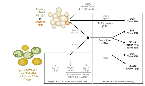
Figure 1: Schematic overview of the experimental design. SUP: supernatant Click here to view larger image.
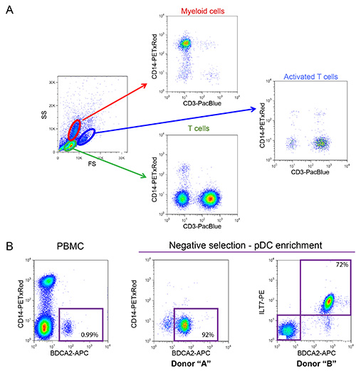
Figure 2: Phenotypic characterization of freshly isolated PBMCs and enriched pDCs. (A) Normal distribution of myeloid cells and T cells observed in PBMCs isolated from a healthy donor (Percentage of myeloid cells should range between 10-20% and T cells between 55-65%). PBMCs sample were stained using fluorophore-conjugated antibodies that recognize surface CD14 (myeloid lineage marker) and CD3 (T cell marker). (B) Phenotypic characterization of pDCs. Prior to enrichment pDCs represent between 0.2-1.2% of the total number of cells within the PBMCs (0.99% in the example shown). These cells can be identified based on expression of specific markers, such as BDCA2 and ILT7, present at the cell surface. Negative selection can be used to significantly enrich the number of pDCs and eliminate potentially detrimental contaminations, such as CD14+ myeloid cells (enrichment reaching 92% and 72% in the two examples shown). Click here to view larger image.
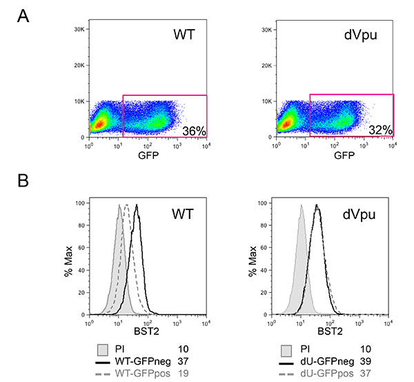
Figure 3: Characterization of HIV-1-infected target MT4 cells. (A) Percentage of infected cells within the MT4 cultures infected with pNL4.3-GFP_ires_Nef WT or dVpu as determined by the percentage of GFP positive cells. (B) BST2 cell surface expression in the infected cells was evaluated after surface staining. The grey filled histograms represent mock-infected cells stained with the pre-immune rabbit serum (unstained control); the remaining histograms represent cells stained with anti-BST2 polyclonal rabbit serum. Histograms with solid lines represent control GFP negative (neg) cells; while histograms with dotted lines correspond to infected GFP positive (pos) cells. Mean fluorescence intensity (MFI) values are indicated for each sample. Flow cytometry was performed and analyzed as described in Figure 2. Click here to view larger image.
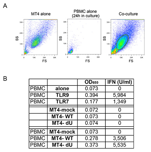
Figure 4: Co culture of freshly isolated PBMCs and infected MT4 T cells. (A) Comparison of flow cytometry FS/SS profiles observed for MT4 T cells alone, PBMCs alone, or co-cultures between PBMCs and MT4 T cells. Due to differences in size and granularity, MT4 T cells can be easily distinguished from PBMCs in co-cultures using flow cytometry. (B) Representative example of the amount of type-I IFN released after stimulation of PBMCs with TLR7 or TLR9 agonist (ago), or after co-cultures with mock or the indicated MT4 cells infected with pNL4.3-GFP_ires_Nef WT or dVpu. Raw data shown as OD650 values and their corresponding converseion to type-I IFN concentration expressed in U/ml. Click here to view larger image.
Discussion
The protocols described here measure sensing of T cells infected with GFP-tagged HIV-1 viruses that differ only in their ability to express Vpu, as described in our recent report 18. However, these methods could easily be modified to study sensing of untagged viruses as well as viruses lacking other viral genes that could potentially modulate innate sensing. If the viruses used do not express GFP, the percentage of infected target cells should be determined by another means prior to co-culture, such as p24 (capsid) intracellular staining and flow cytometry or by p24 ELISA. Furthermore, these protocols can be used with different target T cells, including a variety of available cell lines and primary cells.
The methods described in this manuscript have a number of advantages when compared to early methodologies used to study HIV-1 sensing. First and foremost it has been established by a number of groups that cell-free HIV-1 particles are poor inducers of type-I IFN 2,13,14. Furthermore, early studies relied on older IFNα ELISA-based methods to quantify the amount of type-I IFN produced. A limitation of this detection technique is that most of the older IFNα ELISA kits only measure one type of IFNα, while overlooking the other types of IFNα and IFNβ. The use of the described reporter cell lines overcomes this limitation as the concentration of all bioactive forms of human type-I IFNs can be measured at once. The technique is highly sensitive, less expensive than the new generation of IFN ELISA kit and for many donors large amounts of type-I IFN can easily be detected. Nevertheless, this protocol can be easily adapted for use with other type-I IFN read-out methods such as ELISAs.
Critical steps: It is important that the quality of all cellular components (both targets and effectors) be tightly controlled. Potential contamination, even when asymptomatic, needs to be monitored and controlled. As we previously mentioned, antibiotic-controlled asymptomatic bacterial contamination, and potentially other intracellular pathogens such as mycoplasma, can generate similar immune responses to the ones observed after HIV-1 infection, namely production of type-I IFN. Moreover, all reagents need to be free of LPS or other potential endotoxins. Similarly, monitoring of PBMCs and pDCs is also important since human samples can often be primed by underlying ongoing infections, which may affect normal sensing. We recommend always including the proposed negative controls, such absence of target cells as well as co-cultures with mock-infected cells. Viability of infected cells is also important for proper pDC sensing since type-I IFN is not produced following co-culture with apoptotic cells 2,14.
One important limitation of the technique is the need for freshly isolated primary cells. This procedure was not tested using PBMCs or pDCs that were either previously frozen or kept in culture. The isolation of PBMCs is labor intensive and these cells have a very limited lifespan. Furthermore, standard protocols for protecting against blood-borne pathogen should be used while working with human blood. There are often several sources of variability associated with work involving freshly isolated human cells. Among them are the different procedures implemented to isolate these cells, and the inherited variability encountered from donor-to-donor. While it is impossible to avoid variability among donor, the use of the suggested positive controls, such as measuring response to known agonists of TLR7 and TLR9 pathways, would provide an important internal control for these experiments. We observed that a robust response to agonists of the TLR9 pathway could be correlated with a healthy pDC response, while a responsive TLR7 pathway is required for a proper response against HIV-1 3.
Based on observations made by us and others 2, enrichment of pDCs from PBMCs is often not needed. However, if the experimental design requires it (for instance in the context where depletion of specific pDC surface markers is needed) negative selection is preferable over positive selection, as cells remain free of antibody after the selection process. Isolated pDCs undergo rapid apoptosis in culture. For experiments that require these cells to be cultured for extended period of time, culture media can be supplemented with IL-3. This cytokine induces pDC proliferation and inhibits their apoptosis 16. However, IL-3 usage needs to be tightly controlled since it may lead to pDC maturation or differentiation.
The method described here combines target HIV-1 infected cells with freshly isolated PBMCs or pDCs in order to recreate the initial steps involved during innate sensing. This method will undoubtedly be useful to explore early innate immune events associated with HIV infection. Although the associated protocols are aimed at measuring type-I IFN, they could easily be modified to measure other bioactive molecules released from pDCs upon recognition of infected cells. These could include: type-III IFN (IFN-λ), pro-inflammatory cytokines (such as TNF-α and IL-6) and the chemokines CXCL10, CCL4, and CCL5 17.
Acknowledgements
We thank E. Massicotte and J. Lord for expert technical assistance during flow cytometry and cell sorting experiments. Also, we would like to thank the IRCM clinic staff and all donors for providing blood samples. Peripheral blood samples were obtained from healthy adult donors who gave written informed consent in accordance with the Declaration of Helsinki under research protocols approved by the research ethics review board of the Institut de Recherches Cliniques de Montréal (IRCM). The following reagents were obtained through the NIH AIDS Reagent Program, Division of AIDS, NIAID, NIH: MT4 cells from Dr. Douglas Richman; and Human rIL-2 from Dr. Maurice Gately, Hoffmann - La Roche Inc.
Dr. Cohen is the recipient of the Canada Research Chair in Human Retrovirology. This work was supported by grants from CIHR (CIHR 111226) to Dr. Cohen and from the Fonds de recherche du Québec-Santé (FRQ-S) AIDS network to Dr. Bego and Dr. Cohen.
Materials
| Name | Company | Catalog Number | Comments |
|---|---|---|---|
| MT4 cells | NIH AIDS Reagent Program | 120 | This reagent was obtained through the NIH AIDS Reagent Program, Division of AIDS, NIAID, NIH: MT-4 from Dr. Douglas Richman. |
| HEK-Blu IFN-α/β reporter cells | Invivogen | HKB-IFNAB | |
| RPMI | Wisent | 350-000-CL | |
| DMEM | Wisent | 319-005-CL | |
| FBS | Wisent | 080-150 | |
| Ficoll-Pâque Plus | Myltenyi | 17-1440-03 | |
| CD4+ T cell isolation kit II | Myltenyi | 130-091-155 | |
| Diamond Plasmacytoid Dendritic Cell Isolation Kit II | Myltenyi | 130-097-240 | |
| PHA-L | Sigma-Aldrich | O2769 | |
| Human rIL-2 | NIH AIDS Reagent Program | 136 | This reagent was obtained through the NIH AIDS Reagent Program, Division of AIDS, NIAID, NIH: Human rIL-2 from Dr. Maurice Gately, Hoffmann - La Roche Inc. |
| Imiquimod | Invivogen | TLRL-IMQS | |
| ODN 2216 CpG-A | Hycult Biotec | HC4037 | |
| Human IFNα | PBL Interferon source | 11100-1 | |
| QUANTI-Blue | Cedarlane | REP-QB2 | |
| Flow cytometry (Antibodies/reagents) | |||
| Fc blocking solution (composition below): | Note: make in washing solution (PBS, 5mM EDTA, 5% FBS) | ||
| 10% Goat serum | Sigma-Aldrich | G 9023 | |
| 10% Rabbit serum | Sigma-Aldrich | R 9133 | |
| 10% Mouse serum | Sigma-Aldrich | M 5909 | |
| 2.5 mg/ml human IgG | Sigma-Aldrich | I 4506 | |
| anti-CD3_Pacific Blue | Biolegend | 300417 | |
| anti-CD14_PE-TxRed | Life Technologie | MHCD1417 | |
| anti-BDCA2_APC | Myltenyi | 130-090-905 | |
| anti-ILT7_PE | Biolegend | 326408 | |
| anti-CD4_PerCP/Cy5.5 | Biolegend | 317427 | |
| anti-BST2_Alexa 660 | eBioscience | 50-3179 | |
| Paraformaldehyde | Sigma-Aldrich | P 6148 | |
| Cell strainer | BD Falcon | 352340 | |
| Cyan ADP instrument | Beckman Coulter | CY20030 |
References
- Colonna, M., Trinchieri, G., Liu, Y. J. Plasmacytoid dendritic cells in immunity. Nature immunology. 5, 1219-1226 (2004).
- Lepelley, A., et al. Innate sensing of HIV-infected cells. PLoS pathogens. 7, e1001284(2011).
- Beignon, A. S., et al. Endocytosis of HIV-1 activates plasmacytoid dendritic cells via Toll-like receptor-viral RNA interactions. The Journal of clinical investigation. 115, 3265-3275 (2005).
- Martinelli, E., et al. HIV-1 gp120 inhibits TLR9-mediated activation and IFN-{alpha} secretion in plasmacytoid dendritic cells. Proceedings of the National Academy of Sciences of the United States of America. 104, 3396-3401 (2007).
- Cao, W., et al. Regulation of TLR7/9 responses in plasmacytoid dendritic cells by BST2 and ILT7 receptor interaction. The Journal of experimental medicine. 206, 1603-1614 (2009).
- Bego, M. G., Mercier, J., Cohen, E. A. Virus-activated interferon regulatory factor 7 upregulates expression of the interferon-regulated BST2 gene independently of interferon signaling. Journal of virology. 86, 3513-3527 (2012).
- Neil, S. J., Zang, T., Bieniasz, P. D. Tetherin inhibits retrovirus release and is antagonized by HIV-1 Vpu. Nature. 451, 425-430 (2008).
- Miyagi, E., Andrew, A. J., Kao, S., Strebel, K. Vpu enhances HIV-1 virus release in the absence of Bst-2 cell surface down-modulation and intracellular depletion. Proceedings of the National Academy of Sciences of the United States of America. 106, 2868-2873 (2009).
- McNatt, M. W., Zang, T., Bieniasz, P. D. Vpu binds directly to tetherin and displaces it from nascent virions. PLoS pathogens. 9, e1003299(2013).
- Manel, N., et al. A cryptic sensor for HIV-1 activates antiviral innate immunity in dendritic cells. Nature. 467, 214-217 (2010).
- Yan, N., Regalado-Magdos, A. D., Stiggelbout, B., Lee-Kirsch, M. A., Lieberman, J. The cytosolic exonuclease TREX1 inhibits the innate immune response to human immunodeficiency virus type 1. Nature immunology. 11, 1005-1013 (2010).
- Fonteneau, J. F., et al. Human immunodeficiency virus type 1 activates plasmacytoid dendritic cells and concomitantly induces the bystander maturation of myeloid dendritic cells. Journal of virology. 78, 5223-5232 (2004).
- Ankel, H., Capobianchi, M. R., Frezza, F., Castilletti, C., Dianzani, F. Interferon induction by HIV-1-infected cells: a possible role of sulfatides or related glycolipids. Virology. 221, 113-119 (1996).
- Schmidt, B., Ashlock, B. M., Foster, H., Fujimura, S. H., Levy, J. A. HIV-infected cells are major inducers of plasmacytoid dendritic cell interferon production, maturation, and migration. Virology. 343, 256-266 (2005).
- Bego, M. G., Dube, M., Mercier, J., Cohen, E. A. Effect of calcium-modulating cyclophilin ligand on human immunodeficiency virus type 1 particle release and cell surface expression of tetherin. Journal of virology. 83, 13032-13036 (2009).
- Grouard, G., et al. The enigmatic plasmacytoid T cells develop into dendritic cells with interleukin (IL)-3 and CD40-ligand. The Journal of experimental medicine. 185, 1101-1111 (1997).
- Fitzgerald-Bocarsly, P., Jacobs, E. S. Plasmacytoid dendritic cells in HIV infection: striking a delicate balance. Journal of leukocyte biology. 87, 609-620 (2010).
- Bego MG,, Côté, É, Aschman, N., Mercier, J., Weissenhorn, W. Cohen ÉA. Exploits the Cross-Talk between BST2 and the ILT7 Receptor to Suppress Anti-HIV-1 Responses by Plasmacytoid Dendritic Cells. PLoS Pathog. 11 (7), e1005024(2015).
Reprints and Permissions
