The zebrafish is a powerful model system for developmental biology and human disease research due to their genetic similarity with higher vertebrates. This protocol describes a methodology to create haploid zebrafish embryos that can be utilized for forward screen strategies to identify recessive mutations in genes essential for early embryogenesis.
Method Article
Production of Haploid Zebrafish Embryos by In Vitro Fertilization
Opens in a new tab
In This Article
Summary
Loading...
$$\rightleftharpoonup{xx}$$
$$\longleftharp{xx}$$,
$$\longrightharp{xx}$$,
Abstract
Loading...
$$\rightleftharpoonup{xx}$$
$$\longleftharp{xx}$$,
$$\longrightharp{xx}$$,
The zebrafish has become a mainstream vertebrate model that is relevant for many disciplines of scientific study. Zebrafish are especially well suited for forward genetic analysis of developmental processes due to their external fertilization, embryonic size, rapid ontogeny, and optical clarity – a constellation of traits that enable the direct observation of events ranging from gastrulation to organogenesis with a basic stereomicroscope. Further, zebrafish embryos can survive for several days in the haploid state. The production of haploid embryos in vitro is a powerful tool for mutational analysis, as it enables the identification of recessive mutant alleles present in first generation (F1) female carriers following mutagenesis in the parental (P) generation. This approach eliminates the necessity to raise multiple generations (F2, F3, etc.) which involves breeding of mutant families, thus saving the researcher time along with reducing the needs for zebrafish colony space, labor, and the husbandry costs. Although zebrafish have been used to conduct forward screens for the past several decades, there has been a steady expansion of transgenic and genome editing tools. These tools now offer a plethora of ways to create nuanced assays for next generation screens that can be used to further dissect the gene regulatory networks that drive vertebrate ontogeny. Here, we describe how to prepare haploid zebrafish embryos. This protocol can be implemented for novel future haploid screens, such as in enhancer and suppressor screens, to address the mechanisms of development for a broad number of processes and tissues that form during early embryonic stages.
Introduction
Loading...
$$\rightleftharpoonup{xx}$$
$$\longleftharp{xx}$$,
$$\longrightharp{xx}$$,
The fundamental processes that orchestrate vertebrate development are broadly conserved1. A powerful way to identify genes with essential roles in development is to isolate heritable mutations that lead to phenotypic defects in the process of interest. The zebrafish, Danio rerio, is a small freshwater teleost species belonging to the Cyprinidae fish family that has become a widely used model organism for vertebrate developmental genetics over the last several decades2. Zebrafish eggs are fertilized externally, permitting access to the zygote from the onset of fertilization3. Further, the early embryo is optically transparent, enabling visualization of development with a simple stereomicroscope3. Zebrafish ontogeny is rapid, with the processes of cleavage, gastrulation, and organogenesis of many structures, such as the heart and kidney, largely completed over the first day of development3. The time from larval stages to sexual maturity takes 2-3 months, and the adults are small vertebrates approximately 3-4 cm in length, so many fish can be maintained in minimal space2. In addition, zebrafish adults have high fecundity, with each fish able to produce several hundred embryos per week2. These attributes have rendered the tractability of zebrafish for forward and reverse genetic screens. Zebrafish possess a high degree of conservation in their anatomy, cell biology, and physiology with more advanced vertebrate species like mammals2,4. In fact, recent sequence analysis has demonstrated that the zebrafish genome contains homologs to nearly 70% of human genes5. This conservation has enabled researchers to use zebrafish as a model for numerous human diseases, including both embryonic and adult conditions2,4. Taken together, these factors have made zebrafish an excellent system for screens aimed at identifying genes that are essential for normal vertebrate ontogeny.
A number of large scale screens have been carried out in zebrafish and have identified several thousand mutations in developmental genes6-8. The zebrafish adult male is amenable to mutagenesis, and chromosomal alterations can be generated with relatively high frequency using the chemical mutagen ethylnitrosourea (ENU)6,7 or retroviral insertional mutagenesis9. To date, over 9,000 heritable mutations have been created, and include genes that affect virtually all aspects of embryogenesis. The zebrafish community has established a centralized bank of these mutants at the Zebrafish International Resource Center (or ZIRC)10, which has collected approximately 5,000 mutant and transgenic alleles from around the world. Many of these mutations have been analyzed and cloned, giving researchers across biological disciplines valuable information that has been used to tease apart numerous cellular and physiological pathways through the insights originating with zebrafish experiments.
Although forward screens have identified an impressive number of important genes, much has yet to be appreciated about the genetic regulatory networks that mediate a myriad of processes. Many screens undertaken thus far have not reached saturation, and thus forward as well as reverse genetic screens have remained a cornerstone to identify and analyze mechanisms of embryogenesis. While there are tremendous insights that can be gleaned from any single zebrafish mutant line, a major limiting factor in the field has historically been the time consuming effort involved in the positional cloning. For this reason, the identity of many chemically induced mutations has remained a mystery. However, with the recent advent of whole genome sequencing approaches that facilitate rapid cloning11-14, incredibly efficient identification of genetic mutations is now feasible.
The prototypical forward screen in the zebrafish is a diploid screen that can identify recessive mutations within three generations6,7 (Figure 1, left column). This technique involves generating mutations in the germ cells of the parental (P) male, and then mating this male with a wild type female. Each of the first generation (F1) progeny from this mating will harbor one or more unique mutations due to the chromosomal contributions of the mutated paternal genome. The F1 progeny are raised and outcrossed with wild types to create F2 families. In the F2 family, siblings will be either wild type or heterozygous carriers, and are incrossed randomly to generate F3 clutches that are analyzed for phenotype(s) of interest. The F3 clutches of heterozygous carriers will contain 25% wild type, 50% heterozygous, and 25% homozygous mutant fish. This multi-generational screening schema is effective, however one major drawback to this approach includes the time required to prepare for the screen: at least 1 year of fish husbandry alone is involved, since each generation requires around 3 months to reach sexual maturity. Other limitations of this type of diploid screen are the work, space, and cost of housing these generations.
The haploid screen is one alternative that can be used to identify and subsequently isolate recessive mutations using similar mutagenesis strategies15 (Figure 1, right column). The production of haploid zebrafish embryos was first described over 30 years ago16, and the ability of the normally diploid zebrafish to develop for several days with a haploid chromosomal ploidy has been utilized for numerous genetic studies since this time. The major benefit of the haploid screen is that this method does not involve raising F2 families in order to screen for recessive mutant alleles. Instead, the F1 generation females are evaluated for heterozygosity of recessive mutations in the process of interest by examining their F2 offspring in a haploid condition (Figure 1, right column). Overall, the steps to procure haploid embryos are relatively straightforward (Figure 2). After preparation of the necessary solutions for sperm handling, eggs are obtained from F1 generation females by manual manipulation so as to extrude (or “squeeze”) the eggs from the belly. Once the eggs are obtained, they are fertilized in vitro by exposure to ultraviolet (UV) inactivated sperm. The UV inactivated sperm are incapable of contributing viable DNA to the egg due to the fact that the short wavelength UV crosslinks the paternal DNA. However, the sperm are still capable of triggering egg activity and the events associated with zygotic development. Upon metabolic activation, the egg will complete meiosis II, and proceed to develop with only the one maternally deposited copy of each chromosome. Because of this 1N ploidy, the embryo is subject to the developmental consequences of a recessive mutant allele, if it is present on a maternal chromosome. Thus each F2 haploid clutch will contain 50% wildtype and 50% mutant embryos if the mother is a heterozygous carrier of a fully penetrant recessive allele. This distribution of embryonic genotypes enables relative ease in terms of evaluating the clutch for the presence of mutant phenotype(s) of interest. If such a phenotype is detected, the F1 generation female founder is subsequently outcrossed to wild type male zebrafish to create and raise more heterozygous carriers. Because it is not necessary to raise multiple generations to perform the screen, the researcher can save considerable time, labor, and aquarium space, but still has the power to survey a significant number of genomes based on the number of F1 females examined. Thus, haploid screens are especially feasible for small laboratories or projects initiated in the absence of substantial funding.
However, there are several limitations and drawbacks to the use of haploids. First, haploid zebrafish embryos live for only several days, and typically die between 3 and 5 days post fertilization (dpf)15. Haploid zebrafish embryos can be distinguished from diploids based on several characteristics, foremost among them being a short and stocky body that nevertheless has the normal number of somite segments15,17. As development progresses, the brain exhibits increased cell death and circulation is poor, usually associated with blood pooling by 2-3 dpf and edema15,17. Further, haploids fail to inflate a swim bladder15,17. Regardless of these morphological differences, most major organs and tissues are formed, including the heart, eye, and notochord15,17. One feature noted about haploid clutches is that they contain a range of phenotypes in terms of varieties of defective embryos — a feature observed across zebrafish strains. Thus, one should verify if the tissue(s) and time point of interest show reasonably normal development in the wild type haploid strain and therefore have the potential to be evaluated for genetic defects in a screen or other research project.
Despite these challenges, haploid screens have been implemented successfully to identify genes necessary for early development processes in the zebrafish, and haploid protocols have been well established resources in the community for many years15-19. More recently, the process of generating haploid fish embryos has been used by researchers to create haploid embryonic stem (ES) cell cultures from the medaka fish20,21. After the production of medaka haploid embryos in vitro, the embryos were used to derive primary cell cultures that were passaged for about 15 weeks followed by another 5-8 weeks to generate pure clones of haploid cells with ES cell properties15-19. The generation of fish haploid ES cell lines holds broad future promise for analyzing recessive phenotypes in vertebrate cell lineages, and represents a new approach for genetic analysis in the coming years. Thus, the generation of haploid fish embryos has growing applications in the biomedical research community. This video article provides a visual demonstration of the steps involved with producing haploid zebrafish embryos in vitro using UV-inactivated sperm based on established protocols15-19,22.
Protocol
Loading...
$$\rightleftharpoonup{xx}$$
$$\longleftharp{xx}$$,
$$\longrightharp{xx}$$,
NOTE: The procedures for working with zebrafish embryos described in this protocol were approved by the Institutional Animal Care and Use Committee at the University of Notre Dame. NOTE: Although the following protocol provides a demonstration of sperm isolation that involves euthanasia of males, sperm can be collecting from the genital pore of anesthetized living male zebrafish using gentle abdominal pressure and a microcapillary for sperm collection17,18. Squeezing of live males requires several males to be used in order to collect the equivalent quantity of sperm that can be obtained from one euthanized male17,18. However, males that have undergone squeezing remain quite healthy and can be used for natural matings or subsequent in vitro procedures17. Whenever possible, chosen procedures should minimize unnecessary fish sacrifice.
1. Preparation of Solutions and Zebrafish Mating Chambers
- Prepare Hank’s Stock Solutions (#1, 2, 4, 6) and Hank’s Premix Solution (see Materials table)19, then autoclave and store at 4 °C.
- Select the desired number of adult zebrafish female(s) for haploid clutch collection. NOTE: Based on experience, using a mutagenized F1 comprised of inbred Tübingen strain zebrafish, approximately 50% of sexually mature females were ‘squeezable’ on average after priming them by placing them in a mating cage overnight with a male — i.e. a clutch was obtained via the squeezing procedure — and the majority of these clutches (ranging between 70-90%) produced viable haploids. With two researchers performing the haploid preparation, approximately 80 adult female F1 zebrafish can be squeezed in one morning. To calculate the reagents needed for the haploid experiment, factor in the both the squeeze rate of the F1 generation and the number of researchers available to perform the experiment. Keep in mind that the number of females that will produce high quality haploid clutches is extremely variable, with differences observed between zebrafish females of different strains, ages, and food diet15.
- Prepare mating cages by placing each adult zebrafish female with an adult male zebrafish in a chamber of system water so that they are separated by a divider overnight. NOTE: Females must be exposed to a male overnight to prime, or prepare them, for spawning via squeezing.
2. Dissection of Testes
- Make Hank’s Stock Solution #6 fresh by adding 0.35 g of sodium bicarbonate to 10 ml of distilled water. Mix well to dissolve the sodium bicarbonate powder.
- Combine the following in a tube on ice in order to make the buffered Hank’s Final working solution for the sperm: 9.9 ml of Hanks Premix I with 100 µl of stock solution #6.
- Mix well, then aliquot 500 µl of the Hank’s Final working solution into a microcentrifuge tube and place on ice. NOTE: Each 500 µl aliquot will be used to prepare sperm from the testes of 4-5 male zebrafish.
- Select between 4-5 male zebrafish for each sperm harvest. NOTE: This will harvest enough sperm to be collected, processed, and stored in one 1.5 ml microcentrifuge tube, which can be utilized to fertilize approximately 15 haploid clutches. Select additional cohorts of males in order to prepare additional tubes depending on the scale of the experiment. If the selected male zebrafish are small or the testes dissection is incomplete, typically 5 males will be needed.
- Euthanize each male by using a net to gently transfer the fish into a dish containing 0.2% tricaine for approximately 5-6 min at room temperature. NOTE: Be careful to ensure that the male has been properly euthanized prior to beginning the dissection. Typically, wait several (2-3) min after the gills of the fish stop moving and the fish is no longer responsive to touch.
- Pick the male up gently with a plastic spoon and carefully blot the male dry of liquid. Next, remove the head of the male with a sharp pair of scissors and then cut an incision along the ventral midline. NOTE: Removal of all excess liquid from the fish is essential to prevent triggering activation of the sperm.
- Remove the gut and associated gut organs from the abdominal cavity using fine forceps and place into a biohazard container for disposal.
- Use fine forceps to remove the testes, which are located along the dorsal body wall, lateral to the swim bladder, and have a white opaque appearance when viewed under the stereomicroscope. NOTE: Keeping the testes intact can help to prevent exposure of the sperm to water based bodily fluids and thus prevent premature activation.
- Place the testes into the sperm solution on ice and keep the tube on ice as the other testes are dissected. Repeat steps 2.6-2.9 until all the males have been dissected.
- Place the carcasses and any remaining animal tissues into a biohazard container for proper institutional disposal.
3. UV Sperm Inactivation
- Homogenize the testes by gently grinding the microcentrifuge tube with a sterile microtube pestle.
- Transfer the supernatant to a small petri dish or watch glass on ice. NOTE: Do not transfer tissue debris along with the supernatant solution. This will create shadows and compromise the UV inactivation of sperm.
- Rinse the sperm tube with between 300-400 µl of additional Hank’s Final working solution (prepared in Step 2.2 and stored on ice), and add this supernatant wash to the small Petri dish or watch glass in Step 3.2.
- Expose the dish to UV from a bench lamp (254 nm) for a total of 2 min at a distance from the lamp source of approximately 15 in (38.1 cm). NOTE: Use caution when working with a UV lamp and wear a face shield or other eye personal protective equipment.
- Return the dish to the ice bucket, then transfer the UV inactivated sperm to a fresh microcentrifuge tube and store the sample on ice. NOTE: At this point there will be approximately 800 µl of UV inactivated sperm for haploid clutch fertilization. The sperm should be kept on ice throughout the entire duration of time spent on squeezing females. Sperm in cold Hank’s can be used for up to 6 hr without any reduction in fertilization outcome. Interestingly, other researchers have reported that sperm in cold Hank’s is viable from several hours to several days18.
4. Egg Procurement from the Adult Zebrafish Female and In Vitro Fertilization of the Clutch
- Prepare tricaine bath for anesthesia by combining 20 ml of 0.2% tricaine stock with 80 ml of fish system water.
- Select the female for egg collection, using a net to place her gently in the dish of tricaine.
- Wait for 2-3 min until the female is anesthetized and no longer responds to touch. NOTE: If the female twitches or elicits other movements, give her additional time to succumb to the anesthesia.
- Use a plastic spoon to gently lift the female, decanting the solution before placing the female onto a paper towel to wick away excess fluid. NOTE: Be careful not to leave extra solution on the female, as fluid will trigger egg activation before the addition of sperm.
- Place the female on her slide in a clean Petri dish and visualize her belly under the stereomicroscope.
- Stroke the female’s belly gently for approximately 10-20 sec using one or two fingers from one hand to squeeze eggs from her abdomen, meanwhile supporting the female’s back by positioning one or two fingers from the other hand just behind her dorsal body wall. NOTE: Upon stroking the female’s abdomen, the eggs should come out very easily. If eggs do not emerge with ease it is not advisable to ‘push harder’ and force eggs from the female. This indicates that eggs are not present and/or not ready for fertilization, and continued pushing is associated with considerable risk of harming the female (e.g., deflating the swim bladder or causing other lethal internal injury). If a female is squeezed but provides none or few eggs, return the female to the animal facility. After 1-2 weeks of rest, pair the female with a male for a natural mating to clean out residual egg debris. Segregate females that mate naturally into a tank for another 1-2 weeks of rest before trying a second squeeze. Alternatively, females can be rested for 4 weeks, during which time they can be set up for natural matings15.
- Use a fine probe or small spatula to scoop eggs out from under the female and/or her belly surface.
- Gently lift the female and return her to an appropriately labeled isolation tank containing fish system water.
- Add 50 µl of UV inactivated sperm solution to the clutch of eggs, and incubate at room temperature for 30 sec during which time, affix a corresponding label to the dish to track it with the number assigned to the female parent.
- Add 1 ml of E3 to the eggs, and incubate at room temperature for 1 min.
- Add sufficient E3 solution to fill the dish midway, and place the embryos at 28 °C for subsequent incubation. NOTE: The addition of penicillin/streptomycin (1% (v/v) pen-strep solution containing 5,000 units penicillin and 5 mg streptomycin per ml)19 to the E3 can assist in overall health of the haploid embryos.
5. Observation and Handling of Haploid Embryos
- After 4-8 hr, remove unfertilized embryos from the dish and wash the remaining embryos by replacing the E3 embryo media with fresh E3 solution. NOTE: Unfertilized embryos will display a white, opaque appearance or can display a transparent appearance with a misshapen single cell.
- Incubate until desired time point of interest, and then utilize in the desired screen assay or other research application(s).
Results
Loading...
$$\rightleftharpoonup{xx}$$
$$\longleftharp{xx}$$,
$$\longrightharp{xx}$$,
The production of haploid embryos can be implemented to identify recessive mutations in the F2 generation when the haploids are obtained from mature zebrafish females that are the offspring of mutagenized parents (Figure 1). This saves time and space compared to the traditional diploid screen, in which researchers need to raise F2 families whose diploid offspring are then evaluated for phenotypes of interest (Figure 1).
The major steps in the protocol described in Steps 1-5 above for obtaining zebrafish haploid embryos through in vitro fertilization are schematized as a flowchart (Figure 2). These steps involve preparation of solutions and priming of females by exposure to male hormones in mating tanks (Day 1), followed by sperm and egg procurement and in vitro fertilization (Day 2), and finally rearing of the haploids (Days 3-5) (Figure 2). When combined with a genetic screen, a common mutagenesis procedure in the zebrafish includes previous exposure of the male zebrafish wild type adults to a mutagen (e.g., to a chemical mutagen like ENU, although other procedures, such as retroviral based mutagenesis can be implemented which also facilitate cloning of the chromosomal defect), followed by breeding with wild type females to create a mutagenized F1 generation. Such preparations require several months of work (approximately 6 months) based on the generation times needed to rear wild types, perform mutagenesis, and rear the subsequent mutagenized F115,17,22.
Another major point for consideration in employing haploids for a screen is the choice of zebrafish strain. The AB* (AB star) strain has become well known for producing haploid embryos with better overall characteristics and health than other strains15. However, in the derived Tübingen strain bred in our laboratory, we have recently observed embryological features that have enabled successful screens for developmental anomalies of the renal system (G. Gerlach and R. Wingert, unpublished). Adult zebrafish Tübingen strain males were mutagenized by a series of three ENU treatments, and then crossed to wild type Tübingen females to generate an F1 generation for screening. The F1 females were squeezed to obtain eggs and these F2 eggs were fertilized with UV inactivated sperm. Compared to diploid clutches obtained by natural matings of wild type Tübingen parents (Figure 3A), embryos in these haploid Tübingen strain clutches displayed a shorter, stocky trunk characteristic of the haploid state (Figures 3B,C). Further, haploid clutches typically consist of embryo siblings that display a range of defects, which we observed as well (Figures 3B,C), which is known to vary within strains, as discussed further below15,17.
Previous studies have established a canonical grading series, corresponding to grades A through D, to categorize the range of haploid phenotype defects15,17, and additionally haploids have also been referred to in terms of the adjectives good, intermediate and poor quality22. Grade A haploids, corresponding to high quality haploids, are the most normal in appearance, with a short and stocky body but overall show a morphological appearance similar to diploids15,17,22 (compare diploid siblings (d) to haploid grade A (labeled A) siblings, Figure 3B). Haploid grade A embryos also develop modest pericardial edema, which is a typical feature of haploid zebrafish development15,17,22 (Figure 3B). In comparison to grade A haploids, intermediate or so-called grade B haploid embryos (labeled B, Figures 3B,C) are distinguished by development of a further shortened or kinked/twisted trunk, though features of a head and tail are clearly distinguishable15,17,22. Finally, grade C or poor quality haploids (also listed in some references as C/D)15,17,22 are extremely defective and display a disorganized mass of cells in association of with a yolk ball (labeled C, Figure 3C). Even amongst the same strain, haploid clutches vary in the distribution of A, B, and C phenotypes that one will observe. For example, the haploid clutch from one Tübingen female displayed better development overall, with mostly A and B grade haploid offspring (Figure 3B) compared to haploids obtained from a second Tübingen female, whose clutch consisted of numerous grade C haploid offspring as well as A and B haploid phenotypes (Figure 3C). Similar strain variability of haploid embryo grades have been previously observed in AB and AB* strain fish as well15.
Despite these morphological differences, organogenesis of many structures proceeds relatively normally in the haploid embryo. Although they have a shorter body trunk, development of organs like the eyes and heart is relatively similar to that of wild type diploid embryos, and cell types such as pigment cells (melanophores and xanthophores) can also be evaluated15. In addition, we have documented recently that the pronephros, or embryonic kidney, is developed by 1 day post fertilization in the wild type haploid, similarly to wild type diploids. As evidence of this, pronephros cell type patterning displays the prototypical pattern of segments (Figure 4). Further, the phenotype of recessive mutations that alter pronephros development, such as the lib mutation that reduces retinoic acid production, show a similar pattern in the haploid state compared to the diploid state (Figure 4)23,24. This observation is in keeping with that of other recessive mutations that lead to similar phenotypes in both the haploid and diploid condition, though this is not always the case with all mutations and should be kept in mind if evaluating this type of screen strategy15.
A number of tissues and organs are typically abnormal in haploids. For example, haploids display defects in circulation, commonly exhibiting blood pooling, which may make their ability to study some aspects of vasculogenesis limited15. In addition, it has been noted that haploids can have irregularities in ear development, such that they can be evaluated for mutations that prevent otic vesicle formation entirely, but not for mutations that affect subtle processes involved with morphogenesis of this structure15. As another example of this, brain morphogenesis is abnormal in haploids, and thus haploid screens to identify requisite brain development pathways are limited15. Despite these limitations, our analysis of kidney development in the haploid state (Figure 4) adds this organ to the list of ‘screenable’ embryonic structures. This finding highlights that a haploid screen methodology can be used to dissect the genetic components of renal progenitor patterning and adds emphasis to the sentiment that ingenious screen approaches can circumvent the limitations of haploid embryo ontogeny to uncover valuable new mutant models to study vertebrate development15.

Figure 1. Schematic of diploid and haploid screen strategies in the zebrafish model. Following mutagenesis of the parental generation, the F1 generation can be raised and used either to generate F2 mutant families that are used to screen the F3 generation in a diploid state (left) or directly screened in a haploid strategy (right). In a haploid screen, the sexually mature F1 generation females are used to collect haploid F2 clutches for genetic analysis. Female heterozygous carriers are identified if approximately half of the F2 haploids display a mutant phenotype (red embryos) compared to the wild type haploid siblings (yellow embryos).

Figure 2. Haploid embryo production flow chart. The major steps of in vitro fertilization to produce haploid embryos are schematized as tasks performed on Day 1 (pink boxes) to prepare sperm solutions and prime adult zebrafish females, tasks performed on Day 2 (orange boxes) to collect and UV inactivate sperm, to collect eggs, to fertilize the eggs with UV inactivated sperm to generate haploids, and then to revive the female mother, and finally tasks performed on subsequent Days 3-5 (yellow box) when the clutches are incubated to the desired time point(s) for observation and analysis.

Figure 3. Comparison of diploid and haploid Tübingen strain zebrafish embryos. A) Diploid wild type embryos were produced by natural spawning and then incubated until approximately 30 hpf. B, C) Haploid wild type embryos were produced by in vitro fertilization of clutches obtained from F1 generation mutagenized females with UV-inactivated sperm, and incubated until approximately 30 hpf. Haploids exhibited a range of abnormalities in development compared with diploid wild type embryos (black arrowheads, d to indicate diploid). Relatively normal haploids had a shortened, more stock body analogous to the grading scheme of an A haploid15 (red arrowheads, labeled A), while haploids with gross abnormalities exhibited a severely truncated body axis (purple arrowheads, labeled B) corresponding to a grade of B, and finally grade C (blue arrowheads, labeled C) based on published descriptions15. All embryos were photographed live at a 2.5X magnification.
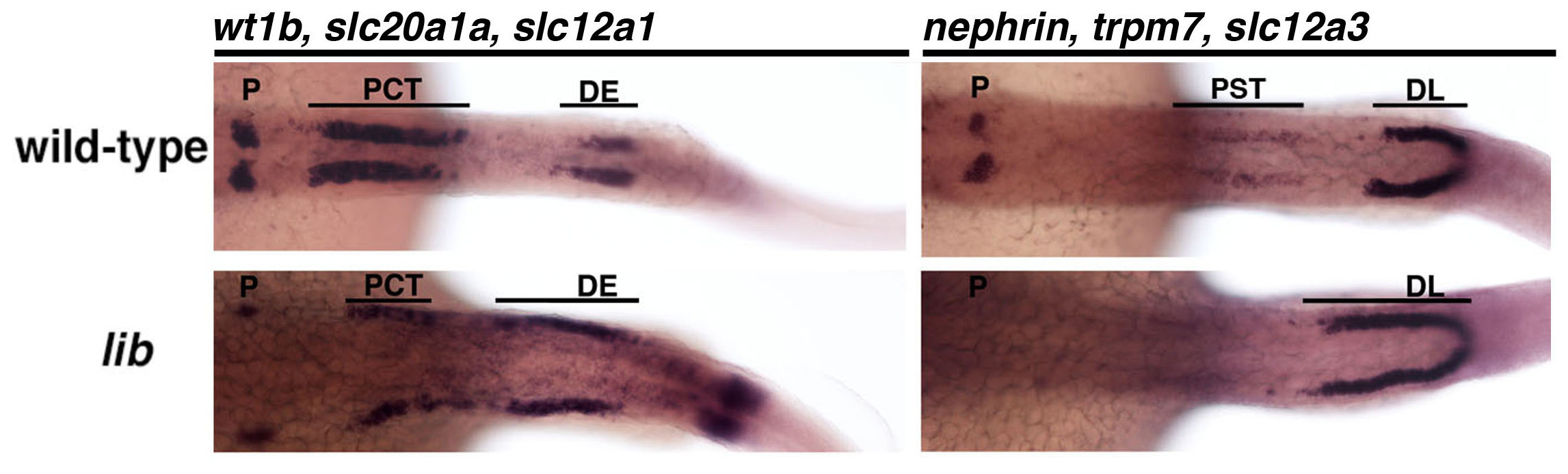
Figure 4. Developmental patterning of cell types that comprise the pronephros embryonic kidney organ in haploid Tübingen strain zebrafish. Haploid wild types displayed a normal pattern of cell types in the embryonic kidney structure (top row). The embryonic kidney consists of a pair of segmented functional units known as nephrons. The segmented pattern of discrete cell types present in nephrons can be visualized based on the whole mount in situ hybridization expression pattern of gene transcripts that are unique to the differentiated nephron cell types, which include the podocytes (P) marked by wt1b and nephrin, the proximal convoluted tubule (PCT) marked by slc20a1a, the proximal straight tubule (PST) marked by trpm7, the distal early (DE) marked by slc12a1, and the distal late tubule (DL) marked by slc12a3. lib haploid mutant embryos (bottom row) display reduced podocytes, PCT and an absent PST, along with an expanded DE and DL segment, similar to diploid lib embryos18. Embryos were photographed in a dorsal view, with anterior to the left, at a 10X magnification.
Discussion
Loading...
$$\rightleftharpoonup{xx}$$
$$\longleftharp{xx}$$,
$$\longrightharp{xx}$$,
Haploid screening is a useful technique to discover essential genes requisite for early developmental stages. Further, culturing of haploid cells from the medaka fish has been used to isolate ES-like cell lines that have numerous research applications and potential, demonstrating that haploid cells may provide a valuable future venue for other types of vertebrate genetic studies20,21. This protocol provides a demonstration of the methods involved with performing the production of haploid zebrafish embryos through in vitro fertilization with UV inactivated sperm. This methodology can be used to perform forward haploid screens with mutagenized F1 fish, which can be made using wild type, transgenic or mutant strains for enhancer/suppressor screening.
Although the screening time using a haploid strategy is vastly shortened compared to diploid screening, it will nevertheless take several months to complete. As we described previously, there are many benefits that exist by performing a screen using a haploid schema, all of which can aid in making novel contributions to the current knowledge about any number of developmental events. The haploid screen methodology is more efficient for the identification of recessive mutant alleles than diploid-based screens due to the reduction of time, space, and the number of fish that are needed. However, there are several pitfalls to a haploid screen. A haploid screen can only identify mutants in genes that are active within the first few days of development, as the embryo can only survive for a limited time with a haploid genome. Genes that are expressed later in development would need to be identified by a different method, such as a diploid screen. Also, some organs do not develop properly in a haploid organism. There may be anatomical abnormalities, or a delayed time of development, which may cause the mutation to be missed by the haploid screening technique. In addition, some strains of fish do not develop as well in a haploid condition15. Haploids can be categorized by a grading series of good, intermediate and poor embryonic features, with A embryos being the most normal, B to designate embryos with defects in the axis but a clear head and tail, and C/D to designate embryos with unrecognizable features (masses of cells atop yolk balls)15,17. The proportions of embryos within these categories varies by zebrafish strain, with increased numbers of better quality haploids more prevalent in particular genetic backgrounds15,17. Due to these factors, it is necessary to find a proper strain of zebrafish to perform the mutagenesis and subsequent crosses. In our recent experience, the wild type Tübingen strain produced viable haploids for screening (as shown in Figures 3 and 4) though historically zebrafish researchers have utilized the AB or the AB* strains for haploid experimentation15.
In addition to these aforementioned challenges, not all primed adult zebrafish females will produce a clutch upon performing the squeezing technique. For example, in working with ENU-mutagenized Tübingen strain females, approximately half of all females produced a clutch upon squeezing, and some fraction of these (typically 10-30%) were poor embryo quality or could not be fertilized. Thus, to perform a haploid screen one must plan to raise at least twice as many fish as needed to screen the desired number of genomes (each female represents one genome screened). Regardless of this consideration, the benefits of the haploid method of screening to cover large numbers of genomes without a massive animal facility are indeed quite significant if the assay is amenable to the haploid state. However, it should be also noted that the further study of the mutation(s) identified in the haploid clutch is fully reliant on successfully raising an outcross from the female founder. As with any screen, ‘hits’ initially identified during the screening process must be re-identified in the diploid state.
Despite the pitfalls of using the haploid screen, it has shown to be very successful in identifying mutants, which have been analyzed in depth with insightful gains to the scientific field15. Haploid screens can be implemented to investigate other poorly understood processes of vertebrate development. Using haploids for enhancer and suppressor screens is one way to gain more insight into the activities and regulatory network of a gene of interest. The haploid screening technique can also be used to identify enhancers and suppressors of mutants that have already been isolated by methods such as chemical or insertional mutagenesis or reverse genetics approaches like TILLING or TALENs. In short, the previously isolated mutant can be mutagenized with ENU and screened for enhancers or suppressors of the original mutant phenotype. The mutant must be able to reach adult stages, so it is necessary to have a heterozygous animal or a weak homozygous mutant fish to perform this screen, however recent advances in transgenesis have allowed researchers to skirt this problem. Learning how to manipulate pathways in development can lead to many exciting possibilities including the prospect of perturbing pathways to treat disease states.
Disclosures
Loading...
$$\rightleftharpoonup{xx}$$
$$\longleftharp{xx}$$,
$$\longrightharp{xx}$$,
The authors have nothing to disclose.
Acknowledgements
Loading...
$$\rightleftharpoonup{xx}$$
$$\longleftharp{xx}$$,
$$\longrightharp{xx}$$,
This work was supported by funding to RAW from the following: NIH grants K01 DK083512, DP2 OD008470, and R01 DK100237; March of Dimes Basil O’Connor Starter Scholar Award #5-FY12-75; start up funds from the University of Notre Dame College of Science and Department of Biological Sciences; and a generous gift to the University of Notre Dame from Elizabeth and Michael Gallagher on behalf of the Gallagher Family to foster stem cell research. The funders had no role in the study design, data collection and analysis, decision to publish, or preparation of the manuscript. We thank the staffs of the Department of Biological Sciences for their support, and the Center for Zebrafish Research at Notre Dame for their outstanding dedication in the care and welfare of our zebrafish colony. We thank G.F. Gerlach for taking the photographs provided in Figure 4. Finally, we thank all the members of our research lab for their comments, discussions and insights about this work.
Materials
| Name | Company | Catalog Number | Comments |
|---|---|---|---|
| Hank's Stock Solution #1 | 8.0 g NaCl, 0.4 g KCl in 100 ml ddH2O | ||
| Hank's Stock Solution #2 | 0.358 g Na2HPO4 anhydrous; 0.60 g K2H2PO4 in 100 ml ddH2O | ||
| Hank's Stock Solution #4 | 0.72 g CaCl2 in 50 ml ddH2O | ||
| Hank's Stock Solution #5 | 1.23 g MgSO4-7H2O in 50 ml ddH2O | ||
| Hank's Premix | Combine in the following order: (1) 10.0 ml HS#1, (2) 1.0 ml HS#2, (3) 1.0 ml HS#4, (4) 86 ml ddH2O, (5) 1.0 ml HS#5. Store all HS Solutions and Premix at 4 °C. | ||
| Hank's Stock Solution #6 | 0.35 g NaHCO3 in 10.0 ml ddH2O; make fresh on day of use. | ||
| Hank's Final working solution | Combine 9.9 ml of Hank's Premix with 0.1 ml HS Stock #6 | ||
| XX-Series UV Bench lamp | UVP | 95-0042-05 | Model XX-15S Bench Lamp |
| XX Bench Stand (Adjustable) | UVP | 18-0062-01 | Model XX-15 Stand |
| Embryo incubation dish | Falcon | 35-1005 | |
| 50 X E3 | 250 mM NaCl, 8.5 mM KCl, 16.5 mM CaCl2, 16.5 mM MgSO4 in distilled water. Supplement with methylene blue (1 x 10⁻⁵ M) (Sigma M9140) to inhibit contamination. | ||
| 1 X E3 | Dilute 50 X E3 in distilled water | ||
| Stereomicroscope | Nikon | SMZ645; SMZ1000 |
References
Loading...
$$\rightleftharpoonup{xx}$$
$$\longleftharp{xx}$$,
$$\longrightharp{xx}$$,
- Large scale genetics in a small vertebrate, the zebrafish. Int J Dev Biol. 40, 221-227 (1996).">Haffter, P., Nüsslein-Volhard, C. Large scale genetics in a small vertebrate, the zebrafish. Int J Dev Biol. 40, 221-227 (1996).
- Animal models of human disease: zebrafish swim into view. Nat Rev Genet. 8, 353-367 (2007).">Lieschke, G. J., Currie, P. D. Animal models of human disease: zebrafish swim into view. Nat Rev Genet. 8, 353-367 (2007).
- Stages of embryonic development of the zebrafish. Dev Dyn. 203, 253-310 (1995).">Kimmel, C. B., et al. Stages of embryonic development of the zebrafish. Dev Dyn. 203, 253-310 (1995).
- Hooked! Modeling human disease in zebrafish. J Clin Invest. 122, 2337-2343 (2012).">Santoriello, C., Zon, L. I. Hooked! Modeling human disease in zebrafish. J Clin Invest. 122, 2337-2343 (2012).
- The zebrafish reference genome sequence and its relationship to the human genome. Nature. 496, 498-503 (2013).">Howe, K., et al. The zebrafish reference genome sequence and its relationship to the human genome. Nature. 496, 498-503 (2013).
- The identification of genes with unique and essential functions in the development of the zebrafish, Danio rerio. Development. 123, 1-26 (1996).">Haffter, P., et al. The identification of genes with unique and essential functions in the development of the zebrafish, Danio rerio. Development. 123, 1-26 (1996).
- A genetic screen for mutations affecting embryogenesis in zebrafish. Development. 123, 37-43 (1996).">Driever, W., et al. A genetic screen for mutations affecting embryogenesis in zebrafish. Development. 123, 37-43 (1996).
- Zebrafish make a big splash. Cell. 87, 969-977 (1996).">Eisen, J. S. Zebrafish make a big splash. Cell. 87, 969-977 (1996).
- A large-scale insertional mutagenesis screen in zebrafish. Genes Dev. 13, 2713-2724 (1999).">Amsterdam, A., et al. A large-scale insertional mutagenesis screen in zebrafish. Genes Dev. 13, 2713-2724 (1999).
- ZFIN: enhancements and updates to the zebrafish model organism database. Nucleic Acids Res. 39, (2011).">Bradford, Y., et al. ZFIN: enhancements and updates to the zebrafish model organism database. Nucleic Acids Res. 39, (2011).
- Efficient mapping and cloning of mutations in zebrafish by low-coverage whole-genome sequencing. Genetics. 190, 1017-1024 (2012).">Bowen, M. E., et al. Efficient mapping and cloning of mutations in zebrafish by low-coverage whole-genome sequencing. Genetics. 190, 1017-1024 (2012).
- Mutation mapping and identification by whole-genome sequencing. Genome Res. 22, 1541-1548 (2012).">Leshchiner, I., et al. Mutation mapping and identification by whole-genome sequencing. Genome Res. 22, 1541-1548 (2012).
- Rapid positional cloning of zebrafish mutations by linkage and homozygosity mapping using whole-genome sequencing. Development. 139, 4280-4290 (2012).">Obholzer, N., et al. Rapid positional cloning of zebrafish mutations by linkage and homozygosity mapping using whole-genome sequencing. Development. 139, 4280-4290 (2012).
- Fast homozygosity mapping and identification of a zebrafish ENU-induced mutation by whole-genome sequencing. PLoS ONE. 7, (2012).">Voz, M. L., et al. Fast homozygosity mapping and identification of a zebrafish ENU-induced mutation by whole-genome sequencing. PLoS ONE. 7, (2012).
- Haploid screens and gamma ray mutagenesis. Meth Cell Biol. 60, 43-70 (1999).">Walker, C. Haploid screens and gamma ray mutagenesis. Meth Cell Biol. 60, 43-70 (1999).
- Production of clones of homozygous diploid zebra fish (Brachydanio rerio). Nature. 291, 293-296 (1981).">Steisinger, G., et al. Production of clones of homozygous diploid zebra fish (Brachydanio rerio). Nature. 291, 293-296 (1981).
- The Zebrafish Book 4th ed. , University of Oregon Press. Eugene, USA. (2000).">Westerfield, M. Chapter 2, Embryo Production by in vitro fertilization.”. The Zebrafish Book 4th ed. , University of Oregon Press. Eugene, USA. (2000).
- Making gynogenetic diploid zebrafish by early pressure. (28), (2009).">Walker, C., et al. Making gynogenetic diploid zebrafish by early pressure. (28), (2009).
- The Zebrafish Book 4th ed. , University of Oregon Press. Eugene, USA. (2000).">Westerfield, M. Chapter 10: Recipes.”. The Zebrafish Book 4th ed. , University of Oregon Press. Eugene, USA. (2000).
- Generation of medaka fish haploid embryonic stem cells. Science. 326, 430-433 (2009).">Yi, M., Hong, N., Hong, Y. Generation of medaka fish haploid embryonic stem cells. Science. 326, 430-433 (2009).
- Derivation and characterization of haploid embryonic stem cell cultures in medaka fish. Nat Protoc. 5, 1418-1430 (2010).">Yi, M., Hong, N., Hong, Y. Derivation and characterization of haploid embryonic stem cell cultures in medaka fish. Nat Protoc. 5, 1418-1430 (2010).
- Undertaking a successful gynogenetic haploid screen in zebrafish.” Zebrafish, Methods and Protocols. 1st ed. , Humana Press. (2009).">Layton, J. E. Undertaking a successful gynogenetic haploid screen in zebrafish.” Zebrafish, Methods and Protocols. 1st ed. , Humana Press. (2009).
- The cdx genes and retinoic acid control the positioning and segmentation of the zebrafish pronephros. PLoS Genet. 3, 1922-1938 (2007).">Wingert, R. A., et al. The cdx genes and retinoic acid control the positioning and segmentation of the zebrafish pronephros. PLoS Genet. 3, 1922-1938 (2007).
- Zebrafish nephrogenesis involves dynamic spatiotemporal expression changes in renal progenitors and essential signals form retinoic acid and irx3b. Dev Dyn. 240, 2011-2027 (2011).">Wingert, R. A., Davidson, A. J. Zebrafish nephrogenesis involves dynamic spatiotemporal expression changes in renal progenitors and essential signals form retinoic acid and irx3b. Dev Dyn. 240, 2011-2027 (2011).
Reprints and Permissions
Request permission to reuse the text or figures of this JoVE article
Request Permission