Here, a procedure is described for the establishment of systemic infection in the neonatal rat with cultures of Escherichia coli K1. This non-invasive procedure permits colonization of the gastrointestinal tract, translocation of the pathogen to the systemic circulation, and invasion of the central nervous system at the choroid plexus.
Method Article
Non-Invasive Model of Neuropathogenic Escherichia coli Infection in the Neonatal Rat
Opens in a new tab
In This Article
Summary
Loading...
$$\rightleftharpoonup{xx}$$
$$\longleftharp{xx}$$,
$$\longrightharp{xx}$$,
Abstract
Loading...
$$\rightleftharpoonup{xx}$$
$$\longleftharp{xx}$$,
$$\longrightharp{xx}$$,
Investigation of the interactions between animal host and bacterial pathogen is only meaningful if the infection model employed replicates the principal features of the natural infection. This protocol describes procedures for the establishment and evaluation of systemic infection due to neuropathogenic Escherichia coli K1 in the neonatal rat. Colonization of the gastrointestinal tract leads to dissemination of the pathogen along the gut-lymph-blood-brain course of infection and the model displays strong age dependency. A strain of E. coli O18:K1 with enhanced virulence for the neonatal rat produces exceptionally high rates of colonization, translocation to the blood compartment and invasion of the meninges following transit through the choroid plexus. As in the human host, penetration of the central nervous system is accompanied by local inflammation and an invariably lethal outcome. The model is of proven utility for studies of the mechanism of pathogenesis, for evaluation of therapeutic interventions and for assessment of bacterial virulence.
Introduction
Loading...
$$\rightleftharpoonup{xx}$$
$$\longleftharp{xx}$$,
$$\longrightharp{xx}$$,
Systemic bacterial infections are a major threat to the well-being and survival of the newborn; preterm infants are particularly vulnerable. Neonatal bacterial meningitis (NBM), frequently associated with bacterial sepsis, continues to be a significant source of mortality and morbidity during the first few weeks of life and the problem is exacerbated by the continuing evolution of resistance to frontline antibacterial drugs1,2. A case of NBM is a medical emergency that carries a high medical, social and economic burden3; consequently, there is an urgent need for new therapeutics and, particularly, novel prophylactic strategies to reduce the burden of infection. Some features of NBM are unusual: in the developed world, Escherichia coli and Group B streptococci are responsible for the large majority of cases and the capacity of these strains to elicit NBM is almost always associated with the presence of a protective polysaccharide capsule that enables the pathogen to evade immune recognition processes4. A very high proportion (80 - 85%) of neuroinvasive E. coli express the K1 capsule5,6, an α-2,8-linked polysialic acid polymer that is structurally identical to host modulators of neuronal plasticity7.
The evaluation of new therapeutics and prophylactics for NBM and associated bacteremia and sepsis would clearly benefit from a robust animal model of infection that mimics the key features of the disease in the human neonate, in particular the strong age-dependency and the natural pathway of infection. A wide range of models for Gram-positive and Gram-negative bacterial meningitis are available8,9 and these have considerably extended our knowledge of pathogenesis, pathophysiology and treatment options in these infections. Thus, experimental infections in rats, mice, rabbits and monkeys have been used to study meningitis in both the neonate and the adult. However, many of these models employ direct intracisternal or subcutaneous injection of bacteria for initiation of infection, creating an artificial pathogenesis by bypassing natural processes of dissemination from the site of colonization. In some cases these methods of inoculation led to significant changes in pathology; for example, subcutaneous administration of E. coli K1 strains abrogated the age-dependency associated with the natural infection, producing bacteremia and invasion of the central nervous system (CNS) in both neonates and adults10. Predisposition to E. coli NBM is critically dependent on vertical transmission of the causative agent from mother to infant at or soon after birth11. Maternally-derived E. coli K1 bacteria colonize the neonatal gastrointestinal (GI) tract11-13, which is sterile at birth but rapidly acquires a complex microbiota14. In colonized neonates, E. coli K1 bacteria have the capacity to translocate from the intestinal lumen into the systemic circulation before entering the CNS across the blood-brain or blood-cerebrospinal fluid barriers9,15. The design of robust models of experimental infection should take these details into account.
Although mice have been widely used for the study of some forms of bacterial meningitis8, they are unsuitable for studies of neonatal infection: they are overwhelmed by systemic infection and do not show the strong age dependency characteristic of human infants16. Further, α-defensins, key peptides of the GI tract providing protection against systemic invasion by E. coli K117, are highly expressed in Paneth cells and neutrophils in humans and rats but not in mice18. There is a remarkable degree of duplication, redundancy and heterogeneity in mouse defensin and related cryptidin genes not found in other animals19. The neonatal rat was initially used by Moxon and coworkers20 to investigate the pathogenesis of Haemophilus influenzae meningitis following intranasal inoculation, replicating the natural site of colonization of this neonatal pathogen in the human, and subsequently adapted for age-dependent E. coli K1 bacteremia and meningitis. Bortolussi et al.21 employed intraperitoneal injection of the bacterial inoculum to initiate infection but the key study of Glode and coworkers22 used oral gastric feeding to parallel the natural route of infection after GI colonization. As the gastric tube may damage mucosal surfaces, the procedure was refined to include feeding of the inoculum to the neonates23. Here, the method for GI tract colonization and procedures for tracking the infection in susceptible rat pups are described; additionally, therapeutic and preventative applications of the model are discussed.
Protocol
Loading...
$$\rightleftharpoonup{xx}$$
$$\longleftharp{xx}$$,
$$\longrightharp{xx}$$,
All animal experiments performed in this study conformed to national and European legislation and were approved by the Ethical Committee of the UCL School of Pharmacy and the UK Home Office (HO). All animal work was conducted under HO project licenses PPL 80/2243 and PPL 70/7773.
1. Rat Preparation
- Carry out all in vivo experiments using pathogen-free Wistar neonatal rats.
- Retain all rat litters (12 neonates per colony) in individual cages with their lactating mothers (9 - 11 week old females), under optimal conditions (19 - 21 °C, 45 - 55% humidity, 15 - 20 air changes/hr and 12 hr light/dark cycle).
2. Bacterial Cell Preparation
- Store E. coli K1 stocks in 20% (v/v) glycerol at -80 °C. For each experiment, plate out stock bacteria onto Mueller-Hinton (MH) agar plates, and incubate at 37 °C O/N.
- The day before feeding, inoculate 10 ml of MH broth with a single E. coli K1 colony and culture O/N at 37 °C, 200 rpm. As a control for medium contamination, prepare 10 ml of uninoculated MH broth and incubate at 37 °C at 200 rpm.
- Transfer 100 µl of O/N bacterial suspension into 9.9 ml of MH broth (1:100 v/v), and incubate at 37 °C, 200 rpm, until mid-exponential-phase is reached, corresponding to an optical density of 0.6 measured at a wavelength of 600 nm (OD600).
3. Feeding of Neonatal Rats with E. coli K1
- Gently hold the animal vertically in one hand from the scruff of the neck, allowing the mouth to open. Slowly insert the sterile pipette tip into the mouth of the animal, and pipette 20 µl of the inoculum (~37 °C) into the mouth of the animal over a 30 sec time period. Return the animal to its mother immediately after feeding.
- Verify that 4 - 8 x 10⁶ bacteria have been fed to each pup by serial dilution of the inoculum in PBS and spotting onto MH agar plates.
4. Assessment of Colonization by E. coli K1
- Gently hold the animal in one hand from the scruff of the neck. Moisten a sterile cotton-tipped swab in sterile PBS, and then gently swab the perianal area. Return the animal to its mother immediately after swabbing.
- Place the swab tip into a tube containing 300 µl of sterile PBS and store on ice.
- Determine the numbers of viable bacteria by serial dilution in PBS and plating onto MacConkey agar plates.
- Determine viable E. coli K1 by bacteriophage susceptibility testing. Pick an individual colony using a sterile microbiological loop, dipped into 200 μl sterile PBS, then subject to vortex mixing.
- Using a sterile microbiological loop, sub-culture onto MH agar in a straight line. Allow the plate to dry for 30 sec, and then pipette 10 µl of bacteriophage K1E (109 plaque forming units/ml (PFU/ml)) onto the center of the line. Incubate the plate O/N at 37 °C.
- Next day, examine the plate for bacteriophage-mediated lysis.
5. Assessment of Disease Severity
- Assess the disease severity of each animal 4 - 5 times daily using the seven-point scoring system outlined in Table 1.
- If an animal scores ≥3 out of 7, immediately cull the animal to minimize suffering. Record the animal as dead.
6. Euthanization of Neonatal Rats and Blood Collection
- Gently hold the animal in one hand, exposing the head and neck. Disinfect the neck of the animal by wiping with an alcohol swab. Disinfect a large pair of scissors using 70% ethanol, before rinsing in sterile PBS to remove trace ethanol.
- Gently hold the animal directly above a Petri dish. Decapitate the neonate using sharp surgical scissors, ensuring that the blood drips into the Petri dish. Avoid contact with the head to prevent contamination of blood with skin flora.
- Immediately collect the blood using a sterile pipette and mix with heparin sodium salt at a concentration of 20 - 50 units/ml in a 0.5 ml microcentrifuge tube.
- Determine the numbers of viable bacteria by serial dilution in PBS and plating onto MacConkey agar plates. Determine the numbers of viable E. coli K1 by testing the susceptibility of viable bacteria to bacteriophage K1E.
7. Dissection
- Following decapitation of the neonatal rat, use aseptic conditions to excise and collect tissues of interest. Wash all instruments (shown in Figure 1) in 70% ethanol and sterile PBS.
- Clean the dissection board and wet the abdomen completely with 70% ethanol. Position the corpse on its back and fix to the dissection board by (i) pinning the left hind leg to the operation table with a sterile needle, (ii) stretching the corpse and pinning the right foreleg to the operation table, (iii) pinning the right hind leg and left foreleg to the operation table.
- Pull the skin along the left hand side of the lower abdomen using forceps, then cut along the left hand side of the corpse from the lower abdomen to the sternum using small dissection scissors.
- Extend the excision across the sternum, and then down the right hand side of the corpse from the sternum to the lower abdomen using forceps and small dissection scissors, ensuring that none of the underlying structures are damaged.
- Finally, gently pull down the skin flap from the sternum to the lower abdomen using iris curved dissection forceps to expose the peritoneum.
- Gently raise the peritoneum with forceps and cut vertically to expose the internal organs, ensuring that none of the underlying organs are damaged.
8. Collecting the GI Tract
- Identify the stomach, small intestine, cecum, colon and mesenteric lymphatic system.
- Remove the stomach by gently transecting at either side, then place into a bijou containing 1.6 ml of sterile PBS using sterile forceps.
- Transect the colon at the rectum. Carefully pull the entire intestinal mass from the corpse using dissection forceps and place in a sterile Petri dish. Ensure that this is done gently so that the intestinal structure and mesenteric lymphatic structures do not separate.
9. Separation of the GI Tract and the Mesenteric Lymphatic System (Figure 2)
- Pour 30 ml of sterile PBS into the Petri dish, ensuring the GI tract is completely submerged.
- Identify the central mass of the mesenteric lymphatic system, and pinch using fine dissection forceps. Identify the most proximal part of the small intestine, and pinch using fine dissection forceps.
- Slowly pull the central mass of mesenteric lymphatic system and the distal small intestine in opposite directions until the two tissues are completely separated from each other. Perform this process with care to ensure that the small intestines are not stretched, as this prevents reproducible collection of proximal, middle and distal regions. Place the mesenteric lymphatic system in 300 - 500 μl sterile PBS and place on ice.
10. Sectioning of the GI Tract
- Transect the GI tract at the cecum to separate the small intestine from the colon.
- Place the colon in 300 - 500 μl sterile PBS and place on ice.
- Collect representative tissues from proximal, middle and distal regions of the small intestine. Align the small intestine from the mid-point.
- Then, (i) collect the last 2 cm of tissue prior to the caecum as the distal small intestine, (ii) collect the tissue from 5 - 7 cm above the mid-point as the proximal small intestine, and (iii) collect the tissue from 3 - 5 cm below the mid-point as the middle small intestine.
11. Collecting the Liver
- Identify the three lobes of the rat liver after removal of the stomach.
- Pull lobes away from the abdominal cavity using fine dissection forceps.
- Remove the liver by cutting any attaching ligaments using fine scissors. Place liver in 300 - 500 μl sterile PBS and place on ice.
12. Collecting the Spleen
- Identify the spleen.
- Pull spleen away from the stomach and use fine scissors to cut the gastrosplenic ligament.
- Place spleen in 300 - 500 μl sterile PBS and place on ice.
13. Collecting the Kidneys
- Identify the kidneys that will become visible at the back of the abdominal cavity upon removal of the gastrointestinal tract and other organs.
- Cut ureters and any attaching ligaments using fine dissection scissors and remove kidneys using fine forceps. Remove adrenal glands and any fatty material (if still attached) using fine forceps and dissection scissors.
- Place both kidneys in 300-500 μl sterile PBS and place on ice.
14. Collecting the Brain
- After decapitation, hold the head in an upright position using curved forceps. Pinch skin on top of head longitudinally using another set of curved forceps, and make an incision using fine dissection scissors. Cut remaining skin near neck, again longitudinally with fine dissection scissors.
- Pull skin in an outwards direction using fine forceps on both sides to reveal the skull and remove skin flaps using fine dissection scissors. Place fine dissection scissors in spinal opening and make an incision along the skull towards the rostrum.
- Pull both sections of skull in an outwards direction using fine forceps. Cut to remove skull segments.
- Remove brain using broad forceps, using an upwards scooping motion from the spinal opening towards the rostrum. Wash the brain in PBS twice to remove any blood from decapitation procedure.
15. Tissue Processing and Homogenization
- For experiments requiring viability counts or DNA extraction, place tissues into tubes containing sterile PBS. Immediately determine the numbers of viable bacteria by serial dilution in PBS and plating onto MacConkey agar plates, or store at -20 °C with 20% glycerol for short term. For DNA extraction, store tissues at -20 °C.
- For experiments requiring RNA extraction, place tissues into appropriate volumes of RNAlater solution (10 μl per 1 mg tissue), then store immediately at -20 °C for short term or at -80 °C long term storage.
- For experiments requiring imaging, place tissues in 10 ml Methacarn solution (60% methanol, 30% chloroform and 10% acetic acid), then store at RT (20 - 25 °C) for up to 3 weeks.
- Calculate tissue weight by comparison of weight of tubes before and after addition of tissues. Homogenize tissues using a homogenizer. Wash the homogenizer once in 70% ethanol and twice in sterile PBS in between homogenization of each sample.
NOTE: Methacarn fixation is desirable for fixation of intestinal samples in order to preserve mucin-related mucosal defense structures; formalin fixation may be used for systemic tissues.
Results
Loading...
$$\rightleftharpoonup{xx}$$
$$\longleftharp{xx}$$,
$$\longrightharp{xx}$$,
The E. coli K1 systemic infection model described here replicates many of the features of the natural infection in humans. The bacteria are ingested, colonize the GI tract, translocate into the blood compartment via the mesenteric lymph nodes before establishing organ-specific disease with associated inflammation of the brain24. Importantly, the model displays strong age dependency; as shown in Figure 3, two-day-old (P2) rat pups are highly susceptible to invasive disease but over a seven-day period the animals become progressively more refractory to infection, but not to GI tract colonization17. After transit from the site of GI colonization to the blood compartment, the bacteria can be visualized in blood samples by fluorescence microscopy (Figure 4) before entering the CNS predominantly at the choroid plexus25. In some animals, there is extensive invasion of other major organs such as lung, spleen and kidney25.
Bacterial numbers in tissues can vary substantially between individual pups25 but when the bioburden is scored as either present or absent there is a high degree of reproducibility with regard to organ invasion. With a litter of 12 pups as a single test cohort, power calculations using G* Power Software determined that this sample size equates to a 98.6% probability of finding an effect based on survival using six animals from the cohort and >99% probability if all twelve are taken into consideration. The model is therefore suitable for evaluation of novel agents specifically tailored for the treatment of neonatal bacterial infections and has been used in the procedure to evaluate the therapeutic potential of the capsule depolymerase EndoE that selectively removes the K1 capsule from the bacterial surface24-26. It may also be used to investigate host-bacteria interactions that impact on the pathogenesis of E. coli neuropathogens; within this context it has been employed for studies of E. coli A192PP colonization and dissemination. It has been demonstrated that E. coli A192PP cells persist in the GI tract of P2, P5 and P9 pups in large numbers; temporal aspects of colonization in these three groups were very similar (Figure 5) and reflects the capacity of the bacteria to replicate and maintain population density within the gut.
The virulence of the clinical isolate A192 was enhanced by serial passage in neonatal rats in order to ensure little or no redundancy of animal use. E. coli A192 colonized P2 neonatal rats with 100% efficiency, elicited bacteremia in 35% of animals and produced a lethal effect in 25%27. The passaged derivative A192PP colonizes the GI tract, produces bacteremia and causes lethality in all P2 pups. Thus, the model can be employed to investigate the virulence of different K1 strains with respect to their capacity to invade the CNS and other organ systems from the site of colonization. In this context, Pluschke and co-workers23 used a neonatal rat infection model to determine the capacity of 95 E. coli K1 strains of human origin to cause bacteremia after gut colonization; they observed wide variations in the efficiency of both colonization and invasive capability, underpinning the clonal nature of E. coli K1 neuropathogens.
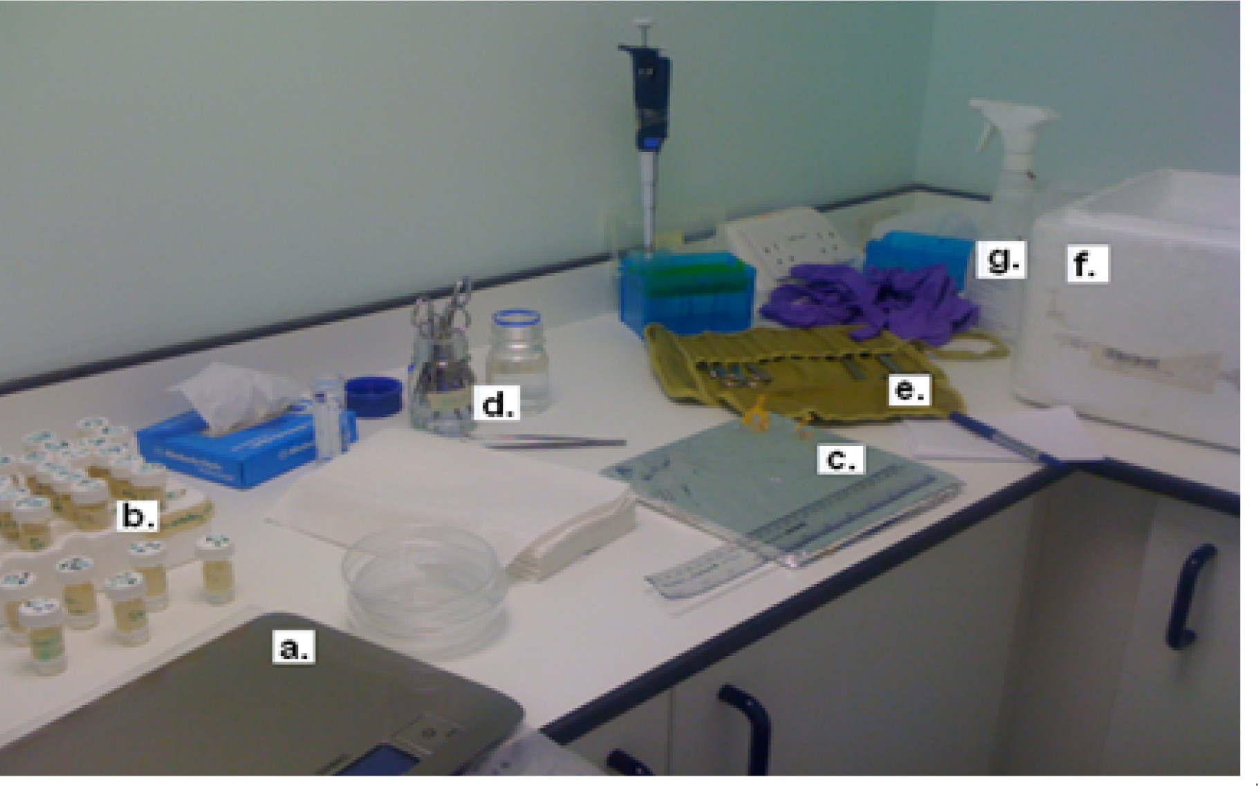
Figure 1. Materials for tissue collection: (A) weighing scale, (B) pre-weighed tubes containing necessary media, (C) operation table, ruler and needles to pin down the animal, (D) 70% (v/v) ethanol and PBS to sterilize the tissue collection equipment, (E) tissue collection kit including a large pair of scissors for decapitation and scissors and tweezers and blades of various size and shapes, (F) ice to preserve the tissues, (G) 70% (v/v) ethanol for sterilizing the operation table and surrounds. Please click here to view a larger version of this figure.
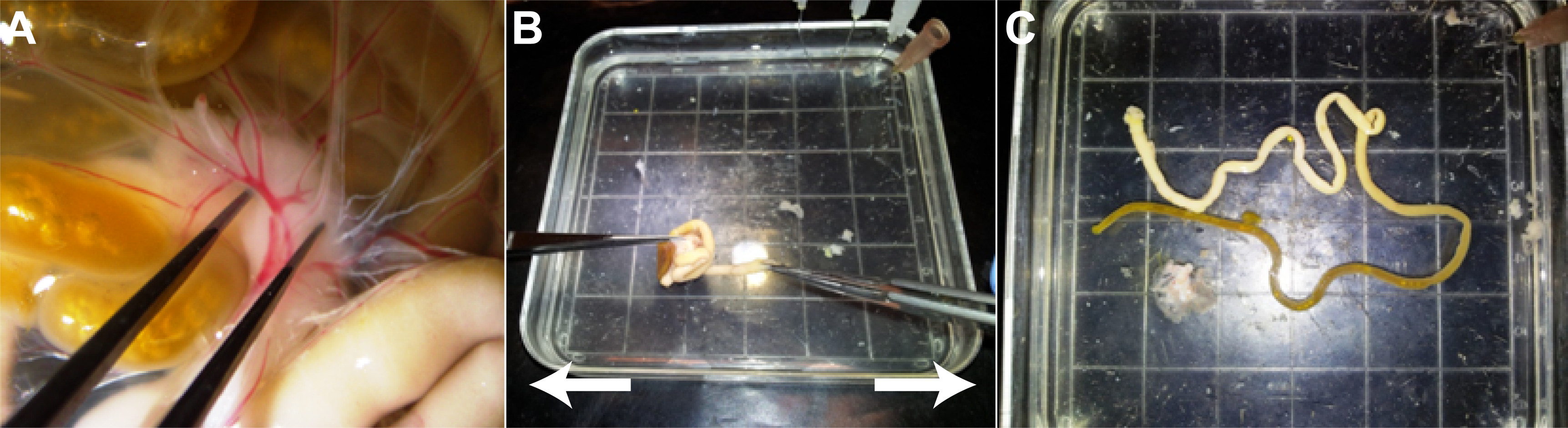
Figure 2. Separation of the GI tract and the mesenteric lymphatic system. (A) Grip the central mass of the mesenteric lymphatic system with fine dissection forceps. (B) Grip the proximal part of the small intestine, and pull in opposite directions. (C) The GI tract and mesenteric lymphatic system will fully separate. Please click here to view a larger version of this figure.
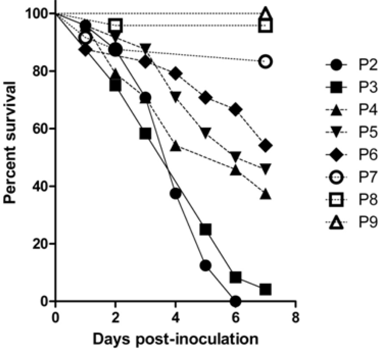
Figure 3. Survival of neonatal rat pups aged from two days (P2) to nine days (P9) following oral administration of E. coli A192PP, illustrating the strong age dependency of systemic infection. Each group represents 24 neonates.
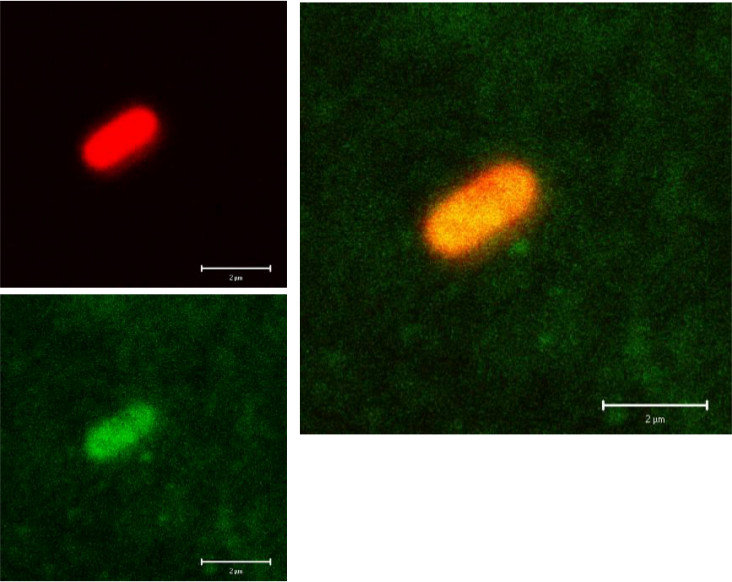
Figure 4. Fluorescence images of E. coli A192PP cells in a blood smear from a P2 pup infected following oral administration of bacteria. The lipopolysaccharide O antigen at the bacterial surface was stained with rabbit anti-O18 polyclonal antibody and Alexa546-conjugated goat-anti-rabbit second antibody. The K1 capsule was visualized with EndoE-GFP reagent. Virtually all bacteria detected in blood samples displayed the protective K1 capsule. Images were captured by Dr Andrea Zelmer. Please click here to view a larger version of this figure.
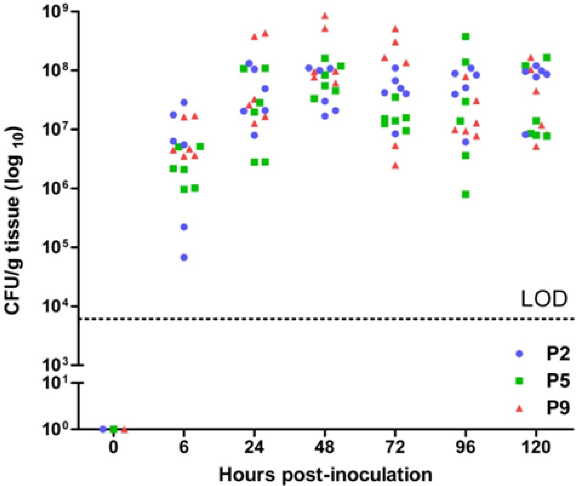
Figure 5. E. coli A192PP intestinal colonization following administration of the bacterial inoculum. DNA was extracted from whole intestine and E. coli K1 colony forming units/g (CFU/g) tissue determined by quantitative polymerase chain reaction (qPCR) targeting the polysialyltransferase (neuS) gene as described elsewhere17. LOD: limit of detection.
| Feature | Healthy | Unhealthy |
| Color of the skin | Pink | Pale/Yellow |
| Agility (righting reflex) | Pup immediately reverses on backward placement | Difficulty in reversing backward placement (> 3 sec) or cannot achieve |
| Gentle pressure on the abdomen | No sound | Sound of agitation |
| Stomach/milk line | Visible and white | Not visible |
| Temperature | Warm | Relatively cold* |
| Weight | Gain of 1.5-2 g per day | No weight gain or weight loss |
| Behavior when placed in cage | Moves towards mother and starts feeding | Cannot move towards mother and shows difficulty feeding |
Table 1. Seven-point scoring system: The first three scores listed are usually the initial signs observed. *Neonates with systemic infection experience elevated body temperature (> 2 °C). However, due to the lack of agility of the animals to reach their mother to maintain body temperature, unhealthy animals may become separated from the litter and feel cold to the bare hand.
Discussion
Loading...
$$\rightleftharpoonup{xx}$$
$$\longleftharp{xx}$$,
$$\longrightharp{xx}$$,
The animal model described here builds on previous work that aimed to reproduce the salient features of naturally occurring infections in humans. Neonatal rats were initially employed to study infant meningitis due to H. influenzae type b as the species satisfied the key criteria for a robust model of infection. Thus, the portal of entry of the relevant pathogen should reflect that of the natural human infection and reproducibly give rise to similar pathology of sufficient duration to allow for therapeutic intervention. The techniques used should not limit the applicability of the procedure and should not contribute to disease outcome20. The model of H. influenzae meningitis in infant rats developed by Moxon and colleagues satisfies these criteria20; the natural infection occurs after colonization of the mucosal membranes of the upper respiratory tract and this important feature was replicated in the rat pups by non-traumatic instillation of the bacteria onto the membranes of the nasal passages. Importantly, the age-dependent nature of the infection was replicated in the model.
The same group were also the first to develop a non-invasive model of E. coli K1 NBM in the neonatal rat22. Pathogen-free Sprague-Dawley pups were colonized by feeding 108 to 1010 bacteria through an oral gastric tube; the inoculum was therefore considerably higher than that employed by us. Colonization with the three K1 strains examined, C94 (O7:K1:H-), EC3 (O1:K1:H-) and LH (O75:K1:H3), occurred in a relatively high proportion (48-74%) of K1-fed animals, but incidences of bacteremia, meningitis and mortality were variable and significantly lower than rates of colonization. The clonal nature of E. coli K1 experimental infection was established later23 and it is now apparent that only O18:K1 and, to a lesser extent, O7:K1 serotypes are able to consistently cause systemic infection. For this reason, these investigations of the pathogenesis of neuropathogenic E. coli K1 were based on the use of the virulence-enhanced O18:K1 strain A192PP. A comparison of E. coli K1 feeding of neonatal rats through a gastric tube, as used by Glode and colleagues22, and a droplet feeding method as employed by Achtman’s group23 revealed excessive numbers of deaths using the former method, almost certainly due to damage to mucosal surfaces by the gastric tube. As rates of colonization are comparable with these two methods, it is recommended to use the less invasive method of feeding the bacteria using a pipette with a sterile tip, as described in this communication.
E. coli strain A192PP used in our studies is O18:K1. It is a more virulent derivative of the clinical strain E. coli A192 that was originally recovered from a patient with septicemia27. The increased virulence of the strain was obtained by serial passage through neonatal rats26. The strain elicits an age-dependent disease severity, with 100% bacteremia and mortality when administered to 2 day old animals28. In contrast, 9 day old animals are completely resistant to disease. K1-specific lytic bacteriophage can be used to differentiate E. coli K1 from other E. coli strains29. In this study, susceptibility of viable bacteria to bacteriophage K1E should be used to (i) check the purity of the E. coli K1 suspensions prepared to be fed to the animals, and (ii) to differentiate E. coli K1 from other coliforms in order to calculate viability in perianal swabs, blood and tissue samples. If the colony is E. coli K1, it will be susceptible to bacteriophage K1E lysis, and bacterial growth will be inhibited at the site of bacteriophage inoculation. If the colony is not E. coli K1, it will be resistant to KIE bacteriophage lysis, and there should be an area of bacterial growth at the site of bacteriophage inoculation. It should be borne in mind that animal models cannot reflect all features of the naturally occurring disease. The current model can be modified to examine the virulence characteristics of neuropathogenic bacteria other than E. coli A192PP and variations in the size of the colonizing inoculum can be accommodated. Future applications of the technique could include the evaluation of much-needed drugs to treat the condition and to uncover details of the host response to colonization and tissue invasion.
The method described here is simple but effective. Single litters of 10 - 12 pups were employed as test or control groups and this within-litter approach ensures a high degree of reproducibility and statistical validity. It is imperative that the pups are returned to their natural mothers as soon as possible after any procedure and litters should not therefore comprise animals undergoing different interventions. It is important that the fed inoculum be warmed otherwise the pups will reject the offered culture. The pups rapidly develop a complex microbiota and within two days of birth the GI tract is colonized with a wide range of bacteria from the phyla established as the most abundant microbes in the infant and adult gut. Pups that have not been fed E. coli A192PP do not carry E. coli K1 in the GI tract17 and so determination of rates of colonization is relatively straightforward. However, the neuS-based qPCR method for detection of colonizing E. coli K1 is far more sensitive that traditional culture methods and is strongly recommended17.
Disclosures
Loading...
$$\rightleftharpoonup{xx}$$
$$\longleftharp{xx}$$,
$$\longrightharp{xx}$$,
The authors declare that they have no competing financial interests.
Acknowledgements
Loading...
$$\rightleftharpoonup{xx}$$
$$\longleftharp{xx}$$,
$$\longrightharp{xx}$$,
This work was supported by research grants G0400268 and MR/K018396/1 from the Medical Research Council, and by Action Medical Research. Further support was provided by the National Institute for Health Research University College London Hospitals Biomedical Research Centre.
Materials
| Name | Company | Catalog Number | Comments |
|---|---|---|---|
| Pathogen-free Wistar rats (12 x neonate, 1 x lactating mother) |  Harlan, UK Harlan, UK | ||
| E. coli K1 A192PP | Taylor lab | Mushtaq et al. 2004 | |
| Bacteriophage K1E | Taylor lab | Mushtaq et al. 2004 | |
| Glycerol | Sigma, UK | G5516 | |
| Mueller-Hinton Agar | Oxoid, UK | CM0037 | |
| Mueller-Hinton Broth | Oxoid, UK | CM0405 | |
| MacConkey Agar | Oxoid, UK | CM0007 | |
| Phosphate buffered saline (PBS) | Sigma, UK | P4417 | |
| Ethanol 100% | Sigma, UK | E7023 | |
| Heparin Sodium Salt | Sigma, UK | 84020 | Prepare 20 - 50 units/ml |
| RNAlater Solution | Sigma, UK | R0901 | 10 μl/mg tissue |
| Acetic Acid | Sigma, UK | 320099 | |
| Chloroform | Sigma, UK | C2432 | |
| Methanol | Sigma, UK | 322415 | |
| Cotton-tipped swabs | Fisher Scientific, UK | 11542483 | |
| Alcotip Swabs | Scientific Laboratory Supplies, UK | SWA1000 | |
| Petri dishes | Sigma, UK | P5856 | |
| 30 ml Universal Tube | AlphaLaboratories, UK | CW3890 | |
| 0.5 ml microcentrifuge tubes | StarLab, UK | I1405-1500 | |
| 1.5 ml microcentrifuge tubes | StarLab, UK | I1415-1000 | |
| 0.1 μl calibrated loops | StarLab, UK | E1412-0112 | |
| L-shaped spreaders | StarLab, UK | E1412-1005 | |
| Cuvettes | Fisher Scientific, UK | 10594175 | |
| Forceps straight with fine points | Fisher Scientific, UK | 12780036 | |
| Forceps straight with blunt tips | Fisher Scientific, UK | 12391369 | |
| Forceps watchmaker's curved with very fine points | Fisher Scientific, UK | 12740926 | |
| Scissors straight with very fine points | Fisher Scientific, UK | 12972055 | |
| Laboratory Scissors | VWR, UK | USBE4251 | |
| 25 G Syringe Needles | Greiner Bio-One Ltd | N2525 | |
| LAMBDA 25 UV/Vis Spectrophotometers | PerkinElmer, UK | L60000BB | |
| Unitemp Incubator | B&T, UK | OP958 | |
| Multitron shaking incubator | INFORS HT, UK | AJ118 | |
| Ultra-Turrax T-10 homogenizer | IKA Werke | 0003737000 |
References
Loading...
$$\rightleftharpoonup{xx}$$
$$\longleftharp{xx}$$,
$$\longrightharp{xx}$$,
- Harvey, D., Holt, D. E., Bedford, H. Bacterial meningitis in the newborn: a prospective study of mortality and morbidity. 23 (5), 218-225 (1999).
- Brouwer, M. C., Tunkel, A. R., van de Beek, D. Epidemiology diagnosis, and antimicrobial treatment of acute bacterial meningitis. Clin. Microbiol. Rev. 23 (3), (2010).
- Silva, L. P. A., Cavalheiro, L. G., Queirós, F., Nova, C. V., Lucena, R. Prevalence of newborn bacterial meningitis and sepsis during the pregnancy period for public health care system participants in Salvador, Bahia, Brazil. J. Infect. Dis. 11 (2), 272-276 (2007).
- Levy, O. Innate immunity of the newborn: basic mechanisms and clinical correlates. Nat. Rev. Immunol. 7 (5), 379-390 (2007).
- Robbins, J. B., et al. Escherichia coli K1 capsular polysaccharide associated with neonatal meningitis. N. Eng. J. Med,, et al. 290 (22), 1216-1220 (1974).
- Korhonen, T. K., et al. hemolysin production, and receptor recognition of Escherichia coli strains associated with neonatal sepsis and meningitis. Infect. Immun. 48 (2), 486-491 (1985).
- Rutishauser, U. Polysialic acid in the plasticity of the developing and adult vertebrate nervous system. Nat. Rev. Neurosci. 9 (1), 26-35 (2008).
- Koedel, U., Pfister, H. W. Models of experimental bacterial meningitis. Infect. Dis. Clin. North Am. 13 (3), 549-577 (1999).
- Tunkel, A. R., Scheld, W. M. Pathogenesis and pathophysiology of bacterial meningiits. Clin. Microbiol. Rev. 6 (2), 118-136 (1993).
- Kim, K. S., et al. The K1 capsule is the critical determinant in the development of Escherichia coli meningitis in the rat. J. Clin. Invest. 90 (3), 897-905 (1992).
- Obata-Yasuoka, M., Ba-Thein, W., Tsukamoto, T., Yoshikawa, H., Hayashi, H. Vaginal Escherichia coli share common virulence factor profiles, serotypes and phylogeny with other extraintestinal E. coli. Microbiology. 148 (9), 2745-2752 (2002).
- Sarff, L. D., et al. Epidemiology of Escherichia coli K1 in healthy and diseased newborns. Lancet. 305 (7916), 1099-1104 (1975).
- Schiffer, M. S., et al. A review: relation between invasiveness and the K1 capsular polysaccharide of Escherichia coli. Pediatr. Res. 10 (2), 82-87 (1976).
- Palmer, C., Bik, E. M., DiGuilio, D. B., Relman, D. A., Brown, P. O. Development of the human infant intestinal microbiota. PLoS Biol. 5 (7), 1556-1573 (2007).
- Nassif, X., Bourdoulous, S., Eugène, E., Couraud, P. O. How do extracellular pathogens cross the blood-brain barrier. Trends Microbiol. 10 (5), 227-232 (2002).
- Moxon, E. R., Glode, M. P., Sutton, A., Robbins, J. B. The infant rat as a model of bacterial meningitis. J. Infect. Dis. 136, 186-190 (1977).
- Birchenough, G. M. H., et al. Altered innate defenses in the neonatal gastrointestinal tract in response to colonization by neuropathogenic Escherichia coli. Infect. Immun. 81 (9), 3264-3275 (2013).
- Ganz, T. Defensins: antimicrobial peptides of innate immunity. Nat. Rev. Immunol. 3 (9), 710-720 (2003).
- Bevins, C. L., Salzman, N. H. Paneth cells, antimicrobial peptides and maintenance of intestinal homeostasis. Nat. Rev. Microbiol. 9 (5), 356-368 (2011).
- Moxon, E. R., Smith, A. L., Averill, D. R., Smith, D. H. Haemophilus influenzae meningitis in infant rats after intranasal inoculation. J. Infect. Dis. 129 (2), 154-162 (1974).
- Bortolussi, R., Ferrieri, P., Björkstén, B., Quie, P. G. Capsular K1 polysaccharide of Escherichia coli: relationship to virulence in newborn rats and resistance to phagocytosis. Infect. Immun. 25 (1), 293-298 (1979).
- Glode, M. P., Sutton, A., Moxon, E. R., Robbins, J. B. Pathogenesis of neonatal Escherichia coli meningitis: induction of bacteremia and meningitis in infant rats fed E. coli K1. Infect. Immun. 16 (1), 75-80 (1977).
- Pluschke, G., Mercer, A., Kusećek, B., Pohl, A., Achtman, M. Induction of bacteremia in newborn rats by Escherichia coli K1 is correlated with only certain O (lipopolysaccharide) antigen types. Infect. Immun. 39 (2), 599-608 (1983).
- Zelmer, A., et al. Administration of capsule-selective endosialidase E minimizes changes in organ gene expression induced by experimental systemic infection with Escherichia coli K1. Microbiology. 156 (7), 2205-2215 (2010).
- Zelmer, A., et al. Differential expression of the polysialyl capsule during blood-to-brain transit of neuropathogenic Escherichia coli K1. Microbiology. 154 (8), 2522-2532 (2008).
- Mushtaq, N., Redpath, M. B., Luzio, J. P., Taylor, P. W. Prevention and cure of systemic Escherichia coli K1 infection by modification of the bacterial phenotype. Antimicrob. Agents Chemother. 48 (1), 1503-1508 (2004).
- Achtman, M., et al. Six widespread bacterial clones among Escherichia coli K1 isolates. Infect. Immun. 39 (1), 315-335 (1983).
- Mushtaq, N., Redpath, M. B., Luzio, J. P., Taylor, P. W. Treatment of experimental Escherichia coli. infection with recombinant bacteriophage-derived capsule depolymerase. J. Antimicrob. Chemother. 56 (5), 160-165 (2005).
- Gross, R. J., Cheasty, T., Rowe, B. Isolation of bacteriophages specific for the K1 polysaccharide antigen of Escherichia coli. J. Clin. Microbiol. 6 (6), 548-550 (1977).
Reprints and Permissions
Request permission to reuse the text or figures of this JoVE article
Request Permission