Here we present a method to stabilize sternal fractures by using locked titanium plates in a low profile design. Performing subperiosteal dissection along the sternum while reducing the fracture, using depth limited drilling, and fixing the plates provides a safe surgical way.
Method Article
Surgical Fixation of Sternal Fractures: Preoperative Planning and a Safe Surgical Technique Using Locked Titanium Plates and Depth Limited Drilling
In This Article
Summary
Abstract
Different ways to stabilize a sternal fracture are described in literature. Respecting different mechanisms of trauma such as the direct impact to the anterior chest wall or the flexion-compression injury of the trunk, there is a need to retain each sternal fragment in the correct position while neutralizing shearing forces to the sternum. Anterior sternal plating provides the best stability and is therefore increasingly used in most cases. However, many surgeons are reluctant to perform sternal osteosynthesis due to possible complications such as difficulties in preoperative planning, severe injuries to mediastinal organs, or failure of the performed method.
This manuscript describes one possible safe way to stabilize different types of sternal fractures in a step by step guidance for anterior sternal plating using low profile locking titanium plates. Before surgical treatment, a detailed survey of the patient and a three dimensional reconstructed computed tomography is taken out to get detailed information of the fracture’s morphology. The surgical approach is usually a midline incision. Its position can be described by measuring the distance from upper sternal edge to the fracture and its length can be approximated by the summation of 60 mm for the basis incision, the thickness of presternal soft tissue and the greatest distance between the fragments in case of multiple fractures.
Performing subperiosteal dissection along the sternum while reducing the fracture, using depth limited drilling, and fixing the plates prevents injuries to mediastinal organs and vessels.
Transverse fractures and oblique fractures at the corpus sterni are plated longitudinally, whereas oblique fractures of manubrium, sternocostal separation and any longitudinally fracture needs to be stabilized by a transverse plate from rib to sternum to rib. Usually the high convenience of a patient is seen during follow up as well as a precise reconstruction of the sternal morphology.
Introduction
Sternal fractures are rare and occur in about 3-8% of all trauma victims1. Usually, these fractures are caused by blunt trauma. Most can be treated conservatively with a sufficient consolidation of the fracture. Some fractures show prolonged healing or even a lack of consolidation with the consecutive development of a pseudarthrosis and persistent painful instability2,3. In these cases surgical stabilization has to be considered. Respecting the different trauma-mechanisms responsible for sternal fractures such as the direct impact to the anterior chest wall or the flexion-compression injury of the trunk, a primarily stabilization of the fractures should be considered4-6. Possible indications for surgical treatment are: severe or persistent pain; respiratory failure or dependency on mechanical ventilation; shifted, overlapping or impacted fractures, as well as deformity or instability of the sternum; hunched posture and restricted movement of the trunk7.
There is a need to retain each sternal fragment in the correct position while neutralizing shearing forces to the sternum in order to restore anatomical shape and normal function of the anterior chest wall. In this context anterior sternal plating provides the best stability and is therefore increasingly used in the majority of cases. The advantage in stability by using a plate instead of wires has already been described in sternum closure after median sternotomy8. The use of locked plates gains importance due to their advantages in biological internal fixation. The principle of a locked plate is the fixation between the threaded screw head and the threaded screw hole of the plate. Thereby the locked plate acts as an internal fixator with the advantage of a minimized plate-bone contact preserving the periosteal blood supply below the plate9.
However, many surgeons are reluctant to perform sternal osteosynthesis due to possible complications. Difficulties in preoperative planning, severe injuries to mediastinal organs or failure of the performed method may be possible reasons10. Only case reports or small series are described for each procedure. Table 1 shows different surgical methods and a selection of trails describing and analyzing them.
| OP-technique | Year | Author | Study | Number of patients with surgery | Outcome |
| locked plate fixation | |||||
| 3.5/4.0 mm fixed angle plate (LCP) | 2010 | Gloyer et al. [10] | Osteosynthesis of traumatic manubriosternal dislocations and sternal fractures with a 3.5/4.0 fixed angle plate (LCP) | 3 | no functional restrictions, no pain |
| locked plate (TiFix) | 2010 | Queitsch et al. [3] | Treatment of posttraumatic sternal non-union with a locked sternum-osteosynthesis plate (TiFix). | 12 | consolidation in all cases |
| low profile titanium plate (MatrixRib) | 2013 | Schulz-Drost et al. [11] | Surgical fixation of sternal fractures: locked plate fixation by low-profile titanium plates - surgical safety through depth limited drilling | 10 | after 12 weeks consolidation in all cases, no dislocation, patient satisfaction 1.4, no complications in follow up |
| SternaLock | 2005 | Wu et al. [12] | Sternal nonunion: a review of current treatments and a new method of rigid fixation | 2 | good functional outcome |
| Steel wires | |||||
| Stainless steel wires | 2002 | Athanassiadi et al. [1] | Sternal fractures: retrospective analysis of 100 cases | 2 | good functional outcome |
| Stainless steel wires | 2002 | Potaris et al. [13] | Management of sternal fractures of 239 cases | 4 | good functional outcome |
| Sternal wire, bone graft | 2002 | Coons et al. [15] | Sternal non union: Case report | 2 | one patient with non union |
| Steel wires | 2009 | Abdul Rahman et al. [16] | Comminutes sternal fracture - a sternotomy wire fixation: report of 2 cases | 2 | good functional outcome |
| Steel wires | 2009 | Celik et al. [17] | Sternum fractures and effects of associated injuries | 2 | good functional outcome |
| non locking plate | |||||
| Ant. 6 hole plate, bone graft | 2004 | Bonney et al. [18] | Sternal fractures: anterior plating rationale | 3 | plate removal after 12 months for personal resons |
| Ant. Cervical plates with 4 holes | 2009 | Ciriaco et al. [6] | Early repair of isolated traumatic sternal fractures unsing a plate system | 6 | one plate removed for sternal pain |
| Plate with 3 screws on eachs side of fracture | 1993 | Kitchensens and Richardson [19] | Open fixation of sternal fractures | 2 | good functional outcome |
| T-shaped compression steel plate, non-locking screws | 2006 | Al-Qudah [20] | Operative treatment of sternal fractures | 4 | 2 plates removed no reasons named |
| two 8 hole one-third tubular plates; H-plate | 2006 | Kälicke et al. [21] | Traumatic manubriosternal dislocation | 2 | no functional restrictions, no pain |
| other devices | |||||
| 2 threaded Steinmann pins, sternal wires | 2005 | Molina [22] | Evaluation and operative technique to repair isolated sternal fractures | 12 | pin migration in one patient |
| Blount staples | 2011 | Abdelhalim El Ibrahimi et al. [23] | Traumatic manubriosternal dislocation: A new method of stabilization postreduction | 1 | good functional outcome |
| Titanium mandibular plates | 2007 | Richardson et al. [24] | Operative fixation of chest wall fractures: an understood prcedure? | 35 | 3 plates removed (1 cardiac surgery, 1 clicking sensation, 1 insurance reasons) |
Table 1: Fixation options – a selection of trails. Modified from HARSTON7.
Recently published surveys usually describe the successful anterior plating with a good outcome11.
The use of low-profile-locked titanium plates guarantees a proper stabilization with high patient comfort. Furthermore the fixing of those plates provides surgical safety, as depth limited drilling is used12.
Hence, this manuscript describes one option to stabilize different types of sternal fractures in a step-by-step guidance for anterior sternal plating using low profile locking titanium plates. In addition, preoperative planning is described step by step.
Diagnosis, Assessment, and Plan:
Any patient admitted to the emergency department primarily is managed by performing the ABCDE-rules, known from the Advanced Trauma Life Support, ATLS25. Thereby life-threatening injuries should be detected and immediately treated or ruled out. Afterwards, a detailed survey of the whole patient needs to be performed to detect any injury. If the patient suffers from pain in the chest or even shows an unstable chest wall with paradox respiratory movements, a sternal fracture needs to be ruled out.
Any patient with suspected sternal fracture receives helical computed tomography of the thorax. In context with suspected concomitant injuries, all patients are examined by a whole body multi-slice CT. A three-dimensional reconstruction of CT data can describe the fracture’s morphology in detail6. The affected region needs to be described as well as the fracture’s direction and possible dislocation of the fragments.
As indications for surgical treatment the unstable anterior chest wall needs to be considered as well as fracture displacement and a persistent, painful instability of more than seven days7,12. Once the decision for surgery of the sternal fracture is made, a reassessment of any concomitant injury should be performed, in order to put the treatment of multiple injuries in an appropriate sequence.
The following protocol shows one possible standard for surgical treatment of (isolated) sternal fractures whereby the possibility of a conservative treatment in most types of fractures needs to be emphasized at this point. In case of concomitant rib fractures additional considerations, which are not shown here, become necessary.
Protocol
NOTE: The following procedures were approved by the head of department and meet all other institutional requirements.
1. Pre-operative Computed Tomography
NOTE: A helical computed tomography of the entire chest wall is taken following the local standards before the protocol begins.
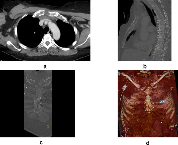
Figure 1: Preoperative CT-Scan. (a) Axial view shows oblique and longitudinal fractures. (b) Sagittal view shows anterior-posterior dislocation or sternal kinking. (c) Coronal view gives an overview over oblique and longitudinal fractures. (d) Volume rendering technique (VRT) gives an overview over the entire anterior chest wall showing rib fractures, manubrium and corpus sterni fracture
- Scan the chest wall for injuries. Inspect the complete anterior chest wall including examination of soft tissue, cartilage and bones.
- Look for bone fractures at the manubrium, corpus sterni and adjacent ribs I to VII on both sides. Look for cartilage disruption. Be aware of cartilage disruption and sternocostal separation not to miss any cause of instability.
- Perform a three dimensional evaluation. See the whole chest wall in the modus of three dimensional reconstructed pictures by evaluating each axial, coronal and sagittal picture in detail.
- Pinpoint the fracture:
- Take the distance from the upper sternal edge (jugulum) to the fracture. Use the sagittal view for measuring.
- In case of oblique fracture or multiple fractures measure both distances to jugulum, the cranial and the caudal edge of the fractures. Take the distance between those edges. The additional use of the coronal pictures might be helpful in this case.
- Determine the thickness of presternal soft tissue. Evaluate and measure the thickness of presternal soft tissue at the point of the fracture.
- Estimate the length of the possible surgical incision, performed in the midline.
NOTE: The basic approach needs to be at least 60 mm plus thickness of presternal soft tissue to get a proper access to a transverse fracture.- In case of oblique fracture or multiple fractures add the distance between the cranial and the caudal edge of the fractures to the basic approach.
NOTE: There might be a need of a longer incision in obese patients, if there are scars in the determined region or for other reasons.
- In case of oblique fracture or multiple fractures add the distance between the cranial and the caudal edge of the fractures to the basic approach.
2. Surgical Treatment
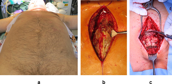
Figure 2: Surgical approach. (a) Positioning on the back gives access to the entire anterior chest wall and both axles; chest tube were already inserted bilaterally in this case. (b) After drawing the landmarks such as jugulum and sternoclavicular joints (above) the midline is identified and the incision performed. Pectoral muscle was dissected from the anterior sternum. A raspatory is inserted subperiostally. (c) The approach gives full access to any fracture identified. In this case two transverse fractures are shown.
2.1) Initial surgical procedures:
- Perform the operation under general anesthesia following the local standards while the patient is intubated. Place the patient in a supine position. Rest the arms on an arm board which must be placed less than 90° to the midline of the body and level the board with the floor. This might be useful if insertion of a chest tube becomes necessary.
- Remove superficial soil, debris, jewelry and transient microbes before applying antiseptic agents. Leave hair at the surgical site in place whenever possible.
- Commence disinfection following the local standards. Start from the cleanest area, usually the operative and/or incision site and proceed in a concentric fashion to the least clean area. Allow the disinfectant solution to dry completely naturally. Ensure that the antiseptic solution remains in contact with the skin for the required period of time as recommended by the manufacturer.
- Secure free access at least to the jugulum, the umbilicus and the axillary midline on both sides. Mark anatomical landmarks such as sternoclavicular joints on both sides, the upper sternal edge at the jugulum, the lower sternal edge and xiphoid, the seventh rib bilaterally and the umbilicus using a sterile pen.
- Draw the midline now between jugulum and umbilicus and mark the cranial and caudal edge of the fractures as measured in step 1.2. Add half of the basic approach cranial and the other half caudal to the edges of the fractured region of sternum.
- Double check the correct position of the fracture(s) and of the midline, then perform the midline incision respecting the anatomical layers in soft tissue.
- Remove presternal haematoma by a vertical incision along the length of the haematoma and wash out as much as possible with 0.9% saline.
- Cautiously take away the pectoral muscle bilaterally, beginning from the midline to the margin of the sternum whilst respecting the anatomical layers. Be aware of bleeding from smaller vessels and stop them. Identify the fracture.
- Expose the intercostal margin of the sternum, both, cranial and caudal of the fracture. Perform an incision of the sternal periosteum longitudinally along the front edge of the sternum. Cautiously dissect the periosteum from the bone laterally and on the posterior surface of the sternum using an elevatory or raspatory device.
NOTE: Preparation very close to the bone during this procedure is very important to prevent injuries to the adjacent internal thoracic vessels and the mediastinum. - Expose the fracture and clear the fracture cautiously. Usually retrosternal haematoma will appear at this time, wash out as much as possible with 0.9% saline.
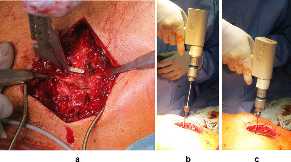
Figure 3: Surgical safety. (a) Measuring the sternal thickness and insert an elevator device. (b, c) Depth limited drilling.
- Measure the thickness of the sternum with a blunt instrument. Do not use a common “lot” due to possible retrosternal injuries. Prepare a drilling machine with a drill of the length of sternal thickness or shorter if not clear.
- Reduce the bone fragments by elevating the posteriorly depressed fragments with an elevator device which can be inserted through the same subperiosteal approach shown in step 2.1.7. Assemble the laterally dislocated fragment should be by lateral compression. Use a pointed ball forceps, weller clamp or compression wires if necessary.
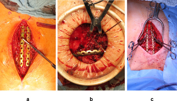
Figure 4: Fixation options of the plates. (a) Transverse fracture at corpus sterni: fixation with a pointed ball forceps and locked screws. (b) Oblique fracture at manubrium: reduction with compression wires , the first plate has been fixed yet. (c) Multiple transverse fracture; fixation of the plate using compression wires at the manubrium (above) and pointed ball forceps (below).
2.2) Transverse fractures and oblique fractures of sternal body:
- Mark the midline of the sternum using a pen or electrocautery.
- Insert a small pointed ball forceps to the posterior surface of the sternum using the intercostal space next to the fracture, again keeping very closed to the sternal bone. Use the intercostal space of the depressed fragment first.
- Put a longitudinal plate next to the midline and fix it with the pointed ball forcipes.
NOTE: At least three screw holes of the plate should cover the cranial and the caudal main fragment. In case of a single fracture, a seven or eight-hole plate offers the correct fixation. Using longer plates is necessary in multiple fractures, where a longer distance needs to be fixed.
- Put a longitudinal plate next to the midline and fix it with the pointed ball forcipes.
- Fix the plate with locked screws, drilling each hole using a drilling guide and the drill as chosen under step 2.1.9 thus ensuring depth limited drilling and a secured locked fixation of each screw to the plate. Again, fix each main fragment with at least three screws.
- Put a second plate on the other side of the midline and repeat the steps 2.2.2-2.2.3.
2.3) Longitudinal fractures, oblique manubrium fractures and sternocostal separation:
- Reduce the fracture as shown under step 2.1.10.
- Insert one small pointed ball forceps to the posterior surface of the sternum on each side using the intercostal space next to the fracture, again keeping very close to the sternal bone. Put a transverse plate from rib to rib, bridging the sternum with its fragments and fix it with the pointed ball forcipes. Place at least three screw holes laterally of each fracture.
NOTE: In case of a single fracture, a seven or eight- hole-plate offers the correct fixation. The use of longer plates usually becomes necessary for multiple fractures. - Fix the plate with locked screws; use a drilling guide and the drill as chosen under step 2.1.9 thus ensuring depth limited drilling. Again, fix each of the main fragments with at least three screws.
- For osteosynthesis of each injured level, repeat steps 2.3.1-2.3.3.

Figure 5: Operative results. (a) Long parallel plates, each fragment was fixed with 3 screws. (b) Postoperative chest x-ray showing longitudinal sternal plate and three transverse plates fixing oblique manubrium fracture, sternocostal separation of second rib and fracture of third rib on the right side. (c) Lateral view of x-ray shows correct position of two parallel plates fixing a fracture located at sternal corpus.
- Double check the correct position of all plates. Examine the anterior chest wall for sufficient stability. Look for any bleeding and stop it. Rule out pleural lesions. If a pleural lesion is indicated or suspected, insert a chest tube into the pleural cavity on the affected side using an axillary approach.
- Insert a subcutaneous drainage if necessary. Place the tube in front of the sternum and percutaneously divert about 5 cm away from the lower edge of the wound.
- Close the wound respecting the anatomical layers. Suture the periosteal incisions to preserve sternal blood supply. Approximation of the pectoral muscle to the midline is recommended to ensure a proper coverage of the implants with soft tissue.
- Take a chest x-ray after surgical treatment to rule out pneumothorax and hemothorax. The additional lateral view will show the position of the sternal fragments and of the implants.
Results
The preoperatively performed computed tomography provides a detailed survey of the whole sternum and the adjacent ribs. Using different windows of the CT data allows a detailed analysis of the bones as well as the adjacent cartilage of the ribs and the surrounding tissue. All fractures of the injured anterior chest wall should be detected. Axial pictures show injuries to cartilage in soft tissue window and disruption of sternocostal joints. Furthermore, the bone window shows longitudinal and oblique fractures of sternum, whereas transverse fractures are rarely detected in this plane. Coronal reconstructed pictures describe the fracture’s direction from a similar point of view to the one during surgical treatment. The dislocation of the fragments in relationship to the midline can be described properly. The sagittal view shows the dislocation of fragments in the anterior – posterior direction. A possible disruption of angulus sterni, the synchondrosis between manubrium and corpus sterni is shown best in this plane. These steps are shown in Figure 1a-d.
Following the steps 1.2-1.4 allows a concise planning of the surgical approach. The supine positioning allows full access to anterior chest wall and both axles (Figure 2a). Marking the landmarks simplifies the identification of the midline as shown in step 2.3 (Figure 2b).
All injured parts of the sternum and the adjacent ribs can be reached without problems while performing a median approach following steps mentioned above (Figure 2c).
Performing an incision of the sternal periosteum longitudinally along the front edge of the sternum and inserting an elevator device to the posterior surface allows the reduction of any dislocated fragment and prevents injuries to the adjacent internal thoracic vessels and the mediastinum as shown in Figure 4a. Moreover, depth limited drilling can be carried out by measurement of the sternal thickness at first (Figure 3a) and the choice of a drill bit with the corresponding length (Figure 3b, c).
The insertion of a pointed ball forceps might be difficult in some cases, e.g., at the manubrium. In those cases anteriorly placed compression wires (length = maximum sternal thickness) will help to reduce the fracture and fix the plate temporarily (Figure 4a-c).
The fixation of each fragment using at least three screws usually shows a proper stabilization for transverse as well as for oblique and longitudinal fractures (Figure 5a, b). Fixing separated ribs to the sternum can be performed by sterno-costal plating as well leading to a stable conjunction26 (Figure 5b).
At the time, all unstable parts of the anterior chest wall are fixed by plates. The anterior chest wall appears as a stable sternocostal union with physiologic respiratory movement. Respecting anatomical layers obtains the most precise wound closure; wound complications are usually rare.
Postoperatively taken chest x-rays in two planes show the correct reduction of the fracture and the correct positioning of the implants which were administered (Figure 5c). The 6 week follow up usually shows first callus surrounding the fracture and after twelve weeks consolidation usually can be observed.
Discussion
Although most of the sternal fractures are treated conservatively, sometimes a surgical fixation becomes necessary. The sternal anatomy and the stability of the anterior chest wall are restored by a stable fixation employing a locked plate osteosynthesis. Using very slim plates (1.5 mm or 2.0 mm thickness) provides a proper stability on the one hand and high convenience for the patient on the other hand12. Very good results are also described for the application of any locked plate, for example LCP System or plates developed for other bones like the cervical spine or the distal radius6,27.
Locked plates are considered to provide the advantage of a biological fixation to be functional as an internal fixator with significantly reduced friction between plate and bone9. The positioning of such an internal fixator on the anterior sternal surface provides a sufficient fixation of the convex surface of the fracture and thus reducing traction forces to the fracture. At the same time each respiratory movement induces compression to internal sternal cortex and to the fracture thus stimulating bone healing. Each screws needs to be locked securely to enable the efficiency of the internal fixator.
As we recommend the fixation of each fragment with at least three screws, there could be limitations in case of very short fragments. Possible solutions are the bridging of those fragments and the use of special plates, which allow longitudinal and transverse fixation rolled into one. In the future, “T” or “H”-shaped plates could be helpful in those situations.
Some surgeons show a reluctance to operate on the chest wall, probably due to a lack of experience in this particular region. There is a need for a simple and safe method for sternal stabilization1,12. The above described method represents one possible standard and reduces the risk of complications due to the operative procedure by a precise preoperative planning, subperiosteal dissection of soft tissue, and depth limited drilling. There lies the difference in the use of any other plates not providing the possibility of depth limited drilling (Table 1), whereas the preoperative planning, the steps of preparation, and reduction of the fracture could be performed analogously to the described way of this manuscript.
As a three dimensional reconstructed CT scan allows the diagnosis of fractures to sternum and concomitant fractures with high sensitivity the surgeon gets detailed information about the injury28,29. The CT shows the particular position and the direction of the fracture as well as any dislocation. This important information simplifies the preoperative planning of the intended surgical approach as shown in steps 1.1-1.5. Furthermore, it allows a precise planning of the number and design of the plates which should be employed for fixation as shown in the steps 2.2-2.3. A possible limitation of a CT scan lies in difficulties showing undislocated fractures and disruption of cartilage although they could contribute to the instability of the chest wall.
The subperiosteal dissection and the depth limited drilling are the two most important steps for surgical safety.
Reducing the sternal fragments to their anatomic position usually necessitates an approach to the posterior wall of the sternum. This procedure may injure mediastinal organs, surrounding vessels and the sternal blood supply7,18. Performing strictly subperiosteal dissection around the sternum as shown in step 2.1.7 will minimize the risk of complications. Fractures which show a lateral dislocation, could have their fragments move very closely to the internal mammary vessels. In those cases the surgeon needs to be aware of any injury to those vessels which could cause severe bleeding. At the time a bleeding in this particular region is identified, the surgeon needs to stop it immediately through an intercostal approach to the internal mammary vessel. The dissection of intercostal muscles and the insertion of a spreader in the affected intercostal space provides a quick and proper access to the injured vessel.
Drilling too deep has to be avoided, as it may cause life-threatening injuries to the mediastinum. This is ensured by depth limited drilling in combination with the measurement of the sternal thickness and the choice of a drill bit of corresponding length12.
Summarizing the discussed procedure and possible limitations, performing anterior sternal plating with low profile locked titanium plates is to be expected successful, if a preoperative planning by computed tomography, subperiostal dissection, and depth limited drilling is performed.
Disclosures
The first author has a consultant agreement with Synthes CMF. No funds were received in connection to the presented study. The coauthors are not involved in any conflict of interest.
Acknowledgements
This article is dedicated to the specialist in pediatric surgery, Professor Dr. R.T. Carbon (Erlangen, Germany) with heartfelt gratitude for his educational impact. We thank the Radiological Institute of the Erlangen University Hospital (Director: Prof. Michael Uder) for providing the X-rays and CT scans.
Materials
| Name | Company | Catalog Number | Comments |
|---|---|---|---|
| MatrixRIB | DePuySynthes CMF | ||
| LCP Forefood/Middlefood 2.4/2.7 | DePuySynthes CMF | only compression wires are employed |
References
- Athanassiadi, K., Gerazounis, M., Moustardas, M., Metaxas, E. Sternal fractures: retrospective analysis of 100 cases. World J. Surg. 26 (10), 1243-1246 (2002).
- Severson, E. P., Thompson, C. A., Resig, S. G., Swiontkowski, M. F. Transverse sternal nonunion, repair and revision: a case report and review of the literature. J. Trauma. 66 (5), 1485-1488 (2009).
- Queitsch, C., et al. Treatment of posttraumatic sternal non-union with a locked sternumosteosynthesis plate (TiFix). Injury. 42 (1), 44-46 (2011).
- Fowler, A. W. Flexion-compression injury of the sternum. J. Bone Joint Surg Br. 39 (3), 487-497 (1957).
- Bailey, J., et al. Thoracic hyperextension injury with complete 'bony disruption' of the thoracic cage: Case report of a potentially life-threatening injury. World J. Emerg Surg. 7 (1), 14(2012).
- Ciriaco, P., et al. Early surgical repair of isolated traumatic sternal fractures using a cervical plate system. J. Trauma. 66 (2), 462-464 (2009).
- Harston, A., Roberts, C. Fixation of sternal fractures: a systematic review. J. Trauma. 71 (6), 1875-1879 (2011).
- Fawzy, H., et al. Sternal plating for primary and secondary sternal closure; can it improve sternal stability. J. Cardiothorac Surg. 4, 19(2009).
- Murphy, W. M. AO Principles of Fracture Management. , Thieme. New York, NY. 25-165 (2000).
- Mayberry, J. C., Ham, L. B., Schipper, P. H., Ellis, T. J., Mullins, R. J. Surveyed opinion of American trauma, orthopedic, and thoracic surgeons on rib and sternal fracture repair. J. Trauma. 66 (3), 875-879 (2009).
- Gloyer, M. A., Frei, H. C., Hotz, T. K., Käch, K. P. Osteosynthesis of traumatic manubriosternal dislocations and sternal fractures with a 3.5/4.0 mm fixed-angle plate (LCP). Arch. Orthop. Trauma Surg. 131 (9), 1261-1266 (2011).
- Schulz-Drost, S., Mauerer, A., Grupp, S., Hennig, F. F., Blanke, M. Surgical fixation of sternal fractures: locked plate fixation by low-profile titanium plates--surgical safety through depth limited drilling. Int. Orthop. 38 (1), 133-139 (2014).
- Wu, L. C., Renucci, J. D., Song, D. H. Sternalnonunion: a review of currenttreatments and a newmethod of rigidfixation. Ann. Plast. Surg. 54 (1), 55-58 (2005).
- Potaris, K., et al. Management of sternal fractures: 239 cases. Asian Cardiovasc. Thorac. Ann. 10 (2), 145-149 (2002).
- Coons, D. A., Pitcher, J. D., Braxton, M., Bickley, B. T. Sternal nonunion, case report. Orthopedics. 25, 89-91 (2002).
- Abdul-Rahman, M. R., et al. Comminuted sternal fracture—a sternotomy wire fixation: report of 2 cases. Heart Surg. Forum. 12 (3), E184-E186 (2009).
- Celik, B., Sahin, E., Nadir, A., Kaptanoglu, M. Sternum fractures and effects of associated injuries. Thorac. Cardiovasc. Surg. 57 (8), 468-471 (2009).
- Bonney, S., Lenczner, E., Harvey, E. J. Sternal fractures: anterior plating rationale. J. Trauma. 57 (6), 1344-1346 (2004).
- Kitchens, J., Richardson, J. D. Open fixation of sternal fracture. Surg. Gynecol. Obstet. 177 (4), 423-424 (1993).
- Al-Qudah, A. Operative treatment of sternal fractures. Asian Cardiovasc. Thorac. Ann. 14 (8), 399-401 (2006).
- Frangen, T. M., Müller, E. J., Muhr, G., Hopf, F. Traumatic manubriosternal dislocation. Arch. Orthop. Trauma Surg. 126 (6), 411-416 (2006).
- Molina, J. E. Evaluation and operative technique to repair isolated stemal fracture. J Thorac Cardiovasc Surg. 130 (2), 445-448 (2005).
- El Ibrahimi, A., et al. Traumatic manubriosternal dislocation: A new method of stabilization postreduction. J. Emerg. Trauma Shock. 4 (2), 317-319 (2011).
- Richardson, J. D., Franklin, G. A., Heffley, S., Seligson, D. Operative fixation of chest wall fractures: an underused procedure. Am. Surg. 73 (6), 591-596 (2007).
- Münzberg, M., Mahlke, L., Bouillon, B., Paffrath, T., Matthes, G., Wölfl, C. G. Six years of Advanced Trauma Life Support (ATLS) in Germany: the 100th provider course in Hamburg. Unfallchirurg. 113 (7), 561-566 (2010).
- Schulz-Drost, S., Syed, J., Besendoerfer, M., Carbon, R. T. Sternocostal Dislocation Following Open Correction of Pectus Excavatum-'Stairway Phenomenon': Complication Management by Means of Sternocostal Locking Titanium Plate Osteosynthesis. Thorac Cardiovasc Surg. 62 (3), 245-252 (2013).
- Ergene, G., Tulay, C. M., Anasız, H. Sternal fixation with nonspecific plate. Ann Thorac Cardiovasc Surg. 19 (5), 364-367 (2012).
- Kehdy, F., Richardson, J. D. The utility of 3-D CT scan in the diagnosis and evaluation of sternalfractures. J. Trauma. 60 (3), 635-636 (2006).
- Alkadhi, H., Wildermuth, S., Marincek, B., Boehm, T. J. Accuracy and time efficiency for the detection of thoracic cage fractures: volume rendering compared with transverse computed tomography images. Comput. Assist. Tomogr. 28 (3), 378-385 (2004).
Reprints and Permissions
