Here we present a protocol that generates large amounts of murine monocytes from heterogeneous bone marrow for translational applications. In comparison to others, this new method helps reduce the number of sacrificed animals and lowers costs by avoiding expensive methods such as high gradient magnetic cell separation (MACS).
A subscription to JoVE is required to view this content. Sign in or start your free trial.
Method Article
Isolation and Intravenous Injection of Murine Bone Marrow Derived Monocytes
In This Article
Summary
Abstract
As a subtype of leukocytes and progenitors of macrophages, monocytes are involved in many important processes of organisms and are often the subject of various fields in biomedical science. The method described below is a simple and effective way to isolate murine monocytes from heterogeneous bone marrow.
Bone marrow from the femur and tibia of Balb/c mice is harvested by flushing with phosphate buffered saline (PBS). Cell suspension is supplemented with macrophage-colony stimulating factor (M-CSF) and cultured on ultra-low attachment surfaces to avoid adhesion-triggered differentiation of monocytes. The properties and differentiation of monocytes are characterized at various intervals. Fluorescence activated cell sorting (FACS), with markers like CD11b, CD115, and F4/80, is used for phenotyping. At the end of cultivation, the suspension consists of 45%± 12% monocytes. By removing adhesive macrophages, the purity can be raised up to 86%± 6%. After the isolation, monocytes can be utilized in various ways, and one of the most effective and common methods for in vivo delivery is intravenous tail vein injection.
This technique of isolation and application is important for mouse model studies, especially in the fields of inflammation or immunology. Monocytes can also be used therapeutically in mouse disease models.
Introduction
The isolation of monocytes is important and critical for many in vitro and in vivo studies. These cells are targets for diseases such as peripheral arterial disease, coronary heart disease, or other ischemic diseases, since collateral vessel growth is strongly driven by local inflammation. Inflammatory responses include endothelial activation and local recruitment of leukocytes, mainly monocytes, which then mature to macrophages and create a highly arteriogenic environment by secreting multiple growth factors to induce the remodeling of an arteriole into a functional collateral artery1-3. Monocytes also mature to dendritic cells, which are frequently used for immunological studies4,5 and cancer research6,7.
Problematic in the approach for monocyte isolation from peripheral blood8 is the high number of donor animals needed to produce a sufficient amount of monocytes for most analyses. Former protocols describe methods such as density gradient centrifugation and cell depletion via MACS9 when isolating monocytes; however, these techniques can alter the characteristics and functionality of monocytes which can lead to difficulties in interpretation10,11. Moreover, these methods are difficult and can reduce experimental reproducibility.
Our aim with this protocol is to provide a simple and cost effective method to generate large amounts of bone marrow-derived monocytes. Due to the high cell yield of 11 x 106 ± 3 x 10⁶ cells obtained by this protocol, we can substantially reduce the number of mice required during the isolation of bone marrow-derived monocytes. The procedure can be completed within a minimal amount of time, and without using expensive and complicated techniques as referenced above. Here, we extract monocytes from native bone marrow suspension of donor mice, cultivate the suspension on ultralow attachment plates, and supplement the solution with 20 ng/ml M-CFS. On day 5 of incubation, cells are harvested and characterized to confirm functional and phenotypic properties.
For experiments in the field of arteriogenesis, intravenous transplantation of these bone marrow-derived monocytes into mice is an effective method of systemic drug delivery, which can be combined with femoral artery ligation in common peripheral arterial disease models.
Access restricted. Please log in or start a trial to view this content.
Protocol
This study was performed with permission of the State of Saxony and Saxony-Anhalt, Regierungspraesidium Dresden/Halle, according to Section 8 of the German Law for animal protection (24D-9168.11-1/2008-24).
1 Cell Isolation
1.1 Preparation of Femur and Tibia
- Anesthetize the mouse using a 5% isoflurane concentration by vaporizing isoflurane in a closed bin. After the mouse has stopped moving for 3 sec, perform the cervical dislocation.
- Disinfect the hind limbs with ethanol (96%). Remove skin and muscles and separate the bones with a sterile scalpel.
- Start with an inguinal skin incision, and extend the incision toward the medial malleolus and perform a circular dissection of the skin around the lower calf. Remove the skin of the legs completely by peeling it proximally.
- Detach the quadriceps muscle from the femur with a sharp scalpel, remove both the anterior peroneus and the gastrocnemius muscle groups and disarticulate the leg at the hip joint by locating and dissecting the ligaments.
- Start anteriorly and proceed around the joint by carefully rotating the leg and transecting the ligaments, thereby disarticulating the joint.
- Wash femur and tibia with 96% ethanol for a minimum of 90 sec to guarantee aseptic cell preparation. Continue the washing process with sterile PBS to rinse off remaining ethanol.
Note: All subsequent steps must be sterile to avoid contamination.
1.2 Harvesting of Bone Marrow
- Cut the proximal and distal end of each bone with a pair of fine scissors to gain access to the femoral and tibial shafts. Be careful with this step, since these bones break very easily.
- Flush the bones with warm medium (M199 + 10% fetal calf serum (FCS), + 1% Penicillin/Streptomycin). Use a sterile 28-G needle, a 1 ml syringe, and between 10-15 ml medium per bone for rinsing.
- Hold the bone with a fine forceps and rinse one by one until it turns semi-translucent. Carefully apply pressure to the end of the syringe plunger in order to minimize cell stress.
- Filter the bone marrow through a 70 µm nylon web and gather the filtrate. To extract the maximum quantity of bone marrow, rinse repeatedly from the distal and proximal ends.
- Rinse the sieve with PBS and gather the filtrate. Carefully dislodge debris and cellular conglomerates by gentle stirring and pipetting.
1.3 Cultivation
- Centrifuge the cell suspension at 250 x g for 10 min at RT. Discard the supernatant and resuspend the cells with approximately 25 ml of medium. Repeat once.
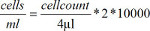
- Resuspend the cells in 6 ml of medium after the second washing step. Mix 50 µl of the cell solution with 50 µl of trypan blue. Count the cells in a counting chamber under a light microscope. Calculate the number of cells in the solution using this formula:
- Seed the cells on 6-well ultra-low-attachment surface plates to prevent permanent adhesion to the bottom of the plate. Use a concentration of 106 cells per ml with up to 6 ml per well.
- Supplement the suspension with 20 ng/ml M-CSF to promote cell differentiation.
- Culture the cells for 5 days at 37 °C and 5% carbon dioxide and observe daily.
1.4 Harvesting of Cells
Note: These steps should be performed on ice. Proceed with either 1.4.1 or 1.4.2 accordingly.
- At this point, harvest the entire culture, including differentiated macrophages that will adhere to the culture-plates, even if they are ultra-low-attachment surface plates.
- Harvest the entire culture by gentle pipetting and detaching with a solution of 4 °C PBS, 0.5% bovine serum albumin (BSA) and 2 mM EDTA. Repeat until the plates are clear of remaining adherent cells - confirm under a microscope if unsure.
- Alternatively, discard adherent macrophages by harvesting the supernatant with the cells in suspension, which predominantly contain monocytes.
- Harvest the non-adherent cells with a solution of EDTA-free PBS and 0.5% BSA. Do not apply too much force in order to avoid dislodging mildly adherent macrophages.
1.5 FACS-Analysis (Optional)
Note: It is possible to deplete the cell suspension of CD117+ stem- and progenitor cells by the use of MACS. Use manufacturer protocols for this procedure.
- Harvest the cells according to either steps 1.4.1 or 1.4.2.
- Centrifuge the cells at 250 x g for 10 min at 4 °C and repeat this step again.
- Resuspend at least 250,000 cells in 300 µl of FACS buffer per probe.
- Transfer cell solution onto a 96-well-plate (30 µl per well).
- Centrifuge the cell suspension at 250 x g for 5 min at 4 °C.
- Remove the supernatant and add 25 µl of antibody to each well (use manufacturer dilution).
- Stain the cells with antibodies (following manufacturer protocols for cell phenotyping after steps 1.5.1-1.5.6. Commonly used markers for monocyte / macrophage identification include CD11b, F4/80 (Figure 3), CD115 (Figure 2), and Gr-1.
Note: For a further description of cell solutions, the use of markers like CD4, CD8a, CD11c, CD25, CD45, CD45R, CD62L, CD80, CD86, CD145, CD117, MHCⅡ, Ly6C, Ly6G, and CD192 is recommended. Cellular characteristics were previously described12. - Incubate the probes for 20 min at 4 °C.
- Centrifuge the probes at 250 x g for 5 min at 4 °C and resuspend with 200 µl of FACS buffer. Repeat this step twice.
- Transfer the probes into FACS tubes and perform the FACS analysis12.
- Perform FACS analysis12 on day 0, 3 and 5 to analyze cell differentiation.
2. Tail Vein Injection
2.1 Preparation
- Warm the animals on a heating plate at 37 °C for about 10 min. Observe the animals while warming to recognize signs of overheating.
- Resuspend the monocytes, isolated in steps 1.1- 1.4, in 150 µl of NaCl, and load the cells into a 1 ml insulin syringe with a 30G needle. Lightly vortex the solution before loading the syringe to ensure all monocytes are injected. Always handle cells on ice to avoid heat-mediated attachment and activation of monocytes.
2.2 Restraining
- Avoid disturbing other mice in the cage when choosing a mouse for intravenous injection.
- Place the mouse into the restrainer. Avoid too much pressure and be careful with the hind limbs. (Figure 4)
Note: Alternatively, use general anesthesia to guarantee stress free handling of the animals. - Ensure that the mouse has space for breathing before closing the restrainer.
2.3 Injection
- Disinfect the injection site with disinfection agent. Spray on the tail of the mouse and let stand for at least 1 min.
- Stop venous blood flow of the tail by applying gentle pressure to the lateral tail side above the injection site, for better visibility of the veins.
- Turn the tail 90 degrees, before injection. (Figure 5)
- Inject at a 45 degree angle. Inject slowly and no more than 5 µl per g to avoid harming animals.
- If there is a blister, which is a sign of a failed injection, discontinue immediately and repeat the injection more proximally.
- Stop the bleeding at the injection side by applying gentle pressure for about 1 min.
- At the end of the procedure, open the retractor and place the mouse in its cage.
- Observe the mouse for 20 min to ensure that the animal was not harmed by the injection and sternal recumbency is maintained. Extend the time of observation until the mouse regains sufficient consciousness. Return the mouse to the company of other animals only after it has fully recovered.
Access restricted. Please log in or start a trial to view this content.
Results
The cell solution extracted from the murine bone marrow consists of various cell types. The major cell types are lymphocytes, granulocytes and monocytes. Cell types can be estimated by size and granularity, which is shown in Figure 1 for both native suspensions and cells harvested after 5 days of differentiation. Note the shifting cellular composition during cultivation. However, accurate classification of populations must rely on distinctive expression of cellular markers.
Th...
Access restricted. Please log in or start a trial to view this content.
Discussion
We describe a simple and cost-effective method to isolate large amounts of murine monocytes from bone marrow. In comparison to other protocols using peripheral blood, which obtain monocyte yields5 of 1.4 x10⁶, we are able to obtain higher yields of 11 x 106 ± 3 x 10⁶ monocytes from a single donor mouse.
When considering challenges with this method, it is important to mention the potential for contamination when working under non-sterile conditio...
Access restricted. Please log in or start a trial to view this content.
Disclosures
The authors declare that they have no competing financial interests.
Acknowledgements
This work was supported by the DFG (Deutsche Forschungsgemeinschaft, German Research Foundation) SFB 854 (Sonderforschungsbereich, collaborative research center).
Thanks to Hans-Holger Gärtner, Audiovisuelles Medienzentrum, Otto-von-Guericke University Magdeburg, Magdeburg, Germany, for technical support.
Access restricted. Please log in or start a trial to view this content.
Materials
| Name | Company | Catalog Number | Comments |
|---|---|---|---|
| 6-well-ultra-low-attachment plate | Corning Incorporated, NY, USA | 6-well-ultra-low-attachment plate, with cap, sterile | |
| 8- 12 week old, male, balb/c mice | Charles River, Sulzfeld, Germany | ||
| 96-well-plate | Greiner bio one GmbH, Frickenhausen, Germany | ||
| Blue dead cell stain | Life technologies GmbH, Darmstadt, Germany | ||
| Bovine serum albumine | GE Healthcare, Freiburg, Germany | Fraction V, pH 7.0 | |
| Canules | B. Braun, Melsungen AG, Melsungen, Germany | 28G, 30G | |
| CD115 | eBioscience, San Diego, USA | 12-1152 | |
| CD11b | eBioscience, San Diego, USA | 53-0112 | |
| Cell culture dish | Greiner Bio-One GmbH, Frickenhausen, Germany | With cap, steril | |
| Centrifuge | Beckman Coulter GmbH, Krefeld, Germany | Allegra® X-15R centrifuge | |
| Depilatory cream | Veet, Mannheim, Germany | ||
| Disinfection agent | Schülke&Mayr GmbH, Norderstedt, Germany | Kodan Tinktur forte | |
| Disposable scalpel No.10 | Feather safety razor Co.Ltd, Osaka, Japan | ||
| EDTA | Sigma Aldrich, Hamburg, Germany | ||
| Ethanol 96% | Otto Fischar GmbH und Co KG, Saarbrücken, Germany | ||
| Extraction unit Pipetus | Hirschmann Laborgeräte GmbH & Co.KG, Eberstadt, Germany | ||
| F4/80 | AbD Serotec, Düsseldorf, Germany | MCA497APC | |
| FACS buffer | Manufactured by our group with single components | PBS, 0.5% BSA, 0.1% NaN3 | |
| FACS device | Becton, Dickinson and Company, Franklyn Lakes, New Jersey, USA | BD FACS Canto II | |
| FACS tubes | Becton, Dickinson and Company, Franklyn Lakes, New Jersey, USA | ||
| Falcon® pipette | Becton Dickenson Labware, NY, USA | ||
| Fetal calf serum | Sigma Aldrich, Hamburg, Germany | ||
| Fine forceps | Rubis, Stabio, Switzerland | ||
| Gloves | Rösner-Matby Meditrade GmbH, Kiefersfelden, Germany | ||
| Gr1 | eBioscience, San Diego, USA | 53-5931 | |
| Heating plate | Labotect GmbH, Göttingen, Germany | Hot Plate 062 | |
| Incubator | Ewald Innovationstechnik GmbH, Bad Nenndorf, Germany | Incu safe | |
| Isofluran | Baxter Deutschland GmbH, Unterschleißheim, Germany | ||
| Light microscope | Carl Zeiss SMT GmbH, Oberkochen, Germany | Axiovert 40 °C | |
| Macrophage-Colony Stimulating Factor | Sigma Aldrich, Hamburg, Germany | SRP3110 | |
| Mechanical shaker | IKA, Staufen, Germany | ms2 minishaker | |
| Medium 199 | PAA Laboratories GmbH, Pasching, Austria | Warm in 37 °C water bath before use | |
| Micro test tubes | Eppendorf AG, Hamburg, Germany | ||
| Microbiological work bench | Thermo Electron, LED GmbH, Langenselbold, Germany | Hera safe | |
| Monocyte wash buffer | Manufactured by our group with single components | PBS, 0.5% BSA, 2 mM EDTA | |
| Mouse restrainer | Various | ||
| NaCl | Berlin Chemie AG, Berlin, Germany | ||
| NaN3 (sodium acide) | Sigma Aldrich, Hamburg, Germany | ||
| Neubauer counting chamber | Paul Marienfeld GmbH und Co.KG, Lauda-Königshofen, Germany | ||
| Nylon cellsieve | Becton, Dickinson and Company, Franklyn Lakes, New Jersey, USA | Cell strainer, 70 µm mesh size | |
| Penicillin/Streptomycin | Sigma Aldrich, Hamburg, Germany | ||
| Phosphate buffered saline | Life technologies GmbH, Darmstadt, Germany | pH 7.4, sterile | |
| Pipettes | Eppendorf AG, Hamburg, Germany | 10µl/100µl/200µl/1,000µl | |
| Pipetting heads | Eppendorf AG, Hamburg, Germany | ||
| Serological pipette | Greiner Bio-One GmbH, Frickenhausen, Germany | Cellstar 5 ml, 10 ml | |
| Suction unit | Integra bioscience, Fernwald, Germany | Vacusafe comfort | |
| Surgical scissors | Word Precision Instruments, Inc., Sarasota, USA | ||
| Syringe | B. Braun, Melsungen AG, Melsungen, Germany | 1 ml Omnifix® -F insuline syringe | |
| Tubes with cap | Greiner bio one GmbH, Frickenhausen, Germany | 15 ml/50 ml Cellstar tubes | |
| Warm water bath | Julabo Labortechnik GmbH, Seelbach, Germany | Julabo SW22 |
References
- Herold, J., et al. Transplantation of monocytes: a novel strategy for in vivo augmentation of collateral vessel growth. Hum Gene Ther. 15 (1), 1-12 (2004).
- Herold, J., Tillmanns, H., Xing, Z., Strasser, R. H., Braun-Dullaeus, R. C. Isolation and transduction of monocytes: promising vehicles for therapeutic arteriogenesis. Langenbecks Arch Surg. 391 (2), 72-82 (2006).
- Francke, A., Weinert, S., Strasser, R. H., Braun-Dullaeus, R. C., Herold, J. Transplantation of bone marrow derived monocytes: a novel approach for augmentation of arteriogenesis in a murine model of femoral artery ligation. American Journal Of Translational Research. 5 (2), 155-169 (2013).
- Leon, B., et al. Dendritic cell differentiation potential of mouse monocytes: monocytes represent immediate precursors of CD8- and CD8+ splenic dendritic cells. Blood. 103 (7), 2668-2676 (2004).
- Timmerman, J. M., Levy, R. Dendritic cell vaccines for cancer immunotherapy. Annu Rev Med. 50, 507-529 (1999).
- Salem, M. L. The use of dendritic cells for Peptide-based vaccination in cancer immunotherapy. Methods Mol Biol. 1139, 479-503 (2014).
- Timmerman, J. M., et al. Idiotype-pulsed dendritic cell vaccination for B-cell lymphoma: clinical and immune responses in 35 patients. Blood. 99 (5), 1517-1526 (2002).
- Berthold, F. Isolation of human monocytes by Ficoll density gradient centrifugation. Blut. 43 (6), 367-371 (1981).
- Houthuys, E., Movahedi, K., De Baetselier, P., Van Ginderachter, J. A., Brouckaert, P. A method for the isolation and purification of mouse peripheral blood monocytes. Journal Of Immunological Methods. 359 (1-2), 1-10 (2010).
- Seeger, F. H., Tonn, T., Krzossok, N., Zeiher, A. M., Dimmeler, S. Cell isolation procedures matter: a comparison of different isolation protocols of bone marrow mononuclear cells used for cell therapy in patients with acute myocardial infarction. European Heart Journal. 28 (6), 766-772 (2007).
- Auffray, C., et al. CX3CR1+ CD115+ CD135+ common macrophage/DC precursors and the role of CX3CR1 in their response to inflammation. The Journal Of Experimental Medicine. 206 (3), 595-606 (2009).
- Francke, A., Herold, J., Weinert, S., Strasser, R. H., Braun-Dullaeus, R. C. Generation of mature murine monocytes from heterogeneous bone marrow and description of their properties. J. Histochem. Cytochem. 59 (9), 813-825 (2011).
- Sneider, E. B., Nowicki, P. T., Messina, L. M. Regenerative medicine in the treatment of peripheral arterial disease. Journal Of Cellular Biochemistry. 108 (4), 753-761 (2009).
- Van Royen, N., Piek, J. J., Schaper, W., Fulton, W. F. A critical review of clinical arteriogenesis research. J Am Coll Cardiol. 55 (1), 17-25 (2009).
- Lawall, H., Bramlage, P., Amann, B. Treatment of peripheral arterial disease using stem and progenitor cell therapy. J Vasc Surg. 53 (2), 445-453 (2011).
- Herold, J., et al. Tetanus toxoid-pulsed monocyte vaccination for augmentation of collateral vessel growth. Journal of the American Heart Association. 3 (2), e000611(2014).
Access restricted. Please log in or start a trial to view this content.
Reprints and Permissions
