We demonstrate a method to generate 3D breast cancer surrogates, which can be cultured using a perfusion bioreactor system to deliver oxygen and nutrients. Following growth, surrogates are fixed and processed to paraffin for evaluation of parameters of interest. The evaluation of one such parameter, cell density, is explained.
A subscription to JoVE is required to view this content. Sign in or start your free trial.
Method Article
Preparation and Analysis of In Vitro Three Dimensional Breast Carcinoma Surrogates
In This Article
Summary
Abstract
Three dimensional (3D) culture is a more physiologically relevant method to model cell behavior in vitro than two dimensional culture. Carcinomas, including breast carcinomas, are complex 3D tissues composed of cancer epithelial cells and stromal components, including fibroblasts and extracellular matrix (ECM). Yet most in vitro models of breast carcinoma consist only of cancer epithelial cells, omitting the stroma and, therefore, the 3D architecture of a tumor in vivo. Appropriate 3D modeling of carcinoma is important for accurate understanding of tumor biology, behavior, and response to therapy. However, the duration of culture and volume of 3D models is limited by the availability of oxygen and nutrients within the culture. Herein, we demonstrate a method in which breast carcinoma epithelial cells and stromal fibroblasts are incorporated into ECM to generate a 3D breast cancer surrogate that includes stroma and can be cultured as a solid 3D structure or by using a perfusion bioreactor system to deliver oxygen and nutrients. Following setup and an initial growth period, surrogates can be used for preclinical drug testing. Alternatively, the cellular and matrix components of the surrogate can be modified to address a variety of biological questions. After culture, surrogates are fixed and processed to paraffin, in a manner similar to the handling of clinical breast carcinoma specimens, for evaluation of parameters of interest. The evaluation of one such parameter, the density of cells present, is explained, where ImageJ and CellProfiler image analysis software systems are applied to photomicrographs of histologic sections of surrogates to quantify the number of nucleated cells per area. This can be used as an indicator of the change in cell number over time or the change in cell number resulting from varying growth conditions and treatments.
Introduction
Three dimensional (3D) culture models that more accurately mimic the tumor architecture and microenvironment in vivo are important for studies aimed to dissect the complex interactions between cells and their microenvironment and to test the efficacy of candidate therapies. Tumor dimensionality impacts oxygen and nutrient gradients, the uniformity of drug exposure, interstitial pressure/blood flow, and 3D architecture1-4. The presence of an appropriate stromal microenvironment contributes to tumor dimensionality and influences cell-ECM signaling and paracrine signaling between stromal cells and malignant epithelial cells. The effects of tumor dimensionality and the microenvironment on cellular function are well established, with both factors altering drug response1,3,5-8. Additionally, cellular growth kinetics, metabolic rates, and cell signaling differ between two dimensional (2D) culture and culture in 3D, with these factors affecting cellular response1,3,8-10.
In vitro, the tumor surrogate microenvironment can be modulated by including representative ECM constituents and stromal cell populations. Malignant epithelial cells are influenced by the ECM and cancer-associated stromal cells either in a synergistic/protective manner to promote tumor progression or in a suppressive manner to inhibit further tumor propagation5,6,10. In either context, the stroma can affect therapeutic response and drug delivery via paracrine signaling and/or by increasing interstitial pressure in the tumor resulting in decreased drug delivery1,6. Therefore, the addition of ECM and stromal cells into preclinical models will help recapitulate aspects of the tumor that cannot be modeled well in 2D culture.
Herein a method to establish breast cancer surrogates that incorporate a recapitulative microenvironment, including ECM constituents and stromal cells, in a 3D volume is described. In breast carcinoma, the stromal cell population is predominately comprised of cancer associated fibroblasts (CAF) and the stromal ECM is largely composed of collagen type I with a smaller proportion of matrix components that are found in the basement membrane, including laminin and collagen type IV1,4,11-13. Therefore, these components of the breast carcinoma microenvironment (i.e., CAF, collagen I, and basement membrane) have been incorporated into the surrogates. This method can be used to generate solid, un-perfused 3D surrogates (Figure 1A) or can be adapted to include perfusion of medium through the surrogate via a bioreactor system (Figure 1B). Both approaches are described here. This method could also be modified to include other stromal elements, such as tumor-associated macrophages, or to model other solid tumors by adjusting the cellular and ECM components, as appropriate.
For the breast carcinoma surrogate described here, we have utilized the MDA-MB-231 (231) breast cancer cell line, CAF previously isolated from human breast carcinoma14, and an ECM composed of 90% collagen I (6 mg/ml) and 10% growth factor reduced basement membrane material (BM). The surrogate is either grown in an 8-well chamber slide (solid surrogate) or a bioreactor system is utilized to provide continuous nutrient perfusion (perfused surrogate). Any perfusion bioreactor system that can accommodate a volume of ECM containing cells can be used15. As an example, we describe the preparation of the tissue surrogates in our bioreactor system. This system was developed in-house and is not commercially available. Because our focus here is on the preparation and analysis of the 3D tissue surrogates, we have not gone into extensive detail regarding the specifics of manufacture and assembly of our bioreactor system. However, a detailed description of this system and its development has been published16. In this bioreactor system, a polydimethylsiloxane (PDMS) flow channel is used to house the surrogate, which is supported by a PDMS foam (formed using methods similar to those described by Calcagnile et al.17). This volume is penetrated by 4 microchannels (each 400 µm in diameter) which are continuously perfused by medium via a microphysiologic pump to supply oxygen and nutrients to the surrogate.
Appropriate analysis of the surrogates is crucial to gain pertinent information regarding cellular function in response to treatment or other manipulations. Surrogates can be analyzed by various methods including direct imaging of intact surrogates using confocal microscopy or other means of non-invasive imaging, indirect cellular analysis by assaying the conditioned media, or perfusate, for secreted products, or analysis on histologic sections after fixation and processing to paraffin. One such parameter that can be evaluated on histologic sections is cell density. We present one approach to measure cell density (i.e., the number of nucleated cells per section area) using semi-automated image processing techniques applied to photomicrographs of surrogate histologic sections stained with hematoxylin and eosin (H&E). The cell density can be used as an indicator of the relative change in cell number over time or that results from varying growth conditions and treatments.

Figure 1. 3D volume and bioreactor system. A) Schematic of the process to generate solid 3D surrogates. Top: cartoon of solid 3D volume containing ECM (pink), epithelial carcinoma cells (yellow), and CAF (orange); Bottom: top view of 8-well chamber slide containing surrogates. B) Schematic of the process to generate perfused 3D surrogates. Top: cartoon of 3D volume with channels to allow for medium perfusion and containing ECM (pink), epithelial carcinoma cells (yellow), and CAF (orange); Middle: image of PDMS flow channel containing PDMS foam (black arrow) to be injected with cell+ECM and penetrated by polymer-coated stainless steel wires (pink arrow) measuring 400 µm in diameter; Bottom: image of the PDMS flow channel containing a surrogate and connected to the bioreactor system to allow for continuous medium perfusion (peristaltic pump and media reservoir not shown). C) Images of processing steps for both solid and perfused surrogates after culture. Left: image of the cryomold containing specimen processing gel and surrogate; Middle: image of a paraffin block containing a fixed and processed surrogate; Right: image of a glass slide with a H&E-stained histologic section of a surrogate. Please click here to view a larger version of this figure.
Access restricted. Please log in or start a trial to view this content.
Protocol
1. Cell Culture
- Thaw BM component overnight at 4 °C, on ice.
- Warm medium to 37 °C. To support growth of both 231 cells and CAF, use Dulbecco's Modified Eagle Medium (DMEM) plus 10% fetal bovine serum (FBS).
Note: The media used will depend upon the cell type and the experimental goals. - Remove medium from the culture dish (10 cm) of near confluent 231 cells and add 1.5 ml Trypsin/EDTA. Incubate for 1 to 3 min at 37 °C, monitoring for cell detachment. Once cells start to round and come off the plate, stop the reaction by adding 3 ml of the medium containing serum. Pipette the medium and cells into a 15 ml conical tube.
- Centrifuge the medium and cells at 150 x g for 5 min. Remove the supernatant, and re-suspend the cell pellet in 2 ml medium.
- Count the number of cells per volume using trypan blue and a hemocytometer. Cell viability should be greater than 90%.
- Repeat the process with other cell types to be included in the surrogate. For the surrogate described here, the process is repeated with the CAF.
- Determine the appropriate volume of each cell suspension to obtain the desired number of cells.
Note: For the surrogate described here, a cell density of 2.1 x 10⁶ cells per 100 µl of ECM, with a 2:1 ratio of epithelial cells to fibroblasts (1.4 x 10⁶ 231 cells and 7 x 10⁵ CAF per 100 µl ECM), is used. This cell density is a good starting point; however, the optimal cell density will depend on the cell type, the duration of culture, and the objectives of the experiment. - Place the appropriate volume of each cell suspension into a 15 ml conical tube (one tube for each cell type). Centrifuge the medium and cells at 150 x g for 5 min.
- Following centrifugation, remove the supernatant, re-suspend one cell type in cell culture grade water (178.8 µl, see Table 1) and the other cell type in 10x DMEM (100 µl, see Table 1, containing phenol red to monitor pH). Place the tubes containing the cells on ice and proceed quickly to preparation of ECM below. Limit the time that the cells remain in water to preserve viability.
Note: Both 10x DMEM and cell culture grade water are required components of the ECM; therefore, we have chosen to re-suspend cells in both. Here we arbitrarily chose to re-suspend the 231 cells in cell culture grade water and CAF in 10x DMEM, although either cell type could be re-suspended in either of these two components.
2. Preparation of Cells in ECM (6 mg/ml Bovine Collagen Type I + 10% BM)
Note: An ECM composed of 90% collagen I + 10% BM was chosen to model invasive breast carcinoma because the tumor stroma in this malignancy is composed primarily of collagen I with components of the BM, such as laminin, collagen IV, and entactin, comprising a smaller portion of the ECM12,13,18,19.
- On ice, add components listed in Table 1, in order, into a 2 ml microcentrifuge tube.
Note: This amount is enough for 8 solid surrogates or, using the bioreactor system described here, 4 perfused surrogates.
| Preparation of cells in ECM (6 mg/ml Bovine Collagen Type I + 10% BM) | |
| 178.8 µl | Cell culture grade water containing the desired number of 231 cells (determined above) |
| 606 µl | Collagen I (10 mg/ml bovine), add drop by drop |
| 100 µl | Basement membrane, thawed |
| 100 µl | 10x DMEM (containing phenol red) with the desired number of CAF (determined above) |
| 15.2 µl | 7.5% (v/v) Sodium Bicarbonate, add drop by drop |
Table 1. Preparation of cells in ECM.
- Gently mix by pipetting, avoiding formation of bubbles. Monitor the pH level using the phenol red in the 10x DMEM. Check that the mixture is an orange/pink color indicating a pH of ~7. If the pH is too low (too yellow), slowly add additional 7.5% sodium bicarbonate one drop at a time (~ 5 µl) until the appropriate color is reached.
- Keep mixture on ice and work quickly to prevent premature ECM polymerization.
3. Surrogate Preparation
- For solid 3D cultures (Figure 1A):
- Working in a cell culture hood using sterile technique, label the lid of a sterile 8-well chamber slide to indicate any experimental variation in surrogates.
- Keeping the chamber slide on ice to prevent premature ECM polymerization, slowly pipette 100 µl of the cell+ECM mixture into each well of the 8-well chamber slide.
Note: Pipetting the cell+ECM mixture around the edges of the well first helps to better distribute the cell+ECM mixture in the well. - Incubate surrogates at 37 °C, 5% CO2 for 45 min to allow ECM polymerization.
- Following ECM polymerization, add 100 µl culture media to each well and incubate at 37 °C, 5% CO2 for the duration of the experiment, changing culture medium every two days.
- For perfused 3D cultures in a bioreactor system (Figure 1B):
- Sterilize all bioreactor components for 3D culture setup (i.e., bioreactor, tubing, forceps, and fittings necessary for bioreactor setup) using the process specific for the bioreactor to be used.
- For the example bioreactor system utilized here, use a combination of autoclaving (e.g., 12 min exposure at 121.1 °C with 15 min drying) and incubating in 70% ethanol for 1 hr.
- Prepare and assemble the portion of the bioreactor system that will house the surrogate.
- For the example bioreactor system used here, insert a PDMS foam backbone into the PDMS flow channel tubing using forceps. Push four (400 µm) polymer-coated stainless steel wires into the PDMS foam to generate parallel microchannels.
- In a cell culture hood, using sterile technique and a 26 gauge needle with syringe, inject the cell+ECM mixture into the area of the perfusion bioreactor designed to contain cells. Proceed quickly to the next step.
- To ensure more even distribution of cells within the surrogates, place the bioreactor component housing the surrogates into a 50 ml conical tube (under the cell culture hood) and continuously rotate at ~18 rpm while incubating at 37 °C for 45 min to allow ECM polymerization.
Note: Rotation can be completed using a rotator in the incubator or an oven with a built in rotator, such as a hybridization oven set at 37 °C. - Connect the bioreactor assembly containing the surrogate to the perfusion pump using manufacturer's instructions.
Note: The specifics of this process will vary depending on the bioreactor and pump being used.- For the example bioreactor system used here, remove the stainless steel wires prior to connecting the bioreactor assembly to the pump.
- Start medium perfusion (bulk flow rate of 167.1 µl/min; microchannel wall shear stress of 1 dyne/cm2) in an incubator at 37 °C, 5% CO2.
Note: The rate of medium perfusion can be adjusted, depending on the bioreactor setup and the goals and the design of the experiment. - Continue medium perfusion for the duration of the experiment, changing culture medium every seven days.
- Sterilize all bioreactor components for 3D culture setup (i.e., bioreactor, tubing, forceps, and fittings necessary for bioreactor setup) using the process specific for the bioreactor to be used.
4. Surrogate Fixation and Processing (Figure 1C)
- Label cryomolds and plastic tissue cassettes for surrogate fixation and processing.
- Next, encase surrogates in specimen processing gel, which is an aqueous material that is liquid at warm temperatures, but solidifies at room temperature. The specimen processing gel assists in keeping the surrogates intact during processing and facilitates histologic sectioning14,20-22.
- Melt specimen processing gel in a 60 °C water bath to liquefy it, keeping at this temperature until ready to use. Move the bioreactor with the surrogate to a biosafety cabinet.
- Pipette approximately 300 µl specimen processing gel into the bottom of the labeled cryomold (Figure 1C, left panel).
- Using a scalpel blade (No. 10 preferred) and forceps carefully remove the surrogate from the bioreactor or from the well of an 8-well chamber slide and place it into the cryomold containing specimen processing gel.
Note: Tissue marking dyes (see Materials/Equipment List for a specific example) of different colors can be used to mark surrogates, thereby allowing multiple samples to be included in one tissue cassette in a distinguishable manner. - Pipette approximately 300 µl specimen processing gel to cover the surrogate in the cryomold and incubate at 4 °C for 30 min to solidify.
- Once the specimen processing gel has solidified, remove the specimen processing gel containing the surrogate from the cryomold, and place it into a tissue cassette.
- Place the tissue cassette containing the surrogate into 10% neutral buffered formalin for 10 to 12 hr at room temperature to allow complete fixation.
- Following fixation, move the tissue cassette containing the surrogate to 70% ethanol until processed to paraffin.
Note: Transferring the surrogate from formalin to ethanol prevents over-fixation with formalin which can cause loss of immunoreactivity of some epitopes23. The length of time in ethanol is not critical. This change of fixative is important if the surrogate will be used for immunohistochemistry or immunofluorescence. The fixed surrogate is now ready for processing to paraffin (Figure 1C, middle panel). This processing is typically performed in a tissue processor located in an appropriately equipped histology laboratory. A shorter program is recommended due to the size and delicate nature of the surrogates.24
5. Sectioning and H&E Staining (Figure 1C, Right Panel)
- Following processing of the surrogates to a paraffin block, section them using a standard microtome for sectioning of formalin-fixed, paraffin-embedded tissues24,25.
Note: This can be performed in a qualified histology laboratory, or in a research laboratory, if properly equipped and experienced. The thickness of the histologic sections can vary depending on the intended use of the sections; however, we typically use sections that are 5 µm in thickness. The PDMS foam used here in the perfused surrogates is easily sectioned with a microtome. - Place sections on plain glass histologic slides.
- After sectioning, bake the histologic sections at 58 °C for 10-12 hr to prepare for staining with hematoxylin and eosin (H&E). Baking melts the paraffin and also allows better adherence of sections to the glass slide.
- H&E staining:
- Set up the stations described in Table 2 in coplin jars or glass staining dishes, depending on the number of slides to stain. Once reagents are set up, move the sections through each station, in order, incubating for the time indicated below24.
- Mount a coverslip to each slide using mounting media.
- Allow mounting media to dry before imaging.
| H&E Staining | ||
| Station | Solution | Time |
| 1 | Xylene | 5 min |
| 2 | Xylene | 5 min |
| 3 | Xylene | 5 min |
| 4 | 100% Ethanol | 5 min |
| 5 | 100% Ethanol | 5 min |
| 6 | 95% Ethanol | 5 min |
| 7 | 95% Ethanol | 5 min |
| 8 | Tap Water | 5 min |
| 9 | De-ionized Water | 5 min |
| 10 | Hematoxylin 7211 | 5 min |
| 11 | Tap Water | 5 min |
| 12 | Clarifier* | 10 dips |
| *Richard Allan #7401 or 70% Ethanol + 0.5% HCl | ||
| 13 | Tap Water | 5 min |
| 14 | Bluing Reagent | 30 sec |
| 15 | Tap Water | 5 min |
| 16 | 95% Ethanol | 10 dips |
| 17 | Eosin-Y | 1 min |
| 18 | 95% Ethanol | 10 dips |
| 19 | 95% Ethanol | 10 dips |
| 20 | 100% Ethanol | 10 dips |
| 21 | 100% Ethanol | 10 dips |
| 22 | 100% Ethanol | 5 min |
| 23 | Xylene | 10 dips |
| 24 | Xylene | 5 min |
Table 2. H&E Staining.
6. Measuring Cell Density
- Image at least one entire H&E stained histologic section of the surrogate using brightfield microscopy at 400X magnification, saving the images as .tif files.
Note: The image processing described has only been completed using color images. While untested, we believe the same processing should also be applicable to grayscale images. - Download CellProfiler from the Broad Institute26 (http://cellprofiler.org/download.shtml) and ImageJ from the National Institutes of Health (http://rsb.info.nih.gov/ij/download.html), both of which are publically available at no charge.
- To measure the area of the surrogate in each image, open ImageJ, select "Set Measurements" (under the "Analyze" tab), select "Area", and then select "Okay".
- Open an image (.tif file) of the surrogate. Using the polygon tool in ImageJ (see Figure 2), outline the area of the surrogate in the image by dragging the mouse and clicking to make anchor points. Use the edges of the ECM as a guide. Once outlined, select "Measure" under the "Analyze" tab.

Figure 2. ImageJ analysis. Screenshot of ImageJ processing. Please click here to view a larger version of this figure.
- Repeat for each image of the tissue surrogate. Save measurements and corresponding image identifiers to a spreadsheet program.
- Upload image files used to measure area in ImageJ into CellProfiler by dragging image files to the "File List". Assign a name to the images imported in the "NamesAndTypes" input module and select the image type (i.e., "Color Image").

Figure 3. CellProfiler example pipeline. Screenshot of the pipeline designed to measure the number of nucleated cells in CellProfiler. Please click here to view a larger version of this figure.
- Create an analysis pipeline that includes the following modules to calculate the number of cells per image, by clicking the "+" sign next to "Adjust Module" (Figure 3, at bottom of left panel). Add each of the modules below.
- Select "ColorToGray" (Figure 4).
- Select the input image name from the drop down menu, name the output image, and select the original image type (i.e., RGB if color image input).
Note: This module would not be needed if using grayscale images
- Select the input image name from the drop down menu, name the output image, and select the original image type (i.e., RGB if color image input).
- Select "ImageMath" (Figure 5).
- Select the "Invert" operation, name the output image, and select the grayscale image in the "select first image" tab.
- Select "IdentifyPrimaryObjects" (Figure 6).
- Select input image (image after math correction), name the primary object to be identified (nuclei), and enter the diameter range for the objects to be measured in pixel units (approximately 25 to 65). Select "adaptive" threshold strategy with "Otsu" thresholding method with "three classes". Do not change any other parameters from the default settings.
Note: Optimal range of diameters should be determined by opening an image in the input image module and measuring the diameter of nuclei (i.e., the primary object) using the measure length tool.
- Select input image (image after math correction), name the primary object to be identified (nuclei), and enter the diameter range for the objects to be measured in pixel units (approximately 25 to 65). Select "adaptive" threshold strategy with "Otsu" thresholding method with "three classes". Do not change any other parameters from the default settings.
- Select "IdentifySecondaryObjects" (Figure 7).
- Select the input image (image after math correction), select the input objects (nuclei), and name the objects to be identified (cells). Choose the "propagation" method to identify secondary objects, use the "adaptive" threshold strategy and "Otsu" method with "two classes" minimizing "weighted variance". Select "no smoothing" and a threshold correction factor of 1, lower and upper bounds of 0 and 1, and a regularization factor of 0.05. Do not change any other parameters from the default settings.
- Select "MeasureObjectSizeShape" (Figure 8).
- Select cells (secondary object) and nuclei (primary object) as the objects to be measured.
- Select "FilterObjects" (Figure 9).
- Name the output objects and select the nuclei (primary object) as the object to filter.
- Keep the next two parameters as per the default settings. Select "AreaShape" as the measurement to filter by category, and "FormFactor" as the measurement.
- Select "Yes" to filter using a minimum measurement value and add a minimum value of 0.52.
- Select "No" to filter using a maximum measurement.
- Select "Yes" to retain the outlines of filtered objects and name the outlined image.
- Select "ExportToSpreadsheet" (Figure 10)
- Select where to save the file and name "Output Files".
- After the analysis pipeline is generated (Steps 6.7.1 to 6.7.7.1), start "Test Mode" in CellProfiler and evaluate each step, including that the nuclei in the test image are appropriately filtered, to ensure optimal parameters were chosen to identify cells. Figure 11 shows an output image from CellProfiler following filtering, where nuclei are correctly identified (circled in green), and the background is filtered out.
- Once parameters are evaluated and determined to be sufficient, save the project and then click "Analyze Images". This project can be used repeatedly for future analysis.
Note: Once parameters have been established, the program is set to analyze all the images in the "File List", sequentially. This will result in multiple windows opening for each image analyzed, which causes a longer processing time.- To avoid a longer processing time, click the eye icon on all of the modules except "ExportToSpreadsheet" under the "Analysis modules" section.
- Once all the surrogate images have been processed by CellProfiler, open the spreadsheet containing the image data generated by CellProfiler and the spreadsheet containing the area measured with ImageJ. Copy the filtered nuclei data (column D in the CellProfiler spreadsheet) and the image identifiers (column R) and paste them into the spreadsheet containing the measured area data.
- Calculate the sum of all of the measurements obtained for the number of nuclei per image of the surrogate.
- Calculate the sum of the measurements obtained for surrogate area from each image of the surrogate. Divide the total area measured by 1x10⁶.
- Divide the total number of nuclei by the total area measurement in the step above to obtain a value for the number of cells per 1x106 pixels2.
- Select "ColorToGray" (Figure 4).

Figure 4. CellProfiler pipeline: changing image to grayscale. Screenshot of "ColortoGray "module. Please click here to view a larger version of this figure.

Figure 5. CellProfiler pipeline: inverting image. Screenshot of "ImageMath" module. Please click here to view a larger version of this figure.

Figure 6. CellProfiler pipeline: identifying nuclei. Screenshot of "IdentifyPrimaryObjects" module. Please click here to view a larger version of this figure.
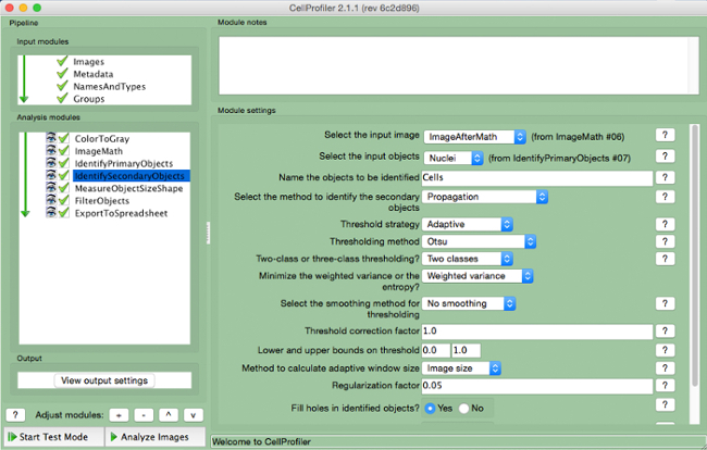
Figure 7. CellProfiler pipeline: identifying cells. Screenshot of "IdentifySecondaryObjects" module. Please click here to view a larger version of this figure.

Figure 8. CellProfiler pipeline: measuring objects. Screenshot of "MeasureObjectSizeShape" module. Please click here to view a larger version of this figure.

Figure 9. CellProfiler pipeline: filtering objects. Screenshot of "FilterObjects" module. Please click here to view a larger version of this figure.

Figure 10. CellProfiler pipeline: exporting data. Screenshot of "ExportToSpreadsheet" module. Please click here to view a larger version of this figure.

Figure 11. CellProfiler output following filtering. Screenshot of output screen in cell profiler following object filtering. Please click here to view a larger version of this figure.
Access restricted. Please log in or start a trial to view this content.
Results
Both solid and perfused 3D breast cancer surrogates were prepared as described above and grown for 7 days. Subsequently, surrogates were fixed, processed to paraffin, sectioned, and stained with hematoxylin and eosin, as described above. The number of nucleated cells per area (both 231 cells and CAF) of each surrogate was measured. As can be seen in Figure 12, representative photomicrographs of the H&E-stained sections demonstrate a higher concentration of cells prese...
Access restricted. Please log in or start a trial to view this content.
Discussion
Herein, a method of 3D culture has been described that incorporates components of the tissue microenvironment, including the extracellular matrix (ECM) and human stromal fibroblasts, in a volume that more closely models human breast cancer to allow for the development of a recapitulative 3D morphology. The 3D culture method described is more representative of human disease than traditional 2D cell culture in that multiple cell types are incorporated into a 3D volume of ECM. It has been noted that these parameters (i....
Access restricted. Please log in or start a trial to view this content.
Disclosures
The authors declare that they have no competing financial interests.
Acknowledgements
The University of Alabama at Birmingham Center for Metabolic Bone Disease performed the histologic processing and sectioning of surrogates. Southern Research (Birmingham, AL) provided support for the manufacture of the bioreactor system. Funding was provided by the United States Department of Defense Breast Cancer Research Program (BC121367).
Access restricted. Please log in or start a trial to view this content.
Materials
| Name | Company | Catalog Number | Comments |
|---|---|---|---|
| Dulbecco's Modified Eagel Medium 1x (DMEM) | Corning CellGro | 10-014-CV | |
| Fetal Bovine Serum (FBS) | Atlanta Biologicals | S11150 | |
| 0.25% Trypsin + 2.21 mM EDTA 1x | Corning | 25-053-CI | |
| Tissue Culture plates, 100 mm | CellTreat Scientific Products | 229620 | Sterile |
| Tissue Culture plates, 35 mm | CellTreat Scientific Products | 229638 | For PDMS foam formation |
| 9" Glass pipette | Fisher | 13-678-20D | Sterile |
| 10 ml pipette | CellTreat Scientific Products | 229210B | Sterile |
| 1,000 µl piptette tips | FisherBrand | 02-717-166 | Sterile Filtered |
| 200 µl pipette tips | FisherBrand | 02-717-141 | Sterile Filtered |
| 10 µl pipette tips | FisherBrand | 02-717-158 | Sterile Filtered |
| 15 ml conical tubes | CellTreat Scientific Products | 229410 | Sterile |
| 50 ml conical tubes | CellTreat Scientific Products | 229422 | Sterile |
| 1.5 ml microcentrifuge tubes | FisherBrand | 05-408-129 | Sterile |
| Trypan blue | Corning Cellgro | 25-900-CI | Sterile |
| Sylgard 184 | Electron Microscopy Sciences | 24236-10 | PDMS elastomer and curing agent. Used for our in-house bioreactor. |
| PDMS Foam | Made in-house for use in our in-house bioreactor. | ||
| High Concentration Bovine Collagen Type I | Advanced Biomatrix | 5133-A | FibriCol ~10 mg/ml |
| Growth Factor Reduced Matrigel (Basement Membrane) | Corning | 354230 | Basement membrane material |
| Sodium Bicarbonate | Sigma | S8761 | |
| Molecular Biology Grade Water | Fisher | BP2819-1 | |
| DMEM 10x | Sigma-Aldrich | D2429 | |
| Nunc Lab-Tek Chamber Slide System | Thermo Scientific | 177402 | 8-well |
| Bioreactor | Made in-house. | ||
| Spring-Back 304 Stainless Steel—Coated with PTFE polymer | McMaster-Carr | 1749T19 | Stainless steel wires to generate microchannels in our in-house bioreactor system. 0.016" Diameter |
| BioPharm Plus platinum-cured silicone pump tubing, L/S 14 | Masterflex | EW-96440-14 | For use in our in-house bioreactor system. Tubing ID: 1.6 mm, Hose barb size: 1/16 in. |
| 2-Stop Tubing Sets, non-flared PVC, 1.52 mm ID | Cole-Parmer | EW-74906-36 | For use in our in-house bioreactor system (with microperistalitic pump). |
| Six Channel precision micro peristaltic pump | Cole-Parmer | EW-74906-04 | For use with our in-house bioreactor system |
| Tuberculin Syringes | BD Medical | 309625 | 26 gauge 3/8 in. needle; Sterile |
| Dissecting Tissue Forceps | FisherBrand | 13-812-36 | 5.5 inch |
| Mini Tube Rotator | Boekel Scientific | 260750 | Equipment option for surrogate rotation. Used with carousel for 50 ml tubes (model number 260753) |
| 50 ml tube carousel | Boekel Scientific | 260753 | Used with mini tube rotator |
| Bambino Hybridization Oven | Boekel Scientific | 230301 | Equipment option for surrogate rotation |
| HistoGel Specimen Processing Gel | Thermo Scientific | HG-4000-012 | Specimen Processing Gel described in Step 5.2 |
| Cryomold | Andwin Scientific | 4566 | 15 mm x 15 mm x 5 mm |
| Tissue Marking Dye | Cancer Diagnostics, inc. | 03000P | Can be used to mark surrogates, allowing multiple samples to be included in one tissue cassette |
| Hinged tissue cassettes | FisherBrand | 22-272-416 | |
| Formalin | Fisher | 23-245-685 | |
| GoldSeal Plain Glass Slides | Thermo Scientific | 3048-002 | |
| Xylene | Fisher | X3P-1GAL | |
| Ethanol, 200 proof (100%), USP | Decon Laboratories, Inc. | 2805M | |
| Hematoxylin | Thermo Scientific Richard-Allan Scientific | 7211 | |
| Clarifier | Thermo Scientific Richard-Allan Scientific | 7401 | |
| Bluing Solution | Thermo Scientific Richard-Allan Scientific | 7301 | |
| Eosin Y | Thermo Scientific Richard-Allan Scientific | 7111 | |
| Cytoseal XYL mounting media | Thermo Scientific Richard-Allan Scientific | 83124 | |
| Coverslips | Fisher Scientific | 12-548-5G |
References
- Hakanson, M., Textor, M., Charnley, M. Engineered 3D environments to elucidate the effect of environmental parameters on drug response in cancer. Integr Biol (Camb). 3 (1), 31-38 (2011).
- Horning, J. L., et al. 3-D tumor model for in vitro evaluation of anticancer drugs. Mol Pharm. 5 (5), 849-862 (2008).
- Dhiman, H. K., Ray, A. R., Panda, A. K. Three-dimensional chitosan scaffold-based MCF-7 cell culture for the determination of the cytotoxicity of tamoxifen. Biomaterials. 26 (9), 979-986 (2005).
- Place, A. E., Jin Huh, S., Polyak, K. The microenvironment in breast cancer progression: biology and implications for treatment. Breast Cancer Res. 13 (6), 227(2011).
- Mao, Y., Keller, E. T., Garfield, D. H., Shen, K., Wang, J. Stromal cells in tumor microenvironment and breast cancer. Cancer Metastasis Rev. 32 (1-2), 303-315 (2013).
- Paulsson, J., Micke, P. Prognostic relevance of cancer-associated fibroblasts in human cancer. Semin Cancer Biol. 25, 61-68 (2014).
- Roskelley, C. D., Desprez, P. Y., Bissell, M. J. Extracellular matrix-dependent tissue-specific gene expression in mammary epithelial cells requires both physical and biochemical signal transduction. Proc. Natl. Acad. Sci. 91, 12378-12382 (1994).
- Pickl, M., Ries, C. H. Comparison of 3D and 2D tumor models reveals enhanced HER2 activation in 3D associated with an increased response to trastuzumab. Oncogene. 28 (3), 461-468 (2008).
- Ivascu, A., Kubbies, M. Rapid generation of single-tumor spheroids for high-throughput cell function and toxicity analysis. J Biomol Screen. 11 (8), 922-932 (2006).
- Lovitt, C. J., Shelper, T. B., Avery, V. M. Advanced cell culture techniques for cancer drug discovery. Biology (Basel). 3 (2), 345-367 (2014).
- Bergamaschi, A., et al. Extracellular matrix signature identifies breast cancer subgroups with different clinical outcome. J Pathol. 214 (3), 357-367 (2008).
- Oskarsson, T. Extracellular matrix components in breast cancer progression and metastasis. The Breast. 22, S66-S72 (2013).
- Kelley, L. C., Lohmer, L. L., Hagedorn, E. J., Sherwood, D. R. Traversing the basement membrane in vivo: A diversity of strategies. JBC. 204 (3), 291-301 (2014).
- Sadlonova, A., et al. Breast fibroblasts modulate epithelial cell proliferation in three-dimensional in vitro co-culture. Breast Cancer Res. 4, (2004).
- Wendt, D., Marsano, A., Jakob, M., Heberer, M., Martin, I. Oscillating perfusion of cell suspensions through three-dimensional scaffolds enhances cell seeding efficiency and uniformity. Biotechnol Bioeng. 84 (2), 205-214 (2003).
- Marshall, L. E., et al. Flow-perfusion bioreactor system for engineered breast cancer surrogates to be used in preclinical testing. J Tissue Eng Regen Med. , (2015).
- Calcagnile, P., Fragouli, D., Mele, E., Ruffilli, R., Athanassiou, A. Polymeric foams with functional nanocomposite cells. RSC Adv. 4, 19177-19182 (2014).
- Naba, A., Clauser, K. R., Lamar, J. M., Carr, S. A., Hynes, R. O. Extracellular matrix signatures of human mammary carcinoma identify novel metastasis promoters. eLife. 3, (2014).
- Lochter, A., Bissell, M. J. Involvement of extracellular matrix constituents in breast cancer. Semin Cancer Biol. 6 (3), 165-173 (1995).
- Joiner, K. S., Spangler, E. A. Evaluation of HistoGel-embedded specimens for use in veterinary diagnostic pathology. J Vet Diagn Invest. 24 (4), 710-715 (2012).
- Varsegi, G. M., Shidham, V. Cell Block Preparation from Cytology Specimen with Predominance of Individually Scattered Cells. J Vis Exp. (29), e1316(2009).
- Sadlonova, A., et al. Human Breast Fibroblasts Inhibit Growth of the MCF10AT Xenograft Model of Proliferative Breast Disease. Am J Pathol. 170 (3), (2007).
- Otali, D., He, Q., Stockard, C. R., Grizzle, W. E. Preservation of immunorecognition by transferring cells from 10% neutral buffered formalin to 70% ethanol. Biotech Histochem. 88 (0), 170-180 (2013).
- Webster, S. S., Jenkins, L., Burg, K. J. L. Histological Techniques for Porous, Absorbable, Polymeric Scaffolds, Used in Tissue Engineering. J Histotechnol. 26 (1), 57-65 (2003).
- Troy, T. -C., Arabzadeh, A., Enikanolaiye, A., Lariviere, N., Turksen, K. Immunohistochemistry on Paraffin Sections of Mouse Epidermis Using Fluorescent Antibodies. J Vis Exp. (11), (2008).
- Carpenter, A. E., et al. CellProfiler: image analysis software for identifying and quantifying cell phenotypes. Genome Biol. 7 (10), R100(2006).
- Kwon, Y. -J., et al. Gli1 enhances migration and invasion via up-regulation of MMP-11 and promotes metastasis in ERa negative breast cancer cell lines. Clin Exp Metastasis. (28), (2011).
- Evilsizor, M. N., Ray-Jones, H. F., Lifshitz, J., Ziebell, J. Primer for Immunohistochemistry on Cryosectioned Rat Brain Tissue: Example Staining for Microglia and Neurons. J Vis Exp. (99), e52293(2015).
- Pal, A., Kleer, C. G. Three dimensional cultures: a tool to study normal acinar architecture vs. malignant transformation of breast cells. J Vis Exp. (86), e51311(2014).
- Hasselbach, L. A., et al. Optimization of High Grade Glioma Cell Culture from Surgical Specimens for Use in Clinically Relevant Animal Models and 3D Immunochemistry. J Vis Exp. (83), e51088(2014).
- Foty, R. A Simple Hanging Drop Cell Culture Protocol for Generation of 3D Spheroids. J Vis Exp. (51), e2720(2011).
- Materne, E. -M., et al. The Multi-organ Chip - A Microfluidic Platform for Long-term Multi-tissue Coculture. J Vis Exp. (98), e52526(2015).
- Sadlonova, A., et al. Identification of Molecular Distinctions Between Normal Breast-Associated Fibroblasts and Breast Cancer-Associated Fibroblasts. Cancer Microenviron. 2, 9-21 (2009).
- Wang, J. D., Douville, N. J., Takayama, S., ElSayed, M. Quantitative analysis of molecular absorption into PDMS microfluidic channels. Ann Biomed Eng. 40 (9), 1862-1873 (2012).
- Halldorsson, S., Lucumi, E., Gomez-Sjoberg, R., Fleming, R. M. Advantages and challenges of microfluidic cell culture in polydimethylsiloxane devices. Biosens Bioelectron. 63, 218-231 (2015).
- Regehr, K. J., et al. Biological implications of polydimethylsiloxane-based microfluidic cell culture. Lab Chip. 9 (15), 2132-2139 (2009).
- Burdett, E., Kasper, F. K., Mikos, A. G., Ludwig, J. A. Engineering tumors: a tissue engineering perspective in cancer biology. Tissue Eng Part B Rev. 16 (3), 351-359 (2010).
- Caruso, R. A., et al. Mechanisms of coagulative necrosis in malignant epithelial tumors (Review). Oncol Lett. 8 (4), 1397-1402 (2014).
- Elmore, S. Apoptosis: A Review of Programmed Cell Death. Toxicol Pathol. 35 (4), 495-516 (2007).
- Majno, G., Joris, I. Apoptosis, oncosis, and necrosis. An overview of cell death. Am J Pathol. 146 (1), 3-15 (1995).
- Ogino, S., et al. Molecular pathological epidemiology of epigenetics: emerging integrative science to analyze environment, host, and disease. Mod Pathol. 26 (4), 465-484 (2013).
- Otali, D., et al. Combined effects of formalin fixation and tissue processing on immunorecognition. Biotech Histochem. 84 (5), 223-247 (2009).
Access restricted. Please log in or start a trial to view this content.
Reprints and Permissions
