An original experimental setup for heating cells in a culture dish using 1.94 µm continuous-wave laser radiation is introduced here. Using this method, the biological responses of retinal pigment epithelial (RPE) cells after different thermal exposures can be investigated.
Method Article
Continuous-wave Thulium Laser for Heating Cultured Cells to Investigate Cellular Thermal Effects
Opens in a new tab
In This Article
Summary
Loading...
$$\rightleftharpoonup{xx}$$
$$\longleftharp{xx}$$,
$$\longrightharp{xx}$$,
Abstract
Loading...
$$\rightleftharpoonup{xx}$$
$$\longleftharp{xx}$$,
$$\longrightharp{xx}$$,
An original method to heat cultured cells using a 1.94 µm continuous-wave thulium laser for biological assessment is introduced here. Thulium laser radiation is strongly absorbed by water, and the cells at the bottom of the culture dish are heated through thermal diffusion. A laser fiber with a diameter of 365 µm is set about 12 cm above the culture dish, without any optics, such that the laser beam diameter is almost equivalent to the inner diameter of the culture dish (30 mm). By keeping a consistent amount of culture medium in each experiment, it is possible to irradiate the cells with a highly reproducible temperature increase.
To calibrate the temperature increase and its distribution in one cell culture dish for each power setting, the temperature was measured during 10 s of irradiation at different positions and at the cellular level. The temperature distribution was represented using a mathematical graphics software program, and its pattern across the culture dish was in Gaussian form. After laser irradiation, different biological experiments could be performed to assess temperature-dependent cell responses. In this manuscript, viability staining (i.e., distinguishing live, apoptotic, and dead cells) is introduced to help determine the threshold temperatures for cell apoptosis and death after different points in time.
The advantages of this method are the preciseness of the temperature and the time of heating, as well as its high efficiency in heating cells in a whole cell culture dish. Furthermore, it allows for study with a wide variety of temperatures and time durations, which can be well-controlled by a computerized operating system.
Introduction
Loading...
$$\rightleftharpoonup{xx}$$
$$\longleftharp{xx}$$,
$$\longrightharp{xx}$$,
Understanding temperature-dependent cell biological responses is of great importance to successful hyperthermia treatments. Retinal laser photocoagulation with a thermal laser, used in ophthalmology, is one of the most established laser treatments in medicine. Visible light, mostly from green to yellow wavelengths, is used in retinal laser treatment. The light is highly absorbed by the melanin in retinal pigment epithelial (RPE) cells, which form the outermost cell monolayer of the retina. There has been recent interest among physicians and researchers in very mild thermal irradiation (sub-visible photocoagulation) as a new therapeutic strategy for different kinds of retinal disorders1,2. Following this trend, our interest is in sub-lethally heating RPE cells under precise temperature control, a technique called temperature-controlled photothermal therapy (TC-PTT).
Recent optoacoustic technology from our institute has allowed for the real-time measurement of temperature increases at irradiated sites in the retina. This enables control over the temperature increase during irradiation3. However, since sub-lethal hyperthermia on the retina, caused by heating RPE cells sub-lethally, has not been previously considered due to the impossibility of measuring and controlling the temperature, the temperature-dependent cell responses of RPE cells following thermal laser irradiation has been studied very little to date. Moreover, not only has the temperature difference not been discussed in detail, but also the difference in the cell behavior of the surviving cells after sub-lethal and lethal irradiation. Therefore, to gather scientific evidence on TC-PTT-based treatments, we aim to elucidate the temperature-dependent RPE cell biological responses and their mechanisms using in vitro experimental setups.
For this purpose, it is necessary to establish a cell-heating setup that meets the following conditions: 1) a possibility for fast temperature increases, 2) a precisely controlled time and temperature, and 3) a relatively high number of examined cells for biological experiments. Regarding the heating method, a clinical laser, such as a frequency-doubled Nd.YAG laser (532 nm), is unfortunately unsuitable for cell culture heating. This is because of the strongly reduced number of melanosomes in cultured RPE cells. The laser light absorption might be inhomogeneous, and the temperature increase at the cellular level is variable between experiments, even when irradiated with same radiation power. Several previous studies have reported the use of black paper beneath the dish bottom during irradiation4 or the use of additional melanosomes that are phagocytized by the culture cells before the experiments5,6. Many of the in vitro biological studies to assess hyperthermia-induced cell responses have been performed using a hot plate, a water bath, or a CO2 incubator with a temperature setting7. These methods require a long heating period because it takes some time (i.e., several minutes) to reach the desired temperature. Furthermore, using these methods, it is difficult to obtain a detailed thermal history (i.e., temperature multiplied by time) at the cellular level. Moreover, the temperature among the cells at different positions in one culture dish may differ due to variable temperature diffusion. In most cases, this temporal and spatial temperature information during hyperthermia has not been taken into consideration for biological analyses, even though biological cell response may be critically affected by the temperature and the time duration of the increased temperature.
To overcome these problems, a continuous-wave thulium laser was used here to heat the cells. Thulium laser radiation (λ = 1.94 µm) is strongly absorbed by water8, and the cells at the bottom of the culture dish are thermally stimulated solely through thermal diffusion. The laser fiber with a 365-µm diameter is set about 12 cm above the culture dish, without any optics in between. The laser beam diameter diverges such that it is almost equivalent to the inner diameter of the culture dish (30 mm) at the surface of the culture medium.With a consistent amount of culture medium, it is possible to irradiate the cells with the temperature increase of high repeatability. Variable power settings enable irradiation with up to 20 W, and the medium temperature at the cellular level may be increased up to ΔT ≈ 26 °C in 10 s.
By modifying the irradiation conditions, it is also possible to change the laser beam profile to vary the temperature distribution in a culture dish. For example, it is possible to investigate with a Gaussian-like temperature distribution, as in the current study, or with a homogeneous temperature distribution. The latter may be advantageous for investigating the effects of temperature-dependent cell responses more specifically for sub-lethal temperature increases, but not for cell death stress or wound healing responses.
Altogether, thulium laser irradiation may enable the investigation of different kinds of biological factors, such as gene/protein expression, cell death kinetics, cell proliferation, and cell functionality development, after different thermal exposures.
Access restricted. Please log in or start a trial to view this content.
Protocol
Loading...
$$\rightleftharpoonup{xx}$$
$$\longleftharp{xx}$$,
$$\longrightharp{xx}$$,
1. RPE Cell Culture
- Isolation of RPE cells from porcine eyes
- Obtain freshly enucleated porcine eyes from the local slaughterhouse. Keep them cool (4 °C) and in a dark environment.
- Remove extracellular tissues with scissors and soak the eyes in an antiseptic solution for 5 min.
- Place the eyes in sterilized phosphate-buffered saline without calcium and magnesium (PBS (-)) until use.
- Using a scalpel, penetrate the sclera at about 5 mm posterior to the corneal limbus. Resect the whole anterior part of the eye by cutting with scissors all way through, parallel to the corneal limbus.
- Remove the anterior part of the eye (i.e., the cornea and lens) and the vitreous. Add 1 mL of PBS (-) and gently remove the neural retina.
NOTE: This "eye cup," consisting of the sclera, choroid, and RPE, is now ready. - Add pre-warmed (37 °C) 0.25% trypsin in PBS (-) to the eye cup. Adjust the volume such that about 80% of the eye cup is filled with this trypsin solution.
- Incubate the eye cup with the trypsin solution in a 5% CO2 incubator at 37 °C for 10 min.
- Remove the eye cup from the incubator and replace the 0.25% trypsin solution with a PBS (-) solution with 0.05% trypsin + 0.2% ethylenediaminetetraacetic acid tetrasodium salt (EDTA·4N). Incubate the eye cup in the incubator for 45 min.
NOTE: After 45 min, the RPE cells will be either just loosely attached to the Bruch's membrane or already detached and floating in the trypsin-EDTA solution. - Collect the RPE cells by gentle pipetting. Collect the cells and solution in a conical tube filled with 10 mL of culture medium (DMEM high glucose with L-glutamine), including 10% porcine serum, antibiotic/antimycotic, and sodium pyruvate (1 mM).
NOTE: The serum may neutralize the effect of trypsin. - Centrifuge the cell suspension at 400 x g for 5 min at room temperature.
- Remove the supernatant and add 10 mL of fresh medium. Centrifuge again under the same conditions for 5 min.
- Remove the supernatant and add new medium, such that the cell concentration results in 5 x 10⁵cells/mL (determined by counting the cells using a hemocytometer). Mix well by gentle pipetting.
- Distribute the cell suspension in cell culture dishes. Use 3 mL per 60 mm-diameter culture dish.
NOTE: This culture is called passage zero (P0). - Maintain the cells in a 5% CO2 incubator at 37 °C. Change half of the conditioned medium to fresh medium every second day.
- Subculture (step 1.2) if it becomes confluent.
- Subculture of the RPE cell culture
- Remove the culture medium and rinse the cells twice with PBS (-).
- Incubate the cells with PBS (-) solution with 0.05% trypsin + 0.2% EDTA in a 5% CO2 incubator at 37 °C for 5 min.
- Detach the RPE cells by gentle pipetting and collect the cell suspension in a conical tube filled with 10 mL of culture medium, including 10% porcine serum.
- Centrifuge the cell suspension at 400 x g for 5 min at room temperature.
- Remove the supernatant and add new culture medium, making the cell concentration 5 x 10⁵ cells/mL (determined by counting the cell number with a hemocytometer). Distribute the cells in new 60 mm-diameter culture dishes, as described in step 1.1.13.
NOTE: The cell culture is now passage 1 (P1). - After confluency is reached, subculture the P1 culture to P2, using the same procedure described in steps 1.2.1-1.2.5. From the P2 culture, seed the cells on smaller culture dishes (30-mm inner diameter) instead of 60 mm-diameter culture dishes.
- For the experiments, use P2 or P3 cultures.
2. Thulium Laser Irradiation
- Construction of the irradiation station
- Connect a thulium laser device (1.94 µm, power range: 0-20 W) to a 0.22-NA, 365-µm core diameter fiber.
- Mechanically fix the fiber tip to the metal arm that is horizontally fixed to the vertical metal post of the irradiation station. Place the vertical post such that the tip of the laser fiber is located above the hot plate on which the cell culture dish is to be placed during irradiation.
- Lay a white paper on the hot plate and turn the aiming beam on (λ = 635 nm, max = 1 mW, diameter at paper level ≈ 30 mm). Mark the circumference of the aiming beam on the white paper so that the position where the culture dish is to be placed during irradiation is known.
NOTE: The z-plane of the fiber tip may be changeable. Without any additional imaging optics, the laser spot diameter on the cell culture plane, placed 12 cm below the fiber tip, is about 30 mm, which is almost equivalent to the inner diameter of the cell culture dish. A schematic drawing of the setup is shown in Figure 1.
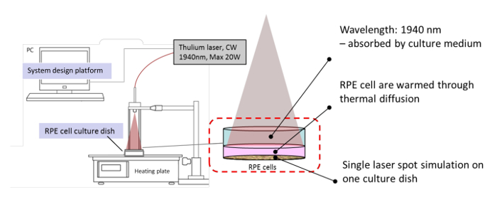
Figure 1: Schematic Image of the Thulium Laser Irradiation Station. A culture dish is placed on the heating plate. The cells are placed 12 cm below the thulium laser fiber tip so that the beam size is almost identical to the inner diameter of the culture dish (about 30 mm). The laser irradiation procedure is controlled by a time-controlled routine of the custom-made system design platform. The power setting must be determined before the irradiation program is started. Please click here to view a larger version of this figure.
- Laser irradiation of the cell culture
- 1 h before irradiation, replace the culture medium completely with 1.2 mL of fresh medium.
NOTE: This is a CRITICAL STEP and must be strictly followed. - Place the irradiation station (i.e., the hot plate and the post with which to fix the laser fiber) on a clean bench.
- Remove the cell culture dish from the incubator and place it on the marked position on the hot plate (step 2.1.3).
- Wear protective glasses. Turn on the thulium laser. Set the power as desired on the laser device (tunable from 0 - 20 W). Turn the emission on.
- Start a system design platform that controls the laser irradiation and timing protocol (supplemental file).
- Immediately after placing the culture dish on the hot plate, click the "pre-heating time" to start the timer for 140 s ("pre-heating time 1"); this will keep the culture medium temperature at 37 °C before irradiation.
NOTE: After 140 s, a beep sound will turn on, and the next timer ("pre-heating time 2") will start automatically counting 8 s. During this 8 s, the examiner may open the culture dish. After 148 s of pre-heating, a 10 s-long laser irradiation on the cell culture will be conducted automatically. In case of emergency, equip the laser device with a force-quit button to stop the laser immediately. This is a CRITICAL STEP and must be strictly followed. A special point of caution relates to opening the cover of the dish just before irradiation, at the beginning of the 8 s pre-heating time. Opening the cover may cool down the medium surface very quickly. - After irradiation, immediately place the cover back onto the culture dish, leave the culture dish on the hot plate for an additional 7 s, and place it back in the 5% CO2 incubator at 37 °C.
- 1 h before irradiation, replace the culture medium completely with 1.2 mL of fresh medium.
- Measurement of the temperature distribution at the cellular level (temperature calibration)
- Make small holes (about 300 µm in diameter) close to the bottom on four sides (every 90°) of a 30 mm diameter culture dish (without cells); use the tip of a needle (20G) heated with a Bunsen burner. Seal the holes with electrical isolation tape from outside and make a small hole with a fine needle so that only a fine thermocouple (200 µm in diameter) may be inserted through this hole under watertight conditions.
- At the outside of the culture dish bottom, draw 2 perpendicular diameters and set the crossing point (i.e., the center of the bottom side) as the coordinate zero (0). Mark every 3 mm radially to the outside out the dish (i.e., 0, 3, 6, 9, 12, and 15 mm) in each direction along the lines (Figure 2, blue dots); the number of points should be 21 in total.
- Fill the cell culture dish with 1.2 mL of new culture medium. Place the culture dish onto a hot plate at 37 °C, insert a fine thermocouple (200 µm in diameter) into the side hole, and place its sensitive tip onto a marked position to be measured.
- Wear protective glasses. Turn on the thulium laser and manually set the power (between 0 and 20 W, in 0.1-W increments) of the laser devise.
NOTE: For temperature calibration, measurements with the power in increments of 3 W should be sufficient. - Turn on the system design platform and click the "Start Temp. Acquisition" button (supplemental file) to start the temperature measurement.
- Conduct same procedure as in step 2.2.6.
NOTE: The control program measures the temperature of the inserted thermocouple every 100 ms and shows the temperature progression during irradiation in the GUI. - Conduct these procedures for all 21 measurement points and at different power settings. Repeat the whole procedure three times for all points and for all power settings to achieve reliable data.
- Export temperature data as csv data, which can eventually be converted to a spreadsheet. Average the maximal temperature at the end of irradiation for the triplicate measurements at every point. Average the values from the points on the same circle (4 points in total, except the central point).
- Plot the obtained averaged maximal temperature on a graph, making the distance from the center of the dish (mm) as the x-axis and the temperature increase (ΔT, °C) as the y-axis. Use the fit function of a mathematical software program to fit a Gaussian model to the raw data. Create a Gaussian fit temperature distribution.
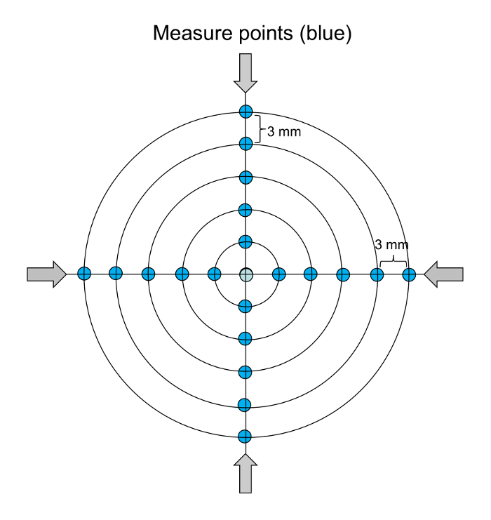
Figure 2: The Points for Temperature Calibration in One Cell Culture Dish. The temperature data was measured in the center and at 5 radial points over 4 different angles (blue dots). Please click here to view a larger version of this figure.
3. Biological Assessments for Cell Responses after Different Thermal Irradiations
- Assessment of cell viability ( i.e., living, apoptotic, and dead) following different power settings and the determination of the cell death threshold
- At the indicated time points (i.e., 3, 24, and 48 h after irradiation), wash the cells with PBS (-) and use a commercially available kit to assess cell viability (i.e., vital, apoptotic, dead) according to the manufacturer's protocol.
- Prepare a staining solution by adding 5 µL of fluorescein isothiocyanate (FITC)-annexin V, 5 µL of ethidium homodimer III, and 5 µL of Hoechst 33342 to 100 µL of 1x binding buffer (all are kit components). Prepare enough staining solution to cover the cells. Incubate the cells for 15 min.
- Wash the cell culture with the binding buffer twice, replace the binding buffer with PBS (-), and set the culture on the stage of a fluorescence microscope.
- Switch the light path to the ocular lens, select the 4',6-Diamidin-2-phenylindol (DAPI) filter, turn on the illumination light, and find the focused plane with the 4x objective.
- Change the light path to the camera, find the image on the computer screen in the microscope imaging software, and adjust the focus.
- Use the stitch function (i.e., the function to record multiple images across the dish and then create a single, large image) of the microscope-specific software to obtain the fluorescence image of the whole cell culture dish. Use 3 different filter sets-DAPI, FITC, and tetramethylrhodamine (TRITC)-to image Hoechst 33342-positive cells (all cell nuclei), FITC-annexin V-positive cells (apoptotic), and ethidium homodimer III-positive cells (dead), respectively.
- Measure the radius (mm) of the dead (ethidium homodimer III-positive) region and the outer/inner radius of the apoptotic (annexin V-positive) band-form region in the stained cell cultures. Apply these radii to the fitted Gaussian function of the temperature distribution for the corresponding power setting. Calculate the exact temperature at the rim of the dead or apoptotic region to clarify the threshold temperatures for cell death and apoptotic change.
Access restricted. Please log in or start a trial to view this content.
Results
Loading...
$$\rightleftharpoonup{xx}$$
$$\longleftharp{xx}$$,
$$\longrightharp{xx}$$,
Temperature distribution after different power settings
All temperature developments for each single irradiation were monitored in the temperature calibration. From this data, the maximal temperature at the measured point was obtained and defined as Tmax (°C). As shown in Figure 3A, the program was executed at the time point when the culture dish was placed on the heating plate. After the 14...
Access restricted. Please log in or start a trial to view this content.
Discussion
Loading...
$$\rightleftharpoonup{xx}$$
$$\longleftharp{xx}$$,
$$\longrightharp{xx}$$,
In discussing temperature-related biological cellular responses, not only the temperature, but also the time duration of the increased temperature, is of importance, since most biochemical processes are time-dependent. Particularly in the field of laser-induced hyperthermia in ophthalmology, due to the short time range-from milliseconds to seconds-it is difficult to investigate cellular thermal effects with precise temperature control. Therefore, a laser irradiation setup suitable for the cell culture model and with an o...
Access restricted. Please log in or start a trial to view this content.
Disclosures
Loading...
$$\rightleftharpoonup{xx}$$
$$\longleftharp{xx}$$,
$$\longrightharp{xx}$$,
The authors have nothing to disclose.
Acknowledgements
Loading...
$$\rightleftharpoonup{xx}$$
$$\longleftharp{xx}$$,
$$\longrightharp{xx}$$,
This work was supported by a research grant from the German Federal Ministry of Education and Research (BMBF) (grant #13GW0043C) and and a European Office of Aerospace Research and Development (EOARD, grant # FA9550-15-1-0443)
Access restricted. Please log in or start a trial to view this content.
Materials
| Name | Company | Catalog Number | Comments |
|---|---|---|---|
| Reagents | |||
| Dulbecco’s Modified Eagle’s Medium - high glucose | Sigma-Aldrich | D5796-500ML | Add (2)-(4) before use. Warm in 37 °C water bath before use. |
| Antibiotic Antimycotic Solution (100 ×) | Sigma-Aldrich | A5955-100ML | Containing 10,000 units penicillin, 10 mg streptomycin and 25 μg Amphotericin B in 1mL. Add 5.5 mL in 500 mL medium bottle (1) before use. |
| Sodium pyruvate (100 mM) | Sigma-Aldrich | S8636-100ML | Add 5.5 mL in 500 mL medium bottle (1) before use (final concentration: 1 mM) |
| Porcine serum | Sigma-Aldrich | 12736C-500ML | Add 50 mL in 500 mL medium bottole (1) before use (final: 10%) |
| Phosphate Buffered Saline (PBS) | Sigma-Aldrich | D8537-500ML | |
| Trypsin from porcine pancreas | Sigma-Aldrich | T4799-25G | |
| Ethylenediaminetetraacetic acid (EDTA) | Sigma-Aldrich | ED-100G | |
| Human VEGF Quantikine ELISA Kit | R&D System | DVE00 | |
| Oxiselect Total Glutathione Assay Kit | Cell Biolabs, Inc | STA-312 | |
| Apoptotic/Necrotic/Healthy Cells Detection Kit | PromoKine | PK-CA707-30018 | |
| Name | Company | Catalog Number | Comments |
| Equipments | |||
| Thulium laser | Starmedtec GmbH | Prototype | 0-20 W |
| 365 mm core diameter fiber | LASER COMPONENTS Germany | CF01493-52 | |
| Thermocouple | Omega Engineering Inc | HYP-0- 33-1-T-G-60-SMPW-M | |
| Heating plate | MEDAX | ||
| Microplate reader (spectrofluorometer) | Molecular Device | Spectramax M4 | |
| cell homogenizer | QIAGEN | TissueLyser LT | |
| Fluorescence microscope | Nikon | ECLIPSE Ti | |
| mathematical software program | The Mathworks. Inc | MATLAB Release 2015b | |
| system-design platform | National Instrument | Labview | Laboratory Virtual Instrument Engineering Workbench |
References
Loading...
$$\rightleftharpoonup{xx}$$
$$\longleftharp{xx}$$,
$$\longrightharp{xx}$$,
- Inagaki, K., et al. Comparative efficacy of pure yellow (577-nm) and 810-nm subthreshold micropulse laser photocoagulation combined with yellow (561-577-nm) direct photocoagulation for diabetic macular edema. Jpn J Ophthalmol. 59 (1), 21-28 (2015).
- Roider, J., et al. Selective retina therapy (SRT) for clinically significant diabetic macular edema. Graefes Arch Clin Exp Ophthalmol. 248 (9), 1263-1272 (2010).
- Brinkmann, R., et al. Real-time temperature determination during retinal photocoagulation on patients. J Biomed Opt. 17 (6), 061219(2012).
- Yoshimura, N., et al. Photocoagulated human retinal pigment epithelial cells produce an inhibitor of vascular endothelial cell proliferation. Invest Ophthalmol Vis Sci. 36 (8), 1686-1691 (1995).
- Denton, M. L., et al. Damage Thresholds for Exposure to NIR and Blue Lasers in an In Vitro RPE Cell System. Invest Ophthalmol Vis Sci. 47 (7), 3065-3073 (2006).
- Shrestha, R., Choi, T. Y., Chang, W., Kim, D. A high-precision micropipette sensor for cellular-level real-time thermal characterization. Sensors (Basel). 11 (9), 8826-8835 (2011).
- Gao, F., Ye, Y., Zhang, Y., Yang, J. Water bath hyperthermia reduces stemness of colon cancer cells. Clin Biochem. 46 (16-17), 1747-1750 (2013).
- Jansen, E. D., van Leeuwen, T. G., Motamedi, M., Borst, C., Welch, A. J. Temperature dependence of the absorption coefficient of water for midinfrared laser radiation. Lasers Surg Med. 14 (3), 258-268 (1994).
- Iwami, H., Pruessner, J., Shiraki, K., Brinkmann, R., Miura, Y. Protective effect of a laser-induced sub-lethal temperature rise on RPE cells from oxidative stress. Exp Eye Res. 124, 37-47 (2014).
- Denton, M. L., et al. Spatially correlated microthermography maps threshold temperature in laser-induced damage. J Biomed Optics. 16 (3), (2011).
- Morgan, C. M., Schatz, H. Atrophic creep of the retinal pigment epithelium after focal macular photocoagulation. Ophthalmology. 96 (1), 96-103 (1989).
Access restricted. Please log in or start a trial to view this content.
Reprints and Permissions
Request permission to reuse the text or figures of this JoVE article
Request Permission