The present protocol describes how Western blotting and immunocytochemistry combined with inhibition of lysosomal enzymes can be used to demonstrate the downregulation of Ciliary Neurotrophic Factor Receptor-α (CNTFRα), which is conveyed by the interaction between Cytokine-like Factor-1 (CLF-1) and the endocytic receptor sorLA.
Method Article
SorLA and CLC:CLF-1-dependent Downregulation of CNTFRα as Demonstrated by Western Blotting, Inhibition of Lysosomal Enzymes, and Immunocytochemistry
In This Article
Summary
Abstract
The heterodimeric cytokine Cardiotrophin-like Cytokine:Cytokine-like Factor-1 (CLC:CLF-1) targets the glycosylphosphatidylinositol (GPI)-anchored CNTFRα to form a trimeric complex that subsequently recruits glycoprotein 130/Leukemia Inhibitory Factor Receptor-β (gp130/LIFRβ) for signaling. Both CLC and CNTFRα are necessary for signaling but so far CLF-1 has only been known as a putative facilitator of CLC secretion. However, it has recently been shown that CLF-1 contains three binding sites: one for CLC; one for CNTFRα (that may promote assembly of the trimeric complex); and one for the endocytic receptor sorLA. The latter site provides high affinity binding of CLF-1, CLC:CLF-1, as well as the trimeric (CLC:CLF-1:CNTFRα) complex to sorLA, and in sorLA-expressing cells the soluble ligands CLF-1 and CLC:CLF-1 are rapidly taken up and internalized. In cells co-expressing CNTFRα and sorLA, CNTFRα first binds CLC:CLF-1 to form a membrane-associated trimeric complex, but it also connects to sorLA via the free sorLA-binding site in CLF-1. As a result, CNTFRα, which has no capacity for endocytosis on its own, is tugged along and internalized by the sorLA-mediated endocytosis of CLC:CLF-1.
The present protocol describes the experimental procedures used to demonstrate i) the sorLA-mediated and CLC:CLF-1-dependent downregulation of surface-membrane CNTFRα expression; ii) sorLA-mediated endocytosis and lysosomal targeting of CNTFRα; and iii) the lowered cellular response to CLC:CLF-1-stimulation upon sorLA-mediated downregulation of CNTFRα.
Introduction
The CLC and CLF-1 constitute the two subunits of the heterodimeric cytokine CLC:CLF-11-3. For cellular signal induction, CLC:CLF-1 first targets the CNTFRα, its primary and GPI-anchored receptor, to form a membrane-bound trimeric complex. The CLC:CLF-1:CNTFRα complex subsequently recruits gp130/LIFRβ which mediates transmembrane signaling via the Janus Kinase/Signal Transducers and Activators of Transcription 3 (JAK/STAT3) pathway4. Mice deficient in any of the three subunits display one and the same phenotype and die within 24 h after birth due to a reduced number of facial neurons and insufficient suckling5-8. Findings in humans likewise underscore the functional coherence of the three subunits. Thus, human mutations (homozygote or compound) causing dysfunction of CLC:CLF-1 or CNTFRα, all result in the so-called cold-induced sweating/Crisponi syndrome, a condition characterized by symptoms such as impaired suckling and swallowing, various dysmorphic features, temperature spikes, and paradoxical and perfuse sweating at low temperatures9-11.
In vitro, the combination of CLC and CNTFRα is both necessary and sufficient for interaction with gp130/LIFRβ and the induction of signaling in cells12. The role of CLF-1 on the other hand is less clear. It is not directly involved in signaling, and has long been regarded as an appendix that mainly serves to facilitate the cellular secretion of CLC1. However, recent findings show that CLF-1 has additional and more important functions implicating both the signaling and turnover of CLC and CNTFRα. Thus, it appears that CLF-1 contains three independent binding sites: one for its well-known binding to CLC; one that mediates direct binding to the CNTFRα; and a third (high affinity) site for interaction with the endocytic receptor sorLA. As both CLC and CLF-1 seem to target CNTFRα with a considerable lower affinity than the CLC:CLF-1 complex, it is conceivable that CLF-1 (via its CNTFRα-binding site) promotes the unification of CLC and CNTFRα and thereby facilitates signaling13.
CLF-1's interaction with sorLA, the main issue of the present presentation, plays a completely different role. SorLA is one of the five type 1 receptors that constitute the Vps10p-domain receptor family14. It is expressed in a variety of tissues but in particular in brain and neuronal tissues15. Similar to the other family members sorLA carries an N-terminal ligand binding Vps10p-domain, but in addition, it also comprises other domain types including ligand-binding elements found in members of the low-density lipoprotein receptor family15. Its cytoplasmic tail interacts with several adaptor proteins, e.g., adaptor protein-1 and -2, and sorLA conveys efficient endocytosis as well as intracellular sorting and transport of bound proteins16-18. The Vps10p-domain comprises a large ten-bladed β-propeller19,20, which binds a series of unrelated ligands including CLF-1 (but notably, not CLC)13. The binding site in CLF-1 is accessible even after complex formation with CLC and CNTFRα which means that sorLA binds not only free CLF-1, but also CLC:CLF-1 and the trimeric complex CLC:CLF-1:CNTFRα13. SorLA conveys rapid endocytosis of CLF-1 and CLC:CLF-1, but these are soluble proteins whereas CNTFRα is fixed to the surface membrane by a GPI-anchor. The question is therefore if binding of CLC:CLF-1:CNTFRα allows sorLA to internalize the entire complex and thereby to alter the surface-membrane expression of CNTFRα (and the subsequent cellular susceptibility to CLC:CLF-1 signal induction), and/or the turnover of CNTFRα.
The experiments described in the present report were designed to clarify the following questions:
Does sorLA mediate CLC:CLF-1-dependent downregulation of surface-membrane CNTFRα? To clarify this, it was initially tried to determine the turnover of CNTFRα (in the absence and presence of sorLA) by means of metabolic labeling and pulse-chase experiments as described in21. However, attempts to label CNTFRα using a mixture of [S35] cysteine and [S35] methionine showed poor incorporation of radioactivity. This suggested a very low degree of new-synthesis, which in all probability would be unable to compensate for a sudden and significant loss of receptors. To determine if CNTFRα was downregulated when interconnected via CLC:CLF-1 to sorLA, it was therefore decided simply to measure (using Western blotting) the total cellular pool of CNTFRα before and after exposure to CLC:CLF-1 - and to compare results in cells transfected or not transfected with sorLA.
Does sorLA target CNTFRα to lysosomes? To answer this question, the localization of CNTFRα — internalized via its CLC:CLF-1-mediated binding to sorLA — was examined using immunocytochemistry, and labeling with antibodies directed towards CNTFRα and the late-endosome/lysosome marker Lysosomal-associated Membrane Protein 1 (LAMP-1). The experiment was performed on cells treated as well as untreated with inhibitors of lysosomal enzymes. The idea was of course that if CNTFRα was degraded in lysosomes, cells treated with enzyme-inhibitors would present CNTFRα-staining accumulating in LAMP-1 positive vesicles, whereas untreated cells would show little or no staining for CNTFRα.
Does CNTFRα downregulation lower the cellular response to CLC:CLF-1 stimulation? The trimeric complex consisting of CLC, CLF-1, and CNTFRα signals via the gp130/LIFRβ heterodimer using the JAK/STAT3 pathway. Accordingly, the capacity of cells to respond to CLC:CLF-1 signaling upon downregulation of CNTFRα was explored by Western blot detection and quantitation of the phospho-STAT3 (pSTAT3)/total STAT3 level in sorLA transfectants.
Protocol
1. Western Blotting of CNTFRα Lysates
- Express full-length sorLA and Myc-tagged CNTFRα constructs in human embryonic kidney 293 (HEK293) cells using pcDNA3.1/zeo(-) and pcDNA3.1/hyg(-), respectively. Transfect the HEK293 cells with the constructs using a commercial transfection reagent according to manufacturer´s protocol. Select stable clones in medium containing 150 µg/mL Zeocin and/or 500 µg/mL Hygromycin B Gold13.
- Seed equal numbers of HEK293 transfectants expressing either CNTFRα-Myc or co-expressing CNTFRα-Myc/sorLA in two 4-well plates as shown in Figure 1. Culture the cells in Dulbecco Modified Eagle´s Medium (DMEM) supplemented with 10% fetal bovine serum (FBS), 100 U/mL penicillin, and 100 μg/mL streptomycin (full medium) in a 5% carbon dioxide incubator at 37 ˚C.
- Wait until the cells are 50 - 80% confluent.
- Remove the medium from the wells, wash once with Dulbecco Phosphate Buffered Saline (DPBS), and add fresh full medium with or without (control) 10 nM CLC:CLF-1 as indicated in Figure 1. Incubate the cells at 37 ˚C.
- After 0 and 5 h of incubation, recover the medium and store it at 4 ˚C in 1.5 mL tubes. Then add 1% Triton X-100 lysis buffer (20 mM Tris-HCl, 10 mM EDTA, pH 8.0) supplemented with a proteinase inhibitor cocktail to each well (1 min, 4 ˚C). Subsequently, transfer the cell lysates to 1.5 mL tubes (4 ˚C).
- Spin the tubes containing the medium and cell lysate (3,000 x g, 3 min, 4 ˚C) and transfer the supernatants to 1.5 mL tubes (4 ˚C). Discard the pellet. Transfer 5 μL of each cell lysate supernatant to new 1.5 mL tubes — use them to measure the protein content later (step 1.7). Immediately, add nonreducing LDS sample buffer to all the supernatants and vortex the samples.
- Use the 5 μL aliquots to measure the protein content in the cell lysate supernatants using a commercial protein assay according to manufacturer's protocol.
- Heat the samples at 95 ˚C for 5 min.
- Subject an equal amount of protein from each sample to reducing SDS-PAGE13 and Western blotting13 and visualize the content of sorLA, β-actin, and CNTFRα-Myc in the medium and in the cell lysates using rabbit anti-sorLA16 (5 μg/mL), mouse anti-β-actin (0.4 μg/mL), and mouse anti-Myc (1 μg/mL) antibodies and the appropriate HRP-linked secondary antibodies (1:2,000).
- Quantify the bands by densitometry according to manufacturer's protocol.
2. Immunocytochemistry in Combination with Lysosomal Inhibitors
- Seed HEK293 cells stably transfected with CNTFRα-Myc/sorLA on cover slides in a 4-well plate as shown in Figure 2. Culture the cells in full medium as above.
- Wait until cell confluence is 20 - 40%.
- Remove the medium from the wells and add full medium with or without 50 μg/mL of leupeptin and of pepstatin A (leu/pep) (inhibits lysosomal hydrolases) to the cells as indicated in Figure 2. Incubate the cells at 37 ˚C. The medium must be replaced every 6 h during the following 24 h of incubation.
- After 24 h of incubation, once again replace the medium (with or without leu/pep) and this time add 10 nM CLC:CLF-1. Incubate the cells at 37 ˚C for 5 h.
- Wash the cells once in PBS and fix them in 4% paraformaldehyde, pH 7 for 15 min at RT.
Caution: Paraformaldehyde is highly toxic, avoid contact with skin, eyes, and mucous. Gloves and safety glasses should be worn and solutions made inside a fume hood. - Wash the cells once in PBS and permeabilize the cells for 5 min at RT in saponin diluted in PBS (0.5% saponin).
- Dilute goat anti-CNTFRα (1 μg/mL) (notably not the mouse anti-Myc used for Western blotting) and mouse anti-LAMP-1 (15 μg/mL) antibodies in 0.5% saponin and incubate the cells with the antibodies for 2 h at RT.
- Wash the cells 4x (5 min) in 0.5% saponin and incubate the cells with donkey anti-goat Alexa 488- and donkey anti-mouse Alexa 568-conjugated secondary antibodies (6 μg/mL) for 2 h at RT.
- Wash the cells 4x (5 min) in 0.5% saponin, twice in deionized water (1 min), and mount the cover slides on microscope slides using mounting media.
- Analyze the antibody-staining of the cells by confocal microscopy using a laser-scanning confocal microscope with a 63X C-apochromat water immersion objective, NA 1.2.
3. Western Blot-detection of the pSTAT3 Level
- Seed HEK293-sorLA cells (which express endogenous levels of CNTFRα) in a 4-well plate as shown in Figure 3. Culture the cells in full medium as above.
- Wait until the cells are 50 - 80% confluent.
- Remove the medium from the wells, wash once in PBS, and add fresh full medium with or without 10 nM CLC:CLF-1 to the cells as indicated in Figure 3. Incubate the cells at 37 ˚C for 5 h.
- Remove the medium, wash the cells twice in unsupplemented DMEM (blank medium; without FBS and antibiotics), and then reincubate the cells for 90 min (37 ˚C) in blank medium.
- Once again, remove the medium and add fresh blank medium with or without 5 nM CLC:CLF-1 (to initiate STAT3 phosphorylation) as indicated in Figure 3. Incubate for 15 min at 37 ˚C.
- Quickly remove the medium and lyse the cells (1 min, 4 ˚C) by adding 1% Triton X-100 lysis buffer (20 mM Tris-HCl, 10 mM EDTA, pH 8.0) supplemented with a proteinase inhibitor cocktail and a phosphatase inhibitor cocktail to each well. Subsequently, transfer the cell lysates to 1.5 mL tubes (4 ˚C).
- Transfer 5 μL of each supernatant to new 1.5 mL tubes — use them to measure the protein content later (step 3.8). Immediately, add nonreducing LDS sample buffer to all the supernatants and vortex the samples.
- Measure the protein content of the 5 μL aliquots using a commercial protein assay according to manufacturer's protocol.
- Sonicate each sample at RT until they are no longer viscous (approximately 1 min) and subsequently heat them at 95 ˚C for 5 min.
- Subject an equal amount of protein from each sample to SDS-PAGE13 and Western blotting13 and visualize the content of pSTAT3 and total STAT3 in the cell lysates using rabbit anti-pSTAT3 (1:2000) and mouse anti-STAT3 (1:1,000) antibodies and the appropriate HRP-linked mouse and rabbit secondary antibodies (1:2,000).
- Quantify the bands by densitometry according to manufacturer's protocol.
Results
Figure 1 shows a schematic representation of the cell-seeding and CLC:CLF-1 incubation in protocol 1. Figure 2 shows a schematic representation of the cell-seeding and leu/pep and CLC:CLF-1 incubation in protocol 2. Figure 3 shows a schematic representation of the cell-seeding and CLC:CLF-1 incubation and stimulation in protocol 3. Figure 4 shows the sorLA-mediated CLC:CLF-1-dependent downregulation of CNTFRα. Figure 5 shows the sorLA-mediated endocytosis and lysosomal targeting of CNTFRα. Figure 6 shows the lower response to CLC:CLF-1 stimulation after CNTFRα downregulation.
Note that bands signifying CNTFRα-Myc have similar density at time zero, whereas downregulation of CNTFRα-Myc is demonstrated by the weaker band in sorLA transfectants after 5 h (Figure 4). Figure 5 shows that CNTFRα-Myc has accumulated in LAMP-1 positive vesicles of leu/pep treated cells. In contrast, no accumulation of CNTFRα-Myc is seen in cells not subjected to leu/pep treatment. This demonstrates that CNTFRα-Myc is sorted to lysosomes and degraded. As can be seen in Figure 6, the level of pSTAT3 is significantly reduced in pre-stimulated cells as compared to the level in cells that has not been exposed to CLC:CLF-1. This is in accordance with the observed sorLA-mediated downregulation of CNTFRα in the pre-stimulated cells (Figure 4).
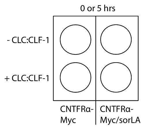
Figure 1: Schematic representation of cell-seeding and CLC:CLF-1 incubation in protocol 1. Seed HEK293-CNTFRα-Myc and HEK293-CNTFRα-Myc/sorLA cells in two 4-well plates (0 and 5 h) and incubate cells with CLC:CLF-1 as indicated. Please click here to view a larger version of this figure.
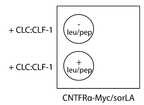
Figure 2: Schematic representation of cell-seeding and leu/pep and CLC:CLF-1 incubation in protocol 2. Seed HEK293-CNTFRα-Myc/sorLA cells on cover slides in a 4-well plate and incubate cells with or without leu/pep and CLC:CLF-1 as indicated. Please click here to view a larger version of this figure.
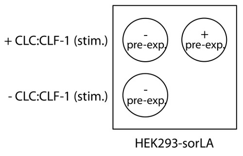
Figure 3: Schematic representation of cell-seeding and CLC:CLF incubation and stimulation in protocol 3. Seed HEK293-sorLA cells in one 4-well plate and incubate cells with or without CLC:CLF-1 (pre-exp.) to downregulate the CNTFRα followed by CLC:CLF-1 stimulation (stim.) to initiate STAT3 phosphorylation as indicated. Please click here to view a larger version of this figure.
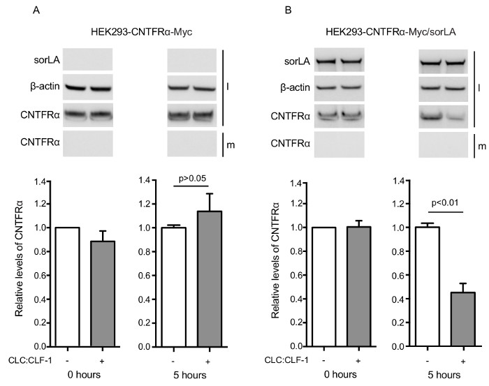
Figure 4: Results showing that SorLA mediates CLC:CLF-1-dependent downregulation of CNTFRα. (A) HEK293-CNTFRα-Myc and (B) HEK293-CNTFRα-Myc/sorLA cells were incubated in the absence (white columns) or presence (grey columns) of 10 nM CLC:CLF-1. After 0 and 5 h, the incubation was stopped and the content of CNTFRα-Myc in the medium (m) and in the cell lysates (l) was detected by Western blotting and quantified by densitometry. The upper panels show Western blot results from representative experiments and the lower panels show the detected levels of CNTFRα-Myc found in the cell lysates. The levels are shown relative to the CNTFRα-Myc level in the single and double transfectants at 0 h. Each column represents mean ±SEM (n = 3). p-values calculated using t-test. Reproduced after original figure13. Please click here to view a larger version of this figure.
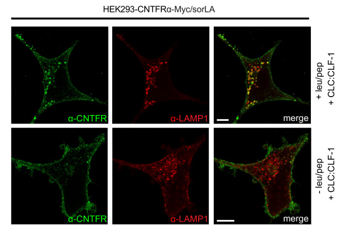
Figure 5: Results showing that SorLA mediates endocytosis and lysosomal targeting of CNTFRα. HEK293-CNTFRα-Myc/sorLA cells were treated with or without leu/pep, incubated with 10 nM CLC:CLF-1 for 5 h, fixed, and finally stained using anti-CNTFRα and anti-LAMP-1 antibodies as described in protocol 2. Scale bars: 5 μm. Reproduced after original figure13. Please click here to view a larger version of this figure.
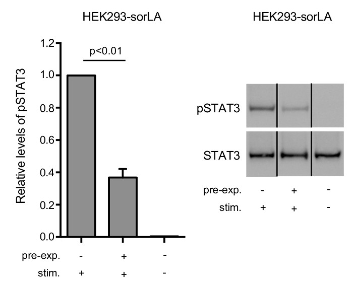
Figure 6: SorLA-mediated downregulation of CNTFRα is accompanied by a lowered cellular response to CLC:CLF-1 stimulation.
HEK293-sorLA cells were pre-incubated in the absence or presence of 10 nM CLC:CLF-1 for 5 h (pre-exp.), starved in blank medium for 90 min, and stimulated with 5 nM CLC:CLF-1 for 15 min (stim.). (left) The columns show the relative levels of pSTAT3 in the cells. Each column represents mean ±SEM (n = 3) relative to the pSTAT3 level in cells preincubated in the absence of CLC:CLF-1 but stimulated with CLC:CLF-1. p-value calculated using t-test. (right) The Western blot shows the response of pSTAT3 and STAT3 obtained in a representative experiment. Reproduced after original figure13. Please click here to view a larger version of this figure.
Discussion
The protocol described here can be used specifically to demonstrate the sorLA-mediated CLC:CLF-1-dependent downregulation and lysosomal targeting of CNTFRα, as well as the accompanying weakened response to CLC:CLF-1 stimulation.
The HEK293 cell line is well-suited for this protocol, as they have only a minor endogenous expression of CNTFRα and sorLA, are easy to transfect, express gp130/LIFRβ, and have a large cell body, which is suited for immunocytochemistry. However, in theory any cell line with similar properties should work as well.
The protocol has two critical steps. The first concerns step 2.3, in which the leu/pep medium must be replaced approximately every 6 h for 24 h. Failure to do so may result in active lysosomal enzymes and less or no detectable accumulation of protein in the lysosomes. The second critical step (3.4) concerns washing upon pre-incubation with CLC:CLF-1. It is important to remove any unbound ligand, and to allow the cells time to recover (avoiding continued stimulation) in unsupplemented DMEM (blank medium). This will ensure a low background level of pSTAT3 during the subsequent re-stimulation with CLC:CLF-1 (step 3.5).
Notably, Figure 6 demonstrates that the cellular response (pSTAT3) decreases in parallel with the sorLA-mediated downregulation of CNTFRα. It should be emphasized however, that although a reduced response is in accordance with (and to be expected upon) a reduction of the CNTFRα pool, it does not prove that the low CNTFRα expression alone accounts for the reduced cellular response. Thus, changes — resulting from pre-stimulation or transfection — in any of the functional elements of the signaling pathway upstream to pSTAT3 may have contributed to the altered response.
The basic concept of the protocol is not limited to this particular receptor system. By modifying the protocol (i.e. another cell line, other antibodies, other ligands, etc.) it can be used to determine the ligand-induced downregulation of other receptors as well — regardless of sorLA´s involvement. However, it should be noted that analysis of receptor-downregulation by Western blotting is mainly suited for receptors, e.g., GPI-anchored receptors, which exhibit a slow turnover and a low degree of new synthesis in unstimulated cells. Thus, in receptors showing a high level of new-synthesis, ligand-induced downregulation may be compensated for by synthesis of new receptors. Under these circumstances, the downregulation of the receptor pool can instead be determined by means of metabolic labeling and pulse-chase experiments as described in21. Alternatively, iodinated antibodies (preferably Fc-fragments) that do not interfere with receptor binding can be used to "tag" the ectodomain of the receptor, and the expected downregulation can then be determined by radioactive counting of cell pellet and medium.
For logistic reasons we transfected our cells with a CNTFRα construct carrying a Myc-tag, and in the present protocol a mouse anti-Myc antibody was used for Western blot-detection of the receptor. In contrast, a goat anti-CNTFRα antibody was used for immunocytochemistry and double labeling as LAMP-1 was detected by a mouse antibody — and obviously use of two primary mouse antibodies would result in unspecific (overlapping) staining with secondary antibodies. Note that since the goat anti-CNTFRα also works in Western blotting (not shown), the protocol could easily be modified and applied to cells transfected with untagged CNTFRα.
Finally, it has recently been shown that also the endocytic receptor sortilin binds CLC:CLF-1 with high affinity22. Thus, it is conceivable that sortilin, like sorLA, interconnects to CLC:CNTFRα via CLF-1 and is able to mediate endocytosis and lysosomal targeting of CNTFRα as well. Using sortilin instead of sorLA, the present protocol may be used to clarify this question.
Disclosures
The authors have nothing to disclose.
Acknowledgements
The authors have no acknowledgements.
Materials
| Name | Company | Catalog Number | Comments |
|---|---|---|---|
| Zeocin | InvivoGen | ant-zn-1 | |
| Hygromycin B Gold | InvivoGen | ant-hg-1 | |
| FuGENE 6 Transfection Reagent | Roche | 11 815 091 001 | |
| 4-well plates (Nunclon Delta Surface) | Thermo Scientific | 176740 | |
| Safe-Lock tubes | Eppendorf | 0030 120.086 | |
| Dulbecco Modified Eagle´s Medium (DMEM) | Lonza | BE12-604F/U1 | |
| Fetal Bovine Serum (FBS) | Thermo Scientific | 10270106 | |
| Penicillin/Streptomycin | Thermo Scientific | 15140122 | |
| Dulbecco Phosphate Buffered Saline (DPBS) | HyClone | SH30028.02 | |
| CLC:CLF-1 | R&D Systems | 1151-CL/CF | |
| Mouse anti-LAMP-1 | DSHP | H4A3 | |
| Rabbit anti-sorLA | Reference 16 | ||
| Mouse anti-b-actin | Sigma | A2228 | |
| Mouse anti-Myc | Genscript | A00704 | |
| Goat anti-CNTFRa | R&D Systems | AF-303 | |
| Rabbit anti-pSTAT3 | Cell signaling Technology | 9145s | |
| Mouse anti-STAT3 | Cell signaling Technology | 9139s | |
| Anti-mouse HRP-linked antibody | Cell signaling Technology | 7076s | |
| Anti-rabbit HRP-linked antibody | Cell signaling Technology | 7074s | |
| Anti-goat HRP-linked antibody | Dako | P0160 | |
| Donkey anti-goat secondary antibody, Alexa Fluor 488 conjugate | Invitrogen | A11055 | |
| Donkey anti-mouse secondary antibody, Alexa Fluor 568 conjugate | Invitrogen | A10037 | |
| Leupeptin | Sigma | L8511-5MG | |
| Pepstatin A | Sigma | P4265-5MG | |
| Triton X-100 | AppliChem | A1388 | |
| Tris-HCl | Merck | 1,082,191,000 | |
| EDTA:Titriplex III | Merck | 1,084,180,250 | |
| cOmplete mini EDTA-free | Roche | 11836170001 | |
| PhosSTOP | Roche | 04 906 837 001 | |
| LDS sample Buffer (4x) | Novex | NP0007 | |
| Bio-Rad Protein Assay | Bio-Rad | 500-0006 | |
| 4% paraformaldehyde (PFA) | VWR | 97,135,000 | |
| Saponin | Sigma | S7900-100G | |
| Mounting Media | Dako | S3023 |
References
- Elson, G. C., et al. CLF associates with CLC to form a functional heteromeric ligand for the CNTF receptor complex. Nat Neurosci. 3 (9), 867-872 (2000).
- Senaldi, G., et al. Novel neurotrophin-1/B cell-stimulating factor-3: a cytokine of the IL-6 family. Proc Natl Acad Sci U S A. 96 (20), 11458-11463 (1999).
- Elson, G. C., et al. Cytokine-like factor-1, a novel soluble protein, shares homology with members of the cytokine type I receptor family. J Immunol. 161 (3), 1371-1379 (1998).
- Lelievre, E., et al. Signaling pathways recruited by the cardiotrophin-like cytokine/cytokine-like factor-1 composite cytokine: specific requirement of the membrane-bound form of ciliary neurotrophic factor receptor alpha component. J Biol Chem. 276 (25), 22476-22484 (2001).
- Zou, X., et al. Neonatal Death in Mice Lacking Cardiotrophin-Like Cytokine (CLC) is Associated with Multifocal Neuronal Hypoplasia. Vet Pathol. 46 (3), 514-519 (2008).
- Forger, N. G., et al. Cardiotrophin-like cytokine/cytokine-like factor 1 is an essential trophic factor for lumbar and facial motoneurons in vivo. J Neurosci. 23 (26), 8854-8858 (2003).
- Alexander, W. S., et al. Suckling defect in mice lacking the soluble haemopoietin receptor NR6. Curr Biol. 9 (11), 605-608 (1999).
- DeChiara, T. M., et al. Mice lacking the CNTF receptor, unlike mice lacking CNTF, exhibit profound motor neuron deficits at birth. Cell. 83 (2), 313-322 (1995).
- Crisponi, L., et al. Crisponi syndrome is caused by mutations in the CRLF1 gene and is allelic to cold-induced sweating syndrome type 1. Am J Hum Genet. 80 (5), 971-981 (2007).
- Crisponi, G. Autosomal recessive disorder with muscle contractions resembling neonatal tetanus, characteristic face, camptodactyly, hyperthermia, and sudden death: a new syndrome. Am J Med Genet. 62 (4), 365-371 (1996).
- Sohar, E., Shoenfeld, Y., Udassin, R., Magazanik, A., Revach, M. Cold-induced profuse sweating on back and chest. A new genetic entity. Lancet. 2 (8099), 1073-1074 (1978).
- Guillet, C., et al. Functionally active fusion protein of the novel composite cytokine CLC/soluble CNTF receptor. Eur J Biochem. 269 (7), 1932-1941 (2002).
- Larsen, J. V., et al. Cytokine-Like Factor 1, an Essential Facilitator of Cardiotrophin-Like Cytokine:Ciliary Neurotrophic Factor Receptor alpha Signaling and sorLA-Mediated Turnover. Mol Cell Biol. 36 (8), 1272-1286 (2016).
- Willnow, T. E., Petersen, C. M., Nykjaer, A. VPS10P-domain receptors - regulators of neuronal viability and function. Nat Rev Neurosci. 9 (12), 899-909 (2008).
- Jacobsen, L., et al. Molecular characterization of a novel human hybrid-type receptor that binds the alpha2-macroglobulin receptor-associated protein. J Biol Chem. 271 (49), 31379-31383 (1996).
- Nielsen, M. S., et al. Sorting by the cytoplasmic domain of the amyloid precursor protein binding receptor SorLA. Mol Cell Biol. 27 (19), 6842-6851 (2007).
- Jacobsen, L., et al. The sorLA cytoplasmic domain interacts with GGA1 and -2 and defines minimum requirements for GGA binding. FEBS Lett. 511 (1-3), 155-158 (2002).
- Klinger, S. C., et al. SorLA regulates the activity of lipoprotein lipase by intracellular trafficking. J Cell Sci. 124 (7), 1095-1105 (2011).
- Kitago, Y., et al. Structural basis for amyloidogenic peptide recognition by sorLA. Nat Struct Mol Biol. 22 (3), 199-206 (2015).
- Quistgaard, E. M., et al. Ligands bind to Sortilin in the tunnel of a ten-bladed beta-propeller domain. Nat Struct Mol Biol. 16 (1), 96-98 (2009).
- Larsen, J. V., et al. Human sorCS1 binds sortilin and hampers its cellular functions. Biochem J. 457 (2), 277-288 (2014).
- Larsen, J. V., et al. Sortilin facilitates signaling of ciliary neurotrophic factor and related helical type 1 cytokines targeting the gp130/leukemia inhibitory factor receptor beta heterodimer. Mol Cell Biol. 30 (17), 4175-4187 (2010).
Reprints and Permissions
Explore More Articles
