This article presents a protocol optimized for the production of microfluidic chips and the setup of microfluidic experiments to measure the lifespan and cellular phenotypes of single yeast cells.
Method Article
Using Microfluidic Devices to Measure Lifespan and Cellular Phenotypes in Single Budding Yeast Cells
In This Article
Summary
Abstract
Budding yeast Saccharomyces cerevisiae is an important model organism in aging research. Genetic studies have revealed many genes with conserved effects on the lifespan across species. However, the molecular causes of aging and death remain elusive. To gain a systematic understanding of the molecular mechanisms underlying yeast aging, we need high-throughput methods to measure lifespan and to quantify various cellular and molecular phenotypes in single cells. Previously, we developed microfluidic devices to track budding yeast mother cells throughout their lifespan while flushing away newborn daughter cells. This article presents a method for preparing microfluidic chips and for setting up microfluidic experiments. Multiple channels can be used to simultaneously track cells under different conditions or from different yeast strains. A typical setup can track hundreds of cells per channel and allow for high-resolution microscope imaging throughout the lifespan of the cells. Our method also allows detailed characterization of the lifespan, molecular markers, cell morphology, and the cell cycle dynamics of single cells. In addition, our microfluidic device is able to trap a significant amount of fresh mother cells that can be identified by downstream image analysis, making it possible to measure the lifespan with higher accuracy.
Introduction
Budding yeast is a powerful model organism in aging research. However, a conventional lifespan assay in yeast relies on microdissection, which is not only labor intensive but also low throughput1,2. In addition, the traditional microdissection approach does not provide a detailed view of various cellular and molecular features in the single mother cells as they age. The development of microfluidic devices has enabled an automated procedure to measure yeast lifespan as well as to follow molecular markers and various cellular phenotypes throughout the lifespan of the mother cells3,4,5,6,7,8. After yeast cells are loaded into a microfluidic device, they can be tracked under a microscope using automatic time-lapse imaging. With the help of imaging processing tools, various cellular and molecular phenotypes can be extracted3,6,8, including lifespan, size, fluorescent reporter, cell morphology, cell cycle dynamics, etc., many of which are difficult or impossible to obtain using the traditional microdissection method. Microfluidic devices have gained prominence in yeast aging research since their successful development a few years ago3,4,6,7. Several groups have subsequently published on variations of the earlier designs5, and many yeast labs have employed microfluidic devices for their study.
In a cell culture undergoing exponential growth, the number of aged mother cells that are available for observation is miniscule. Therefore, the general design principle of the microfluidic device for lifespan measurements is to retain mother cells and to remove daughter cells. One such designs makes use of the fact that yeast undergoes asymmetric cell division. The structures in the device will trap bigger mother cells and allow smaller daughter cells to be washed away. The microfluidic chip described in this article uses a soft polydimethylsiloxane (PDMS) pad (vertical pensile columns) to trap mother cells (Figure 1). Devices of similar design have been reported previously3,4,6,7. This protocol uses a simpler procedure to fabricate microfluidic devices and a straightforward cell-loading method that is optimized for the time-lapse imaging experiments. One of the key parameters in the microfluidic device is the width of the PDMS pads used to trap mother cells. Our device uses wider pads that can keep more mother cells under each pad, including a significant fraction of fresh mother cells that can be tracked throughout their lifespan. In addition to lifespan measurements, this protocol is useful for single cell time-lapse imaging experiments when the cells need to be tracked for many generations or when an observation throughout the entire lifespan is necessary.
Protocol
1. Silicon Wafer Mold Fabrication
NOTE: The photomask is designed with AutoCAD software and manufactured by a commercial company. This design contains three layers of different patterns (Supplementary File 1). The heights of the first, second, and third layers are about 4 µm, 10 µm, and 50 µm, respectively. The silicon wafer mold was created from the photomask using soft lithography9,10.
- Bake a silicon wafer at 200 °C for 10 min to evaporate the water vapor. Spin coat negative photoresist SU-8 onto the silicon wafer.
NOTE: Spin coat SU-8 3005 at 5,000 rpm for 30 s to generate the first layer, SU-8 2010 at 3,000 rpm for 30 s to generate the second layer, and SU-8 3025 at 2,000 rpm for 30 s to generate the third layer. - Soft-bake the coated wafer before the pattern transfer. Align and expose the wafer in "direct contact" mode using a mask aligner.
NOTE: Soft-bake the wafer for 2 min at 95 °C for the first layer, 3 min at 95 °C for the second layer, and 15 min at 95 °C for the third layer. Use the exposure dose at 100 mJ/cm2 for the first layer, 130 mJ/cm2 for the second layer, and 200 mJ/cm2 for the third layer. - After exposure, post-bake the wafer and develop it using SU-8 developer. Dry the wafer using an N2 gun and hard-bake the wafer at 200 °C for 30 min.
NOTE: The silicon wafer mold is fixed to a 15 cm-diameter plastic Petri dish using scotch tape, with the pattern side facing up. Usually, we put several identical chip structures on the same mold, which allows multiple microfluidic chips to be fabricated at the same time. Each mold can be re-used many times to fabricate microfluidic chips.
2. Microfluidic Chip Fabrication
- Place a clean weighing boat on a balance and tare the balance. Pour 50 g of PDMS base into the weigh boat.
- Add 5 g of PDMS curing agent to the weigh boat (w/w ratio of 1:10 to the PDMS base).
NOTE: This volume is based on a 15 cm-diameter Petri dish with the mold. Adjust the amount of reagent if using a Petri dish of a different size. - Stir the PDMS base and the curing agent with a disposable pipette. Start from the edge of the weighing boat and slowly move inwards. Stir thoroughly for several minutes until small bubbles form throughout the mixture; thorough mixing is essential for PDMS polymerization.
- Pour the mixture slowly into the Petri dish; the mixture must completely cover the silicon wafer mold.
- Place the Petri dish in a vacuum for 10 min to remove all the air bubbles from the PDMS mixture. If bubbles remain on the surface of the mixture, use a pipette to blow them out.
- Incubate the silicon wafer mold with PDMS in an oven at 75 °C for about 2 h.
- Gently peel the layer of polymerized PDMS off the silicon wafer mold, avoiding any damage to the construction of the wafer mold. Alternatively, cut the PDMS directly from the silicon wafer mold with a minimum 5-mm margin around the pattern using a single-edge industrial razor blade; gently peel the PDMS layer off the wafer mold.
- Place the PDMS layer on the bench with the pattern side facing up. Carefully cut out the individual chip with a single-edge industrial razor blade, retaining an adequate margin from the edge of each single chip to avoid construction damage.
- With the pattern side facing up, use a punch pen (0.75-mm I.D.) to punch holes straight down through the inlet and outlet circles on each side of the channels.
NOTE: These holes create the pathway for the flow of medium. Therefore, it is crucial to go through the circles and punch all the way through the PDMS layer. Make sure to remove the PDMS columns from the hole. - Check every punched hole by inserting the punch pen needle again into the hole. Make sure the needle can come out from the other side, indicating that there is no blockage. Apply tape to the pattern surface, then gently peel off the tape to clean dust particles. Repeat this step at least three times. Leave a clean piece of scotch tape on the PDMS to maintain sterilization.
- Repeat this procedure on the opposite side of the PDMS and leave the last piece of tape on as well.
- Prepare a 24 mm x 30 mm cover glass with a thickness of 0.13-0.17 mm. Spray 70% ethanol on the glass and dry with dust remover to sterilize the surface; additionally, the glass can be washed with autoclaved water and dried with dust remover.
- Transfer the cover glass and PDMS to a plastic plate. Remove the scotch tape from the PDMS and place the pattern side facing up. Avoid any contact with the pattern surface during the transfer.
- Place the plastic plate in the plasma machine. Apply oxygen plasma treatment to the PDMS and the cover glass to render the surfaces hydrophilic with the following operation parameters: exposure, 75 s; gas stabilization, 20 cc/min; pressure, 200; and power, 100 W.
- Carefully place the PDMS onto the cover glass, connecting both hydrophilic surfaces (the surfaces that faced up during the plasma treatment). Make sure that there are no air bubbles between the PDMS and the cover glass.
- Incubate the PDMS chip in an oven at 75 °C for at least 2 h.
NOTE: Deficient microfluidics may cause liquid leakage onto the microscope during the experiment. Therefore, it is important to check the bond between the PDMS and the cover glass by slightly lifting the PDMS from the edges with tweezers. Gently lifting the PDMS should not separate it from the cover glass, indicating a successful bond.
3. Preparing for the Experiment
- Tube preparation.
- Submerge the input and output tubes in 70% ethanol solution separately 1 day before PDMS chip preparation.
- Fill a 5-mL syringe with autoclaved water to wash the tubes. Wash each tube by plugging the tube on the syringe and flushing the tube with water.
- Repeat the washing step at least 3 times to clean ethanol residue.
- Examine the PDMS channels under an optical microscope with a 10X objective to make sure the structure is consistent and intact. Stabilize the PDMS chip onto the microscope platform using scotch tape.
NOTE: Make multiple chips at one time in step 2 to ensure that there are backup chips, especially if making the device for the first time. - Fill a 5-mL syringe with autoclaved water. Secure the syringe to a syringe pump and insert the corresponding input and output tubes. Set the washing speed to 750 µL/h to rinse the chip for about 10 min. The air bubbles on the branch channels should be washed away during this period; if there are remaining bubbles, manually adjust the speed to eliminate the bubbles.
- Yeast sample preparation.
- Transfer 1 mL of prepared yeast sample (OD600 0.6-0.8) into two 1.5-mL microcentrifuge tubes and centrifuge for 5 min at 3,000 x g.
- Remove the majority of the supernatant from each tube and combine the remainder to form a 0.5-mL sample.
- Pause the syringe pump and remove the input tubes from the PDMS chip.
- Manually reverse the syringe pump to suck a small air bubble (1 to 2 cm in length) into each input tube. Move on to suck in the yeast sample; the air bubble will show the sample boundary and indicate how much sample has been drawn.
NOTE: Avoid sucking the sample into the syringe. - Insert the input tube back into the PDMS chip and restart the pump to load the cells at a speed of 750 µL/h.
- Leave the syringe pump on for 10 min then and examine the cell loading progression under the microscope; a successful loading will trap cells under more than 60% of suspend pillar.
- Switch to nutrition solution and adjust the speed to 400 µL/h for cell culturing.
- Select observation positions for the microscope.
NOTE: For lifespan measurements, images were taken once every 15 min by an optical microscope with a 40x objective. For the fluorescent reporter analysis, images were taken once every 15 min by a fluorescence microscope with a 60X oil objective.
Results
After the experiments, the lifespans of the cells and many cellular and molecular phenotypes can be extracted from the recorded time-lapse images. Since there are a number of different features that can be extracted from each cell, the first step of the analysis is to annotate the cells and events, including the positions and boundaries of the cells and the timing of various events that are being tracked, such as the budding events. These annotations will make it easier to return to the same set of cells and analyze different features in the future. Using image analysis software, such as ImageJ11 and the associated plugins, a list of phenotypes can then be extracted from the image data using the recorded annotations of the cells.
The following are a few examples of phenotypes measured with the microfluidic device. The lifespan phenotype of yeast can be obtained by counting the number of buds produced by each trapped mother cell and estimating with the Kaplan-Meier estimator. The cells initially trapped in the microfluidic devices are of unknown ages. To minimize the bias of lifespan measurements originating from these unknown ages, previous methods used the average number of bud scars on the trapped cells to calibrate the lifespan4,7. However, bud scar measurements require the additional staining of cells and provide only an indirect estimation of the bias. Using this protocol, the device frequently traps fresh daughter cells that budded off from already trapped cells. These cells then turn into mother cells. Such cells can be identified using our downstream image analysis software (Movie 1). These fresh mother cells provide a more accurate lifespan measurement. As shown in Figure 2, the lifespan curves of fresh mother cells were compared with those of the cells of unknown age. In this experiment, the average lifespan of the fresh mother cells was slightly longer (about 2 generations of difference in median lifespan).
In addition to the number of buds, other interesting phenotypes, such as the time interval between two successive budding events, as shown in Figure 3, can be extracted from the imaging data3,7,8. Cells entered a period of faster budding following a few slower initial buddings. The budding slowed down dramatically towards the end of their lifespan, indicating the unhealthy state of these aged cells. The cell cycle dynamics contains very useful information regarding the cellular state of the young and old cells and has been used, for example, to characterize telomerase mutants12. Importantly, this device can be used to measure fluorescence signals throughout the lifespan (Figure 4), allowing for the tracking of molecular markers that may be the driver of the aging process.

Figure 1: A schematic of the design of the microfluidic device. The device consists of 6 independent functional modules that can operate in parallel. Each module is made of one main channel connected to two side channels. Each side channel has 113 pensile columns. An additional bridge is added in the middle to connect the main channel with the two side channels. Mother cells are trapped underneath the pensile columns, while the smaller daughter cells are washed away by the flow. Please click here to view a larger version of this figure.
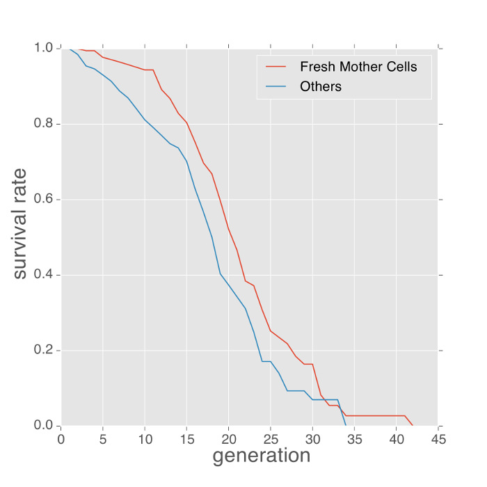
Figure 2: Fresh mother cells live longer than cells with unknown history. The replicative lifespan of fresh mother cells versus cells of unknown history (cells trapped from the beginning of the experiment) in SD media, 30 degrees. On average, fresh mother cells live about 2 generations longer. Please click here to view a larger version of this figure.

Figure 3: The length of budding intervals changes as cell aged. Budding intervals measured as time frames between buddings were color-coded, cells were ordered by their replication lifespan, and fresh mother cells were separated from cells of unknown history. Please click here to view a larger version of this figure.

Figure 4: Measuring the activity of Heat Shock Factor 1 (HSF1) using a microfluidic device. The Hsf1-activity reporter is constructed by a green fluorescent protein (GFP) fused to a crippled CYC1 promoter with HSE upstream13. Fluorescence images were taken every 30 min. GFP intensity was then quantified using a customized MATLAB code14. Data points for each single cell are connected and indicated with a colored line. In this experiment, the strain was grown in SD medium with 2% glucose (wt/vol) at 30 °C overnight; it was then diluted with the same medium for recovery. Afterward, SD medium with 0.05% glucose (wt/vol) was used to grow cells in the microfluidic chip during the fluorescence measurement. Please click here to view a larger version of this figure.

Movie 1: An example of fresh mother cells being trapped within the device for the whole lifespan. The movie shows a fresh mother cell born from a trapped cell. The red arrow at the beginning of the movie marks the position of the fresh mother cell and its first budding. This mother cell was trapped in the microfluidic device for its whole lifespan. Please click here to view this video. (Right-click to download.)
Discussion
The PDMS device needs to be freshly made. Otherwise, the air bubbles caused by inserting tubes into the device will be difficult to remove. Step 3.4 is important to improve the cell loading efficiency by concentrating the cells. To increase the throughput of the experiment, 4 to 6 modules on the same PDMS chip connected to independently operating pumps are typically used to perform 4 to 6 different experiments (different strains or media compositions) simultaneously.
Compared to the conventional yeast replicative aging assay (which uses microdissection), the microfluidic method presented here is less laborious and time-consuming. Moreover, the microfluidic device allows for the detailed quantification of various cellular or molecular parameters, including cell size, cell cycle dynamics, cell morphology, and various molecular markers. This microfluidic method achieves long-term cell tracking with high-resolution microscopy by retaining mother cells under PDMS micro-pads as daughter cells are flushed automatically.
This device uses soft PDMS micro-pads to trap mother cells4, with the basic structure similar, as Huberts et al. described in his work7. There are a number of differences in the device design and experimental protocols. The wider PDMS pad in this device allows some newborn daughter cells to be trapped and analyzed (Figures 2 and 3, Movie 1). To identify such cells, we annotated the daughter cells budded from already trapped mother cells, ignoring the daughter cells that flushed away without budding. On average, we got about 2 fresh mother cells per PDMS pad. The ratio of these fresh mother cells to the cells of unknown history was about 1:2, among which about one third were kept in the device for the whole lifespan. These cells allow a more accurate lifespan measurement and make it possible to analyze correlations between mother and daughter cells. In this device, a deeper main channel is connected to two shallower side channels where the observations are made; this design helps to reduce the chance of the side channels being blocked by big air bubbles. For microfluidic chip production, this protocol also uses a simple method to bond the PDMS and cover glass, just using plasma exposure and oven baking, which increases the success rate.
With this protocol, the microfluidic device is able to robustly trap at least one cell (3-5 cells on average) per PDMS pad at the beginning of the experiment for wild-type haploid yeast strains (i.e., BY4741 or BY4742). About 30% of the cells can be retained throughout their whole lifespan. It is worth noting that the performance of the PDMS device depends on the gap size between the pensile columns and the glass and the size of the yeast cells. For wild-type haploid yeast strains, the suitable size for the gap is 3.5-4.5 µm. Outside this range, the loading efficiency and cell retention rate decline sharply. Thus, new devices for yeast strains with much larger or smaller cell sizes7 must be fabricated by modifying the height of the first layer made in step 1.
In summary, the device and protocol described in this article are not only suitable for yeast aging studies but are also applicable to other experiments that require the tracking of mother cells and the monitoring of molecular markers for many generations or throughout the lifespan.
Disclosures
The authors declare that they have no competing financial interests.
Acknowledgements
This research was supported by NIH Grant AG043080 and the National Natural Science Foundation of China (NSFC), No. 11434001. We thank Lucas Waldburger for proofreading the manuscript.
Materials
| Name | Company | Catalog Number | Comments |
|---|---|---|---|
| 3'' <111> silicon wafer | Addison Engineering | ||
| SU-8 2000 and 3000 Series | MicroChem | ||
| Sylgard® 184 Silicone Elastomer Kit | ellsworth | 2065622 | Include Sylgard® silicone elastomer base and curing agent |
| Petri dishes | VWR | 391-1502 | |
| Harris Uni-core™ punch (I.D. 0.75 mm) | Sigma-Aldrich | 29002513 | |
| 24 mm x 40 mm SLIP-RITE® cover glass | Thermo Fisher Scientific | 102440 | |
| 3M Scotch Tape | ULINE | S-10223 | |
| VWR® Razor Blades | VWR | 55411-050 | |
| Pure Ethanol, Koptec | VWR | 64-17-5 | |
| Whoosh-Duster™ | VWR | 16650-027 | |
| 5 mL BD Syringe (Luer-Lock™ Tip) | Becton, Dickinson and Company | 309646 | |
| PTFE Standard Wall Tubing (100 ft, AWG Size: 22, Nominal ID: 0.028) | Component Supply Company | SWTT-22 | |
| Needle Assortment | Component Supply Company | NEKIT-1 | |
| Desiccator | HACH | 2238300 | |
| Lab Oven | Fisher Scientific | 13246516GAQ | |
| Nikon TE2000 microscope with 40X and 60X objective | Nikon | ||
| Zeiss Axio Observer Z1 with 40X and 60X objective | Zeiss | ||
| Syringe Pump | Longerpump | TS-1B |
References
- Mortimer, R. K., Johnston, J. R. Life span of individual yeast cells. Nature. 183 (4677), 1751-1752 (1959).
- Polymenis, M., Kennedy, B. K. Cell biology: High-tech yeast ageing. Nature. 486 (7401), 37-38 (2012).
- Xie, Z., et al. Molecular phenotyping of aging in single yeast cells using a novel microfluidic device. Aging Cell. 11 (4), 599-606 (2012).
- Zhang, Y., et al. Single cell analysis of yeast replicative aging using a new generation of microfluidic device. PLoS One. 7 (11), e48275(2012).
- Chen, K. L., Crane, M. M., Kaeberlein, M. Microfluidic technologies for yeast replicative lifespan studies. Mech Ageing Dev. , (2016).
- Lee, S. S., Avalos Vizcarra,, Huberts, I., H, D., Lee, L. P., Heinemann, M. Whole lifespan microscopic observation of budding yeast aging through a microfluidic dissection platform. Proc Natl Acad Sci U S A. 109 (13), 4916-4920 (2012).
- Huberts, D. H., Janssens, G. E., Lee, S. S., Vizcarra, I. A., Heinemann, M. Continuous high-resolution microscopic observation of replicative aging in budding yeast. J Vis Exp. (78), e50143(2013).
- Jo, M. C., Liu, W., Gu, L., Dang, W., Qin, L. High-throughput analysis of yeast replicative aging using a microfluidic system. Proc Natl Acad Sci U S A. 112 (30), 9364-9369 (2015).
- Mata, A., Fleischman, A. J., Roy, S. Fabrication of multi-layer SU-8 microstructures. Journal of Micromechanics and Microengineering. 16 (2), 276-284 (2006).
- Xia, Y. N., Whitesides, G. M. Soft lithography. Angewandte Chemie-International Edition. 37 (5), 550-575 (1998).
- Schneider, C. A., Rasband, W. S., Eliceiri, K. W. NIH Image to ImageJ: 25 years of image analysis. Nat Methods. 9 (7), 671-675 (2012).
- Xie, Z., et al. Early telomerase inactivation accelerates aging independently of telomere length. Cell. 160 (5), 928-939 (2015).
- Boy-Marcotte, E., et al. The heat shock response in yeast: differential regulations and contributions of the Msn2p/Msn4p and Hsf1p regulons. Mol Microbiol. 33 (2), 274-283 (1999).
- Yang, X., Lau, K. Y., Sevim, V., Tang, C. Design principles of the yeast G1/S switch. PLoS Biol. 11 (10), e1001673(2013).
Reprints and Permissions
