DFS70 autoantibodies mimic common disease-associated antinuclear antibody patterns making accurate interpretation challenging when using conventional HEp-2 substrates. The protocol describes advantages of novel engineered HEp-2 substrate over conventional HEp-2 in ANA screening and distinguishing DFS70 patterns with high confidence in both monospecific and mixed ANA positive cases.
Method Article
Simultaneous Distinction of Monospecific and Mixed DFS70 Patterns During ANA Screening with a Novel HEp-2 ELITE/DFS70 Knockout Substrate
In This Article
Summary
Abstract
Systemic autoimmune connective tissue disorders are characterized by circulating antinuclear antibodies (ANA). Although there are several technologies available for ANA screening, indirect immunofluorescence (IIF) using Human epithelial cells-2 (HEp-2) substrate remains the primary and recommended method because of its superior sensitivity. HEp-2 substrates can detect a multitude of patterns resulting from autoantibody binding to various protein and nucleic acid autoantigens distributed throughout the nucleus and cytoplasm of the cells. The great diversity of monospecific and mixed patterns resulting from positive reactions on HEp-2 substrate also complicate the interpretation and accuracy of reporting. One specific example which received utmost attention recently is the dense fine speckled 70 (DFS70) pattern resulting from autoantibodies that specifically bind to a protein called lens epithelium derived growth factor (LEDGF). Lack of clear association with a specific systemic autoimmune disease and high prevalence in healthy populations have made accurate interpretation of DFS70 pattern important. Accurate distinction of DFS70 pattern from disease-associated patterns using conventional HEp-2 substrate is challenging. Moreover, frequent co-occurrence of DFS70 pattern along with disease-associated patterns such as homogeneous, speckled, and mixed homogeneous-speckled patterns complicate the IIF interpretation. The goal of this paper is to demonstrate the utility of a novel engineered HEp-2 IIF substrate that retains all advantages of conventional HEp-2 substrate while simultaneously providing the ability to distinguish DFS70 pattern with high confidence in both monospecific and mixed ANA positive examples. The new substrate is further able to unmask disease-associated ANA patterns previously concealed by DFS70 pattern.
Introduction
ANAs are a hallmark of autoimmune mediated systemic connective tissue diseases1. Several methods are employed for screening and confirmation of ANAs. IIF technique using HEp-2 substrate remains the most widely used and frequently recommended method for screening ANAs1,2. Despite advances in solid phase diagnostic assay methodologies such as enzyme linked immunosorbent assay (ELISA), enzyme immunoassay (EIA), chemiluminescence immunoassay (CLIA), and bead-based multiplex assays, IIF by HEp-2 is the most prevalent screening method due to its diagnostic usefulness and cost effectiveness1,2,3. The above-mentioned assay technologies rely on a limited set of purified native or recombinant autoantigens whereas the HEp-2 cell monolayer preparations on glass slide wells present an extensive array of autoantigens in their natural form, distributed across the nucleus and cytoplasm, that react with the autoantibodies from patient sera or positive controls, resulting in a multitude of patterns that are associated with various autoimmune conditions. These patterns are classified into nuclear, cytoplasmic, and mitotic patterns based on the location of the antigen in the cells. Each pattern and the associated antigen/antigens and disease states are well characterized4. In the IIF method, patient and control sera are incubated on glass slide wells that present a monolayer culture of HEp-2 cells suitably fixed to preserve a multitude of autoantigens in their natural configuration. The autoantibodies in the patient sera or positive controls are allowed to bind to the autoantigens presented by HEp-2 cells on the substrate (glass slide well) during incubation. Post incubation, unbound antibodies are washed off and bound antibodies of the IgG class are detected with fluorescein/fluorescein isothiocyanate (FITC) conjugated anti-human IgG reagent. Unbound reported-conjugate is washed away and the glass coverslips are mounted on top of the slides using a mounting medium that preserves the reactions and facilitates observation under a fluorescent microscope. Reactions are observed under a fluorescence microscope equipped with appropriate excitation-emission filter combination that is configured as per the reporter used (for FITC reporter, a diagnostic microscope typically uses filters with excitation range of 467 - 498 nm and an emission range of 513 - 556 nm). The presence of ANA is demonstrated by a bright green fluorescence of specific structures in the cells. Unlike automated assay platforms, IIF methodology relies on the laborious process of serial diluting the sera and the visual positive determination of the staining patterns which requires significant user expertise5,6. Despite these limitations, IIF by HEp-2 remains the most widely used and recommended method for screening of ANAs due to the standardization efforts towards pattern interpretation and nomenclature by the International consensus on ANA patterns (ICAP) committee, increased availability of automation solutions for IIF slide processing, and result interpretation3.
Among the multitude of patterns detected by the HEp-2 IIF method, autoantibodies targeting the psip1 gene product (also referred to as LEDGF/p75/DFS70), have gained significant interest in recent years for several reasons4,5,6,7,8,9,10. DFS70 autoantibodies are present in high titers in healthy controls, and studies so far have failed to demonstrate a distinct clinical association for the presence of DFS70 autoantibodies7,8,9,10,11,12,13. The rates of reported positive results for DFS70 autoantibodies in various disease states and healthy cohorts varied widely between studies6,8,9,10,14,15,16,17,18,19,20,21,22,23,24,25,26,27,28. The monospecific DFS70 pattern is well differentiated from a homogeneous or speckled pattern but interpretation of mixed homogeneous-speckled patterns and anti-DFS70 antibodies co-occurring with other ANAs is challenging even for expert readers. The current generation of IIF screening methods and DFS70 specific solid phase assays are not capable of differentiating a monospecific DFS70 reaction from mixed patterns (Figure 1A, yellow shaded area of the algorithm). Misinterpretation of DFS70 pattern as homogeneous, speckled, or mixed pattern, or vice versa, may have serious implications on patient care. The current commonly used protocol is as follows: the clinical labs employ a number of solid phase confirmatory tests for disease-associated ANAs and/or an immunoadsorption method for DFS70/LEDGF when dense fine speckled (AC-2) pattern is suspected (Figure 1A)4,5.
The goal of this protocol is to demonstrate the utility of a novel engineered HEp-2 IIF substrate (HEp-2 ELITE/DFS70 knockout (KO), will be referred to as HEp-2 ELITE) that retains all the advantages of conventional HEp-2 for screening ANAs while simultaneously distinguishing DFS70 pattern with high confidence in both monospecific and mixed ANA positive cases (Figure 1B, yellow shaded area of the algorithm)5. HEp-2 ELITE substrate is composed of an approximate mixture of unmodified conventional HEp-2 cells and engineered cells devoid of LEDGF/p75/DFS70 antigen in 1:9 ratio5. The IIF procedure for HEp-2 ELITE is identical to the conventional HEp-2 method. The unique configuration of HEp-2 ELITE substrate not only retains all the advantages of conventional HEp2 but can differentiate homogeneous, speckled, and mixed reaction patterns from DFS70 pattern at the initial screening or testing of a sample (Figure 1B)5. Patient sera to be reacted on slides with HEp-2 IIF substrate should be diluted 1:40 with screening dilution or as per the individual laboratory criterion. A specific reaction at 1:40 or higher titer is considered positive.
In this study, positive sera for homogeneous, speckled, centromere, nucleolar, cytoplasmic reticular/AMA, and DFS70 patterns that produced a medium positive signal (relative to the negative control and positive ANA control provided by the kit) at screening dilution of 1:40 were selected for simulating the mono-specific and mixed patterns on HEp-2 substrates (conventional and HEp-2 ELITE). The 1:40 diluted DFS70 positive sera were mixed with 1:40 dilutions of individual disease-associated ANA pattern controls (homogeneous, speckled, centromere, nucleolar, or cytoplasmic reticular-AMA) in various ratios (control:DFS70 in 100:0, 80:20, 50:50, 20:80, and 0:100) and evaluated on conventional and HEp-2 ELITE substrates following standard IIF procedure outlined below.
Protocol
The protocol follows the guidelines of Trinity Biotech institutional human research ethics committee.
1. Sample and Substrate Preparation
- Allow pouches containing substrate slides (glass slides with 12 wells on each slide containing either HEp2 or HEp-2+HEp2 KO cell mixture) to equilibrate to room temperature for optimal performance and prevent condensation of moisture on slide surface prior to the addition of sample. This should take approximately between 10 - 15 min.
- Once equilibrated to room temperature, carefully remove the substrate slides using fingers without them touching the sides of the pouch.
- Label the slides and place them in an incubation chamber (at room temperature) lined with paper towels moistened with water to prevent drying of slides during sample and conjugate incubation.
NOTE: Non-specific staining can result from dry wells; a wet paper towel inside the incubation chamber provides humidity and prevents well dryness.
2. Addition of Controls and Samples
- Dispense approximately 50 µL of the negative/positive controls, combinations of DFS70-ANA positive sera as described in this study, or patient serum samples onto individual slide wells.
- Dispense control(s) and samples into each well on the slide.
- To prevent well cross-contamination, avoid overfilling the wells. While not overfilling wells, ensure complete coverage of well surface and avoid air bubbles.
NOTE: The recommended volume is 50 µL as per step 2.1. - Place the lid on the incubation chamber and incubate slides for 30 min at room temperature. If there is any ANA present in the serum of the patient, it will bind to the cells present in each well during this incubation period.
- Once the incubation period is over, remove the slide from the incubation chamber. Hold the slide at tab end and rinse gently with approximately 10 mL phosphate-buffered saline (PBS) wash buffer provided in the kit, using a pipette or in a beaker filled with PBS for 10 - 15 s. Transfer the slide immediately into a Coplin jar with PBS wash buffer and wash (soak) for 10 min. Repeat the process with all remaining slides.
NOTE: When placing multiple slides in a Coplin jar, ensure that the cell side of the slide does not contact another slide, as this will result in damaging the cells adhered on the glass slide.
3. Addition of Fluorescent Conjugate
- Remove only one slide at a time from the Coplin jar. To remove the excess PBS wash buffer from the slide, gently tap or blot the slide on the paper towel. Each kit is provided with FITC labeled anti-human IgG fluorescent conjugate. On to each well, gently apply 1 drop (approximately 50 µL) of the provided conjugate.
- Incubate the slides in the closed staining incubation chamber for 30 min.
NOTE: The use of Fc specific IgG conjugate is recommended. If there is any ANA present in the serum of the patient, the conjugate will bind to the cells present in each well during this incubation period, which results in the specific fluorescence of the cells present in the wells. The conjugate is sensitive to light. The closed incubation chamber will help to prevent light interference with the conjugate reaction of the cells present in each well. - Once the incubation period is over, remove the slide from the incubation chamber. Hold the slide at the tab end and rinse gently with approximately 10 mL PBS wash buffer provided in the kit, using a pipette or in a beaker filled with PBS. Transfer the slide immediately into a Coplin jar with PBS wash buffer and wash (soak) for 10 min. Repeat the process with all remaining slides.
NOTE: When placing multiple slides in a Coplin jar, ensure that the cell side of the slide does not contact another slide, as this will result in damaging the cells adhered on the glass slide. - Place the coverslips that are provided in the kit on a dry paper towel. The mounting media is provided with the kit. To the bottom edge of the coverslip, apply between 3 - 4 drops of the mounting media in a line.
- Take out one slide at a time from the Coplin jar containing the wash buffer. To remove the excess wash buffer, gently tap or blot the slide on the paper towel.
- Lower the slide gently on to the coverslip by placing the lower edge of the slide to the edge of the coverslip. This allows the mounting media present on the lower edge coverslip to flow to the top edge of the slide, and minimizes the formation of air bubbles, which may interfere with the reading of the each well of a slide.
NOTE: Minimal pressure should be used to mount the cover slide on to the slide. Any excess pressure can cause lateral movement of the cover slips. This can cause damage to the monolayer preparation of the cells and result in distortion. - Store the slides in the dark at 2 - 8 °C. Examine the slides as soon as they are mounted or within 48 h.
NOTE: Longer storage may result in deterioration of the pattern and fluorescent signal.
4. Identification of Positive and Negative Results
- View the slides with a fluorescent microscope using a FITC filter set, located in a dark room.
- Examine for specific bright green fluorescence under a fluorescence microscope at a magnification of 200X or greater to make the final interpretation regarding positivity and pattern. Observe the positive and negative controls.
NOTE: The positive control will display bright green fluorescence in the nucleus (Figure 2A, Homogeneous). The negative control will display no specific green fluorescence under a fluorescence microscope. - Record the positive/negative result for each well and include the ANA pattern for positive cases.
Results
HEp-2 ELITE/DFS70 KO Substrate Preserves Classic ANA Patterns and Facilitates Clear Distinction of DFS70 Pattern:
Conventional and engineered cells on HEp-2 ELITE/DFS70 KO substrate are identical, preserving all autoantigens except for the LEDGF/p75. The negative control provided by the kit establishes the baseline fluorescent signal. Positive controls for homogeneous, speckled, centromere, nucleolar, and mitochondrial patterns on HEp-2 ELITE produce a pattern identical to expected patterns on conventional HEp-2 IIF substrate (Figure 2A). These reference patterns have been described in detail along with each pattern's antigen and disease association previously4,29. A brief description of patterns observed in Figure 2A are provided in Table 1.
DFS70 pattern on HEp-2 ELITE:
Approximately 10% of the cells in each well show a dense fine speckled pattern (ICAP assigned nomenclature: AC-2) that can be observed on the interphase nucleus, and chromatin associated staining is seen on mitotic nuclei (Figure 2B). On a conventional HEp-2 substrate, all the cells will have this pattern when reacted with a DFS70 positive sample. The remaining ~90% of HEp-2 cells have the psip1 gene encoding the LEDGF antigen knocked out and therefore will not produce a signal or pattern when reacted with a monospecific DFS70 positive serum (Figure 2B). If 10% of the HEp-2 cells have brighter fluorescence corresponding to the DFS70 pattern and rest of the cells present additional patterns (e.g., fine speckled or nucleolar), then this indicates the presence of mixed patterns. Closer examination of such patterns is essential to confirm all the autoantibody specificities that are present in addition to the DFS70 pattern. To demonstrate the capability of the HEp-2 ELITE substrate, a panel of sera samples was created with various concentrations of DFS70 and ANA positive control sera and tested on both conventional and HEp-2 ELITE substrates using a standard IIF protocol (Figure 3, Figure 4, Figure 5, Figure 6, Figure 7).
Interpretation of DFS70 Monospecific and Mixed Reactions on Conventional and HEp-2 ELITE Substrates:
For each specific disease-associated ANA pattern (Figure 3, Figure 4, Figure 5, Figure 6, Figure 7), a monospecific ANA pattern on left most column and a monospecific DFS70 pattern on right most column can be observed. The three columns in the middle represent various ratios of disease-associated and DFS70 pattern positive controls. The resulting mixed patterns are challenging to discern on conventional HEp-2 substrate (Figure 3, Figure 4, Figure 5, Figure 6, Figure 7, top row). However, with the novel Hep-2 ELITE substrate, in most of the samples, the difference in intensities between unmodified and engineered cells is distinct which not only confirms the presence of DFS70 autoantibodies (when the ratio of brighter to less bright cells is 1:9) but also reveals a secondary ANA pattern. In the case of homogeneous-DFS70 or speckled-DFS70 mixed reactions, it is impossible to confidently predict the correct pattern which leads to the labs opting to run a number of additional assays (Figure 1A). In the case of centromere-DFS70 and nucleolar-DFS70 mixed reactions, suspecting DFS70 positivity is very challenging (Figure 5, Figure 6). With nucleolar and cytoplasmic reticular/AMA reactions, the DFS70 pattern is not dominant until the sera ratio reaches 50%. In all the examples, the HEp-2 ELITE substrate can present the DFS70 pattern and the underlying secondary pattern over a wider range of intensity levels and thus facilitates a more accurate ANA interpretation.
To closely observe the mixed patterns, results for various 50:50 combinations of ANA and DFS70 positive controls were enlarged (Figure 8) for both conventional and HEp-2 ELITE substrates. It is encouraged to observe both the interphase and mitotic staining for all ANA patterns including DFS70 on HEp-2 substrates to improve interpretation and accuracy of the reported pattern. However, for mixed patterns, the differential pattern presented by dividing cell nuclei may not be sufficient to facilitate accurate interpretation. Red arrows (Figure 8) indicate the mitotic nuclei in both conventional and engineered cell. It can be observed that mixed DFS70 reactions on dividing cells do not comply with the established set of rules for interpretation of disease-associated ANA patterns. Yellow and blue arrows indicate unmodified and engineered (DFS70 KO) HEp-2 cell nuclei, respectively. In all the combinations (Figure 8), a clear intensity difference between the unmodified cell nuclear staining (~10%) and engineered cell nuclear (~90%) pattern in the same microscopic field of view is observed, which enables simultaneous screening and confirmation of the DFS70 pattern.
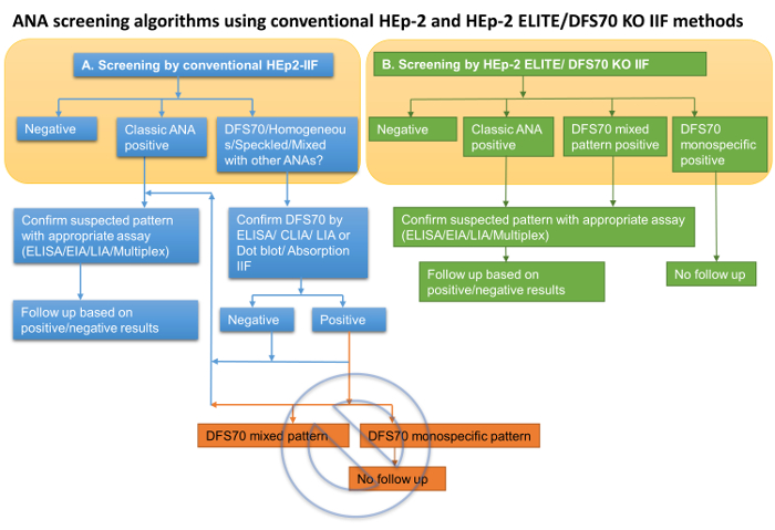
Figure 1: ANA screening algorithms using conventional HEp-2 and HEp-2 ELITE/DFS70 KO IIF methods. (A) Schematic unravels the deficiency of conventional HEp-2 IIF in identifying monospecific and mixed DFS70 positive patterns at the screening stage (yellow shaded area) and its inability to rule out a secondary disease-associated ANA pattern that may be concealed by DFS70 pattern. Further, the current generation of DFS70 specific solid phase confirmatory assays are not able to confirm monospecific DFS70 positivity which leads to additional confirmatory testing for ANAs. (B) Schematic reveals the capabilities of HEp-2 ELITE for screening of ANAs, simultaneous distinction of monospecific and mixed DFS70 reactions, and eliminating the need for solid phase DFS70 confirmation assays. Algorithm using HEp-2 ELITE significantly simplifies the ANA screening and reduces the required number of confirmation assays. Please click here to view a larger version of this figure.
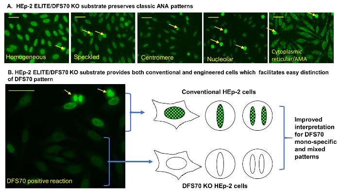
Figure 2: Classic ANA and DFS70 pattern on HEp-2 ELITE/DFS70 KO substrate. (A) HEp-2 ELITE/DFS70 KO substrate preserves classic ANA patterns. Classic homogeneous reaction and DFS70 positive reactions were produced using the positive controls provided by the kit. Other reactions were produced using previously established positive control sera for speckled, centromere, nucleolar, and cytoplasmic reticular/AMA reactions5. (B) HEp-2 ELITE/DFS70 KO substrate provides both conventional and engineered cells, which facilitates easy distinction of DFS70 pattern. Schematic shows the resulting DFS70 monospecific reaction and the staining pattern on interphase and dividing cell nuclei for both conventional and engineered (DFS70-KO) cells. Engineered HEp-2 cells are devoid of LEDGF/p75/DFS70 antigen which enables the simultaneous detection of secondary patterns that are usually concealed by DFS70 autoantibody reaction on conventional HEp-2 substrates. Arrows (yellow) indicate examples of mitotic cell nuclei. Scale bars represent 20 µm. Please click here to view a larger version of this figure.
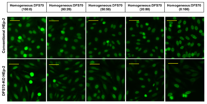
Figure 3: DFS70 and homogeneous sera in combination. DFS70 monospecific sera were combined with a positive homogeneous pattern control in various ratios (100:0, 80:20, 50:50, 20:80, and 0:100) and tested on conventional and engineered (DFS70-KO) HEp-2 substrates. Scale bars represent 20 µm. Please click here to view a larger version of this figure.
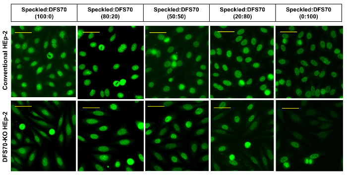
Figure 4: DFS70 and speckled sera in combination. DFS70 monospecific sera were combined with a positive speckled pattern control in various ratios (100:0, 80:20, 50:50, 20:80, and 0:100) and tested on conventional and engineered (DFS70-KO) HEp-2 substrates. Scale bars represent 20 µm. Please click here to view a larger version of this figure.

Figure 5: DFS70 and centromere sera in combination. DFS 70 monospecific sera were combined with a positive centromere pattern control in various ratios (100:0, 80:20, 50:50, 20:80, and 0:100) and tested on conventional and engineered (DFS70-KO) HEp-2 substrates. Scale bars represent 20 µm. Please click here to view a larger version of this figure.

Figure 6: DFS70 and nucleolar sera in combination. DFS 70 monospecific sera were combined with a positive nucleolar pattern control in various ratios (100:0, 80:20, 50:50, 20:80, and 0:100) and tested on conventional and engineered (DFS70-KO) HEp-2 substrates. Scale bars represent 20 µm. Please click here to view a larger version of this figure.
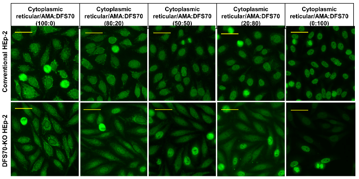
Figure 7: DFS70 and cytoplasmic reticular/AMA sera in combination. DFS70 monospecific sera were combined with a positive mitochondrial pattern control in various ratios (100:0, 80:20, 50:50, 20:80, and 0:100) and tested on conventional and engineered (DFS70-KO) HEp-2 substrates. Scale bars represent 20 µm. Please click here to view a larger version of this figure.
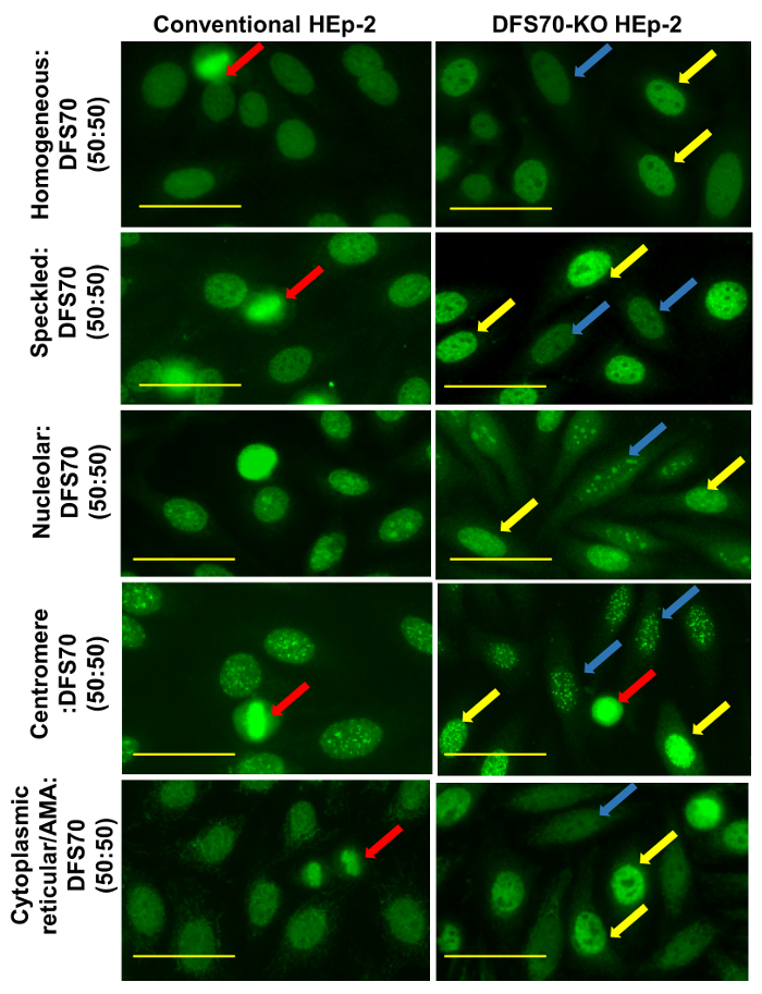
Figure 8: Mixed patterns (50:50 ratio) produced on conventional and HEp-2 ELITE substrates. Differences between the two substrates are revealed. Red arrows indicate the mitotic nuclei in both conventional and engineered cells on both substrates. For these combinations, the differential pattern presented by dividing cell nuclei on conventional HEp-2 substrate is not sufficient for accurate interpretation of mixed patterns. It can be observed that mixed DFS70 reactions on dividing cells can perturb the established set of rules for interpretation of disease-associated ANA patterns. Yellow and blue arrows indicate unmodified and engineered (DFS70 KO) HEp-2 cell nuclei, respectively. Results obtained for all the combinations tested on the engineered substrate: a clear intensity difference between the interphase nuclear pattern belonging to unmodified cells (~10%) and engineered cells (~90%) was observed, which enabled simultaneous detection and confirmation of DFS70 pattern. Scale bars represent 20 µm. Please click here to view a larger version of this figure.
| ANA pattern | Description | |
| Homogeneous | The entire nucleus fluoresces evenly with a diffuse staining pattern. | |
| Speckled | Discrete, coarse to fine speckles of fluoresce throughout the nucleus. Fine speckled and coarse speckled patterns have been described. | |
| Nucleolar | The nucleoli stain as multiple solid or speckled bodies within the nucleus. Subtypes have been described previously. | |
| Centromere | Large speckles of finite number. Reactive antigen segregates with condensed chromosomes in cells undergoing mitosis. | |
| Cytoplasmic reticular/AMA | The cytoplasm appears granular with positive staining of the mitochondria. | |
| DFS70 | •Conventional HEp-2 cells: A dense fine speckled pattern is observed on the interphase nucleus and chromatin associated staining is seen on mitotic nuclei. | |
| •Engineered DFS70 KO cells: Anti-DFS70 antibodies produce negative staining | ||
| HEp-2 Elite/ DFS70 KO has conventional and engineered (DFS70 KO) cells in approximately 1:9 ratio. Conventional cells produce typical DFS70 pattern and engineered cells produce a negative pattern when reacted with a DFS70 monospecific/isolated positive reactions. | ||
Table 1: Description of ANA Patterns.
Discussion
Autoantibodies to nuclear and cytoplasmic antigens are highly prevalent in systemic autoimmune diseases. Although no single assay provides the best possible sensitivity and specificity, IIF by HEp-2 is widely regarded as a highly sensitive and cost-effective screening method for systemic autoimmune diseases2,3,4. Despite the prevalence of IIF protocol for more than 50 years, accuracy of IIF assay is highly dependent on the skill of the technician, careful execution of the individual steps of the protocol, and accurate interpretation of results using a well-maintained fluorescence microscope. Diagnosis of systemic autoimmune disease should not be based on the results of a single serological test such as IIF by HEp-2.
Reconstitution of reagents using high quality water (distilled/deionized), use of standardized kit components (slides with HEp-2 cell monolayers, sample diluent, positive and negative controls, pre-diluted optimized FITC-conjugate, and mounting medium) have a positive impact on the results. Skipping the addition of controls or samples on wells can encourage false negative interpretations. Drying of slides at any point after the addition of sample or control can result in artifactual false positive reactions and/or improper patterns. Too much wash buffer left on slide or drying of slide wells prior to dispensation of mounting medium can result in artifacts. Adding insufficient quantity of mounting medium or generating bubbles on the slide surface prior to placing a coverslip can result in reactions that look blurry or out of focus when observed under the microscope. Mounted slides must be stored at 2 - 8 °C and analyzed within the recommended window of time to minimize false negative or artifactual results.
Using a well-maintained epi-fluorescent microscope which houses an appropriate LED/Hg/Xenon light source and clean optical components including excitation and emission filters that are not damaged is critical to obtain accurate IIF results. End user training for discriminating positive/negative reaction intensities and interpretation of patterns is critical. HEp-2 IIF assays are vulnerable to lack of standardization in procedures, microscope configuration, and end-user training or skill. The consensus nomenclature and representative patterns and examples of images acquired using various manufacturers are made available by ICAP (<www.ANApatterns.org>) for educational purposes4. A HEp-2 cell pattern classification tree provided by ICAP for various nuclear, cytoplasmic, and mitotic patterns is a valuable resource to learn the subtle differences between various patterns that may look similar to the untrained eyes4. One of the primary examples of a challenging pattern is the DFS70 which also lacks a distinct association with an autoimmune condition, and as discussed in previous sections this pattern is highly prevalent in healthy populations5.
Depending on the study, the prevalence of anti-DFS70 autoantibodies vary between healthy cohorts, ANA screening populations, and ANA positive individuals with no evidence of systemic autoimmune disease9,16,20,30,31. DFS70 autoantibodies have been reported to occur in isolation as well as with other disease-associated ANA patterns. Isolated or monospecific DFS70 pattern is reported in 2 - 22% of healthy controls but in less than 1% of the cases with autoimmune rheumatic disease32. Based on these trends, DFS70 mono-positive pattern was proposed as a criterion to exclude in autoimmune rheumatic diseases by ICAP committee3,31,32. The ICAP committee, established to promote harmonization of autoantibody test nomenclature and interpretation of the HEp-2 IIF results, recommends reporting DFS70 positivity while reporting ANA screening results4.
Prior to ruling out a systemic autoimmune disease based on DFS70 mono-positivity, it is vital to review the state of current screening and confirmation methods (particularly those specific for DFS70). Agreement between DFS70 positivity by HEp-2 IIF and solid phase confirmatory assays namely ELISA, CLIA, and line immunoassay/dot blot methods) varied widely between various studies13,22,25,33. IIF interpretation and confirmatory assay parameters that contribute to this disagreement have been discussed previously5,13,34. A recently proposed immunoadsorption approach for anti-DFS70 antibody confirmation addresses some of the deficiencies of current solid phase assays (for DFS70) but requires the labs to perform an additional step in IIF procedure on suspected samples, which increases the cost and turnaround time for reporting results13. This additional step is not required when HEp-2 ELITE/DFS70 KO cells are used for routine ANA screening. This reduces the overall cost and moreover there is no impact on turnaround time as DFS70 pattern is discerned in the initial screening step27. An additional disadvantage of using conventional HEp-2 IIF method is that it is unable to distinguish DFS70 pattern co-occurring with other ANA patterns at the screening stage. However, this disadvantage is overcome by the use of HEp-2 ELITE/DFS70 KO cells27.
Comparative evaluation of conventional HEp-2 and the new engineered HEp-2 (HEp-2 ELITE/DFS70 KO) using ANA positive controls and their combinations with a mono-positive DFS70 control, demonstrate the utility of the new substrate's ability to simultaneously screen for ANAs and confirm DFS70 pattern (Figure 1). The IIF protocol for the new substrate is identical to the conventional HEp-2 IIF method. Interpretation scheme for all disease-associated ANA patterns is identical with the exception of the DFS70 pattern (Table 1). Interpretation of both mono-positive and mixed DFS70 patterns was relatively easier using the new method and it does not warrant additional assays (Figure 1B). Implementation of this new method in clinical lab simplifies the challenge associated with the interpretation of DFS70 pattern and improves the overall accuracy of the ANA screening by further revealing disease-associated ANA patterns previously concealed by DFS70 (mixed patterns).
Disclosures
The authors Kishore Malyavantham and Lakshmanan Suresh are employees of Trinity Biotech, USA.
Acknowledgements
We thank Andrew Zenger for performing the IIF experiments and collecting images.
Materials
| Name | Company | Catalog Number | Comments |
|---|---|---|---|
| HEp-2 ELITE / DFS70-KO 12 Well Slide sealed individually in pouch | Trinity Biotech | part of kit# 1108, 1108-120, 1108-240 | |
| ANA positive control | Trinity Biotech | part of kit# 1108, 1108-120, 1108-240, 1103, 1103-240 | |
| DFS70 antibody positive control | Trinity Biotech | part of kit# 1108, 1108-120, 1108-240 | |
| Negative control | Trinity Biotech | part of kit# 1108, 1108-120, 1108-240, 1103, 1103-240 | |
| Anti-human IgG FITC conjugate containing Evan's blue counterstain | Trinity Biotech | part of kit# 1108, 1108-120, 1108-240, 1103, 1103-240 | |
| Buffer diluent for serum | Trinity Biotech | part of kit# 1108, 1108-120, 1108-240, 1103, 1103-240 | |
| Phosphate buffered saline (PBS) | Trinity Biotech | part of kit# 1108, 1108-120, 1108-240, 1103, 1103-240 | |
| Mounting medium | Trinity Biotech | part of kit# 1108, 1108-120, 1108-240, 1103, 1103-240 | |
| Coverslips | Trinity Biotech | part of kit# 1108, 1108-120, 1108-240, 1103, 1103-240 | |
| ImmuGlo ANA HEp-2 Cells 12 well slide sealed individually in pouch | Trinity Biotech | part of kit# 1103, 1103-240 | |
| Name | Company | Catalog Number | Comments |
| Materials used in the study but not provided by the kits | |||
| Diagnostic fluorescent microscope | Nikon Eclipse 50i | Equipped with a spot dignostic camera, model 2.2.1 | |
| Incubation chamber | Trinity Biotech | provided for customers (no catalog #) | |
| Coplin jar | n/a | General labware | |
| Paper towels | n/a | General lab supplies | |
| distilled/deionized water | n/a | General lab supplies |
References
- American College of Rheumatology. Position Statement. Methodology of testing for antinuclear antibodies. , Available from: https://www.rheumatology.org/Portals/0/Files/Methodology%20of%20Testing%20Antinuclear%20Antibodies%20Position%20Statement.pdf (2011).
- Meroni, P. L., Schur, P. H. ANA screening: an old test with new recommendations. Ann Rheum Dis. 69 (8), 1420-1422 (2010).
- Agmon-Levin, N., et al. International recommendations for the assessment of autoantibodies to cellular antigens referred to as anti-nuclear antibodies. Ann Rheum Dis. 73 (1), 17-23 (2014).
- Chan, E. K., et al. Report of the First International Consensus on Standardized Nomenclature of Antinuclear Antibody HEp-2 Cell Patterns 2014-2015. Front Immunol. 6, 412(2015).
- Malyavantham, K., Suresh, L. Analysis of DFS70 pattern and impact on ANA screening using a novel HEp-2 ELITE/DFS70 knockout substrate. Auto Immun Highlights. 8 (1), 3(2017).
- Sperotto, F., Seguso, M., Gallo, N., Plebani, M., Zulian, F. Anti-DFS70 antibodies in healthy schoolchildren: A follow-up analysis. Autoimmun Rev. , (2017).
- Ayaki, M., et al. Detection of cytotoxic anti-LEDGF autoantibodies in atopic dermatitis. Autoimmunity. 35 (5), 319-327 (2002).
- Ochs, R. L., et al. Autoantibodies to DFS 70 kd/transcription coactivator p75 in atopic dermatitis and other conditions. J Allergy Clin Immunol. 105 (6 Pt 1), 1211-1220 (2000).
- Watanabe, A., et al. Anti-DFS70 antibodies in 597 healthy hospital workers. Arthritis Rheum. 50 (3), 892-900 (2004).
- Yamada, K., et al. Humoral immune response directed against LEDGF in patients with VKH. Immunol Lett. 78 (3), 161-168 (2001).
- Seelig, C. A., Bauer, O., Seelig, H. P. Autoantibodies Against DFS70/LEDGF Exclusion Markers for Systemic Autoimmune Rheumatic Diseases (SARD). Clin Lab. 62 (4), 499-517 (2016).
- Ochs, R. L., et al. The significance of autoantibodies to DFS70/LEDGFp75 in health and disease: integrating basic science with clinical understanding. Clin Exp Med. 16 (3), 273-293 (2016).
- Bizzaro, N., et al. Specific chemoluminescence and immunoasdorption tests for anti-DFS70 antibodies avoid false positive results by indirect immunofluorescence. Clin Chim Acta. 451 (Pt B), 271-277 (2015).
- Bizzaro, N., et al. Antibodies to the lens and cornea in anti-DFS70-positive subjects. Ann N Y Acad Sci. 1107, 174-183 (2007).
- Daniels, T., et al. Antinuclear autoantibodies in prostate cancer: immunity to LEDGF/p75, a survival protein highly expressed in prostate tumors and cleaved during apoptosis. Prostate. 62 (1), 14-26 (2005).
- Dellavance, A., et al. The clinical spectrum of antinuclear antibodies associated with the nuclear dense fine speckled immunofluorescence pattern. J Rheumatol. 32 (11), 2144-2149 (2005).
- Gundin, S., et al. Measurement of anti-DFS70 antibodies in patients with ANA-associated autoimmune rheumatic diseases suspicion is cost-effective. Auto Immun Highlights. 7 (1), 10(2016).
- Kang, S. Y., Lee, W. I. Clinical significance of dense fine speckled pattern in anti-nuclear antibody test using indirect immunofluorescence method. Korean J Lab Med. 29 (2), 145-151 (2009).
- Lee, H., Kim, Y., Han, K., Oh, E. J. Application of anti-DFS70 antibody and specific autoantibody test algorithms to patients with the dense fine speckled pattern on HEp-2 cells. Scand J Rheumatol. 45 (2), 122-128 (2016).
- Mariz, H. A., et al. Pattern on the antinuclear antibody-HEp-2 test is a critical parameter for discriminating antinuclear antibody-positive healthy individuals and patients with autoimmune rheumatic diseases. Arthritis Rheum. 63 (1), 191-200 (2011).
- Marlet, J., et al. Thrombophilia Associated with Anti-DFS70 Autoantibodies. PLoS One. 10 (9), e0138671(2015).
- Mercado, M. V., et al. Detection of autoantibodies to DSF70/LEDGFp75 in Mexican Hispanics using multiple complementary assay platforms. Auto Immun Highlights. 8 (1), 1(2017).
- Muro, Y., Sugiura, K., Morita, Y., Tomita, Y. High concomitance of disease marker autoantibodies in anti-DFS70/LEDGF autoantibody-positive patients with autoimmune rheumatic disease. Lupus. 17 (3), 171-176 (2008).
- Muro, Y., Sugiura, K., Nakashima, R., Mimori, T., Akiyama, M. Low prevalence of anti-DFS70/LEDGF antibodies in patients with dermatomyositis and other systemic autoimmune rheumatic diseases. J Rheumatol. 40 (1), 92-93 (2013).
- Mutlu, E., Eyigor, M., Mutlu, D., Gultekin, M. Confirmation of anti-DFS70 antibodies is needed in routine clinical samples with DFS staining pattern. Cent Eur J Immunol. 41 (1), 6-11 (2016).
- Okamoto, M., et al. Autoantibodies to DFS70/LEDGF are increased in alopecia areata patients. J Autoimmun. 23 (3), 257-266 (2004).
- Pazini, A. M., Fleck, J., dos Santos, R. S., Beck, S. T. Clinical relevance and frequency of cytoplasmic and nuclear dense fine speckled patterns observed in ANA-HEp-2. Rev Bras Reumatol. 50 (6), 655-660 (2010).
- Schmeling, H., et al. Autoantibodies to Dense Fine Speckles in Pediatric Diseases and Controls. J Rheumatol. 42 (12), 2419-2426 (2015).
- Wiik, A. S., Hoier-Madsen, M., Forslid, J., Charles, P., Meyrowitsch, J. Antinuclear antibodies: a contemporary nomenclature using HEp-2 cells. J Autoimmun. 35 (3), 276-290 (2010).
- Mahler, M., Fritzler, M. J. The Clinical Significance of the Dense Fine Speckled Immunofluorescence Pattern on HEp-2 Cells for the Diagnosis of Systemic Autoimmune Diseases. Clin Dev Immunol. 2012, 494356(2012).
- Mahler, M., et al. Anti-DFS70/LEDGF Antibodies Are More Prevalent in Healthy Individuals Compared to Patients with Systemic Autoimmune Rheumatic Diseases. J Rheumatol. 39 (11), 2104-2110 (2012).
- Conrad, K., Rober, N., Andrade, L. E., Mahler, M. The Clinical Relevance of Anti-DFS70 Autoantibodies. Clin Rev Allergy Immunol. , (2016).
- Miyara, M., et al. Clinical Phenotypes of Patients with Anti-DFS70/LEDGF Antibodies in a Routine ANA Referral Cohort. Clin Dev Immunol. 2013, 703759(2013).
- Bizzaro, N., Tonutti, E., Villalta, D., et al. Recognizing the dense fine speckled/lens epithelium-derived growth factor/p75 pattern on HEP-2 cells: not an easy task! Comment on the article by Mariz et al. Arthritis Rheum. 63 (12), 4036-4037 (2011).
Reprints and Permissions
