This protocol describes the round-the-corner technique and the individualized stem-positioning of calcar-guided short stems alongside the medial calcar, depending on the level of the osteotomy. This differs from conventional total hip arthroplasty and includes a learning curve.
Method Article
Individualized Stem-positioning in Calcar-guided Short-stem Total Hip Arthroplasty
In This Article
Summary
Abstract
Bone- and soft-tissue sparing short stems are increasingly used in total hip arthroplasty (THA). However, there are a large variety of models of short stems, differing in design and function. Calcar-guided short stems provide an anatomical curvature in the medial calcar region, thus, positioning is done individually alongside the calcar in the "round-the-corner" technique. Depending on the level of the neck's osteotomy, stems can be aligned individually in a large bandwidth of varus- and valgus anatomies. This differs from conventional total hip arthroplasty and potentially includes a severe learning curve. Given that a great variety of caput-collum-diaphyseal (CCD)-angles can be retained, the reconstruction of femoro-acetabular offsets can be achieved precisely. However, particularly extensive varus- and valgus positioning has raised concerns in regard to stability and bone remodeling. The purpose of the present manuscript is to showcase the implantation technique in calcar-guided short-stem THA and to summarize short-term clinical and radiological results.
Introduction
Modern calcar-guided short-stems have been increasingly used in THA in recent years1. New calcar-guided, metaphyseal anchoring short-stems are emerging that focus on sparing muscles, soft-tissue, and bone2,3, thus allowing minimally-invasive (MIS) techniques and approaches to be successfully applied4.
The possible benefits of calcar-guided short-stem THA can be accomplished through a special implantation technique, which differs from conventional techniques used with traditional straight-stem designs. The most important aspect, in this regard, is the anatomical curvature, which has been adapted from the calcar. The positioning of the stem follows the individual anatomy alongside the calcar curve, and permits individualized implantation5. Applying the so called "round-the-corner" technique, the greater trochanter region and most importantly the gluteal muscles, can be spared almost completely2.
Modern THA is largely dependent on the successful preservation of hip geometry. The accurate reconstruction of the hip joint anatomy is crucial for the clinical outcome. The femoro-acetabular offset has increasingly come into focus5. Reduced offset might lead to gluteal insufficiency along with instability of the hip joint and increased risk of dislocation6,7. On the other hand, a severe increase in offset can cause trochanteric bursitis. Given these findings, it appears that undesired changes of offset have great clinical relevance.
Reconstruction of the femoral offset is highly dependent on the ability to reproduce different CCD-angles8. However, valgization has been found to be the limiting factor in the successful reconstruction of hip geometry in many stem designs, causing reduced offset and increased leg length9. The reconstruction of different CCD-angles, in this regard, seems to be the key to accomplish the retainment of the hip anatomy.
In calcar-guided short-stem THA, stem alignment can be individualized, which supports the successful reconstruction of the femoral offset8. Guiding the stem alongside the calcar, the positioning of the stem in the proximal femur is dependent on the resection level of the femoral neck. Given a preexisting varus anatomy, a high resection also results in a varus position of the implant, maintaining a large femoral offset. On the other hand, given a preexisting valgus anatomy, a low resection results in a valgus position, causing a small femoral offset10 (Figure 1; Figure 2).
Consequently, the presented individualized implantation technique allows a broad reconstruction of CCD-angles and thus enables a precise preservation of hip geometry. The presented technique differs from conventional total hip arthroplasty and potentially includes a severe learning curve.
Protocol
The presented research has been performed in compliance with all institutional, national, and international guidelines for human welfare. Institutional review board approval has been obtained.
- Position the patient in supine position on a standard operating table with two separate leg supports. Allow the ipsilateral legs to remain mobile.
- Apply standard sterile coverage suitable for the antero-lateral approach in supine position.
- Slightly flex the ipsilateral side using a knee roll. In addition, hyperextend the contralateral side about 15° for the femoral preparation.
- Locate the tip of the greater trochanter as well as the anterior superior iliac spine by palpation serving as orientation.
- Perform the skin- incision using a surgical knife (6 - 12 cm, depending on the patient's anatomy) centered on the tip of the greater trochanter aiming at the anterior superior iliac spine above the intermuscular septum between the gluteus medius and the tensor fasciae latae.
- After incision of subcutaneous fat, use two skin retractors anteriorly and posteriorly. Open the fascia without causing damage to the tensor fasciae latae.
- Do a blunt dissection using your index finger, pushing the gluteal muscles posteriorly without damage.
- Expose the joint capsule using three retractors.
- Perform the capsulectomy alongside the femoral neck.
NOTE: No sharp dissection of any muscle, in particular the gluteal muscles, is needed. - After removal of the anterior joint capsule, expose the femoral neck in order to perform the osteotomy by placing the two facing curved retractors intracapsular around the femoral neck (Figure 1).
NOTE: The most important step in the implantation of a calcar-guided short stem is the choice of the individual level of the osteotomy. This way, stem-positioning can be done individually in a great variety, resulting in a large bandwidth of CCD-angles to be reconstructed (Figure 2; Figure 3). Consequently, a preoperative planning is mandatory to display the exact level (Figure 4: red line (OL: level of osteotomy)). - For the surgeon's orientation, in order to determine the level of the osteotomy, perform a palpation of the lesser trochanter and the fossa piriformis. If a valgus position is desired according to the preexisting anatomy, perform the osteotomy distally by resecting most of the femoral neck (Figure 5a). In order to align the stem in a varus position, resect proximally, retaining most of the femoral neck, according to the preoperative planning (Figure 5c). Femoral offset and leg length can thus be maintained precisely (Figure 5).
- Perform the osteotomy (Figure 1) in slight external rotation of the ipsilateral leg according to the preoperative planning using a long stiff bladed oscillating saw.
- Remove the femoral head from the acetabulum using the femoral head extractor. To protect the gluteus medius, place a "Langenbeck"-retractor medially and pull proximally.
- During acetabular preparation, insert a "Steinmann"-pin in the proximal end of the acetabulum to protect the gluteal muscles.
- Expose the acetabulum using two extractors.
- Implant the acetabular component according to the preoperative planning and depending on the patient's individual anatomy.
- For femoral preparation, first remove the knee roll and hyperextend the contralateral leg about 15°. Now perform a 90° external rotation and a maximum of 90° flexion of the knee joint. Have the assistant hold the subject's leg at maximal adduction (about 40°).
- Position two retractors on the medial side of the proximal femoral neck and proximally at the posterior (medial) cortical end of the femoral neck. Avoid contact to the greater trochanter, to minimize the risk of possible damage to the bone and the muscle insertions.
- Apply the "round-the-corner" technique to open the proximal femur alongside the calcar with the curved opening awl.
NOTE: Posterior structures such as the greater trochanter or the gluteal muscles are not affected. - Drive in gently specially curved, implant-shaped rasps in ascending sizes using a hammer in order to prepare the proximal femur and the femoral canal until cortical contact and a stable fit and fill are reached. Note that a double offset minimally-invasive rasp handle is available. Perform the insertion guided by the calcar in the "round-the-corner" technique (Figure 6).
- Choose one of the two available trial cones with different offset versions (standard and lateral offset) according to the preoperative planning.
- Assess an intraoperative fluoroscopy using a digital image intensifier after inserting a trial head and performing a trial reduction to compare the positioning of the rasp (trial implant) to the preoperative planning (Figure 7). Perform an anterior-posterior radiograph. To screen the second plane, also perform an axial view radiograph. Perform adjustments, if necessary.
NOTE: Reaching cortical contact is essential regarding the risk of postoperative subsidence. Avoid undersizing! (Figure 8) - Remove the trial implants and insert the definitive implant containing the chosen offset version using the special implant impactor. Note that the original stem aligns itself exactly like the trial rasp (Figure 9).
- After the final reduction by applying axial tension combined with internal rotation of the leg, complete the procedure by standard wound closure.
- In most cases, allow full weight-bearing after surgery using 2 crutches under physiotherapeutic surveillance starting 4 h after the operation.
NOTE: In heavy patients, primary stability might be impaired, thus, weight bearing protocol must be adjusted accordingly. - Provide analgesics according to the intensity of pain recorded, non-steroidal-anti-inflammatory-drugs (NSAID) to prevent heterotopic ossification, and venous thromboembolic prophylaxis.
Results
Several short-term results of the investigated short-stem have previously been published, mostly arising from an ongoing observational study at the author's institution2,8,11,12,13,14,15,16. Using the presented technique, the reconstruction of different femoro-acetabular offsets can be accomplished precisely5. Particularly the ability to preserve the preexisting CCD-angle allows physiological hip geometry retainment8. After two years, only a few radiological alterations, such as stress-shielding and cortical hypertrophy were obvious14. The minimally-invasive technique caused low incidences of heterotopic ossifications11. Therefore, the blood loss and transfusion rate remains at a lower rate compared to the implantation technique using conventional straight stems17.
Excellent clinical results have led to successful usage of this technique in one-stage bilateral short-stem THA2. Ein-Bild-Roentgen-Analyses "femoral component analysis" (EBRA-FCA) of the presented implant have shown an increased initial axial subsidence especially in heavy and active male patients, given full weight bearing postoperatively, however, without any clinical consequences in the early stage13,15. Particularly stems that have been aligned in the valgus position result in increased early subsidence, but undersizing and insufficient cortical contact laterally, could be identified as the main cause18.
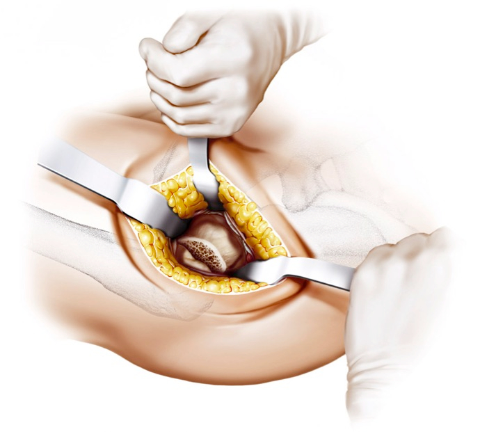
Figure 1: Osteotomy of the femoral neck. The osteotomy is done according to the preoperative planning. The level of the osteotomy is determined by palpating the lesser trochanter and the tip of the greater trochanter, serving as a reference. From2,4.

Figure 2: Valgus positioning. The stem is aligned in valgus position following a low osteotomy.

Figure 3: Varus positioning. The stem is aligned in varus position following a high osteotomy.

Figure 4: Preoperative planning. Blue: Template of the calcar-guided short stem; red: template of the cement-less cup. The level of osteotomy is determined according to the preoperative planning (OL). Intraoperatively, also the height of the lateral shoulder of the implant serves as orientation (SH). Most importantly the stem has to be upsized until cortical contact laterally is reached (CC).
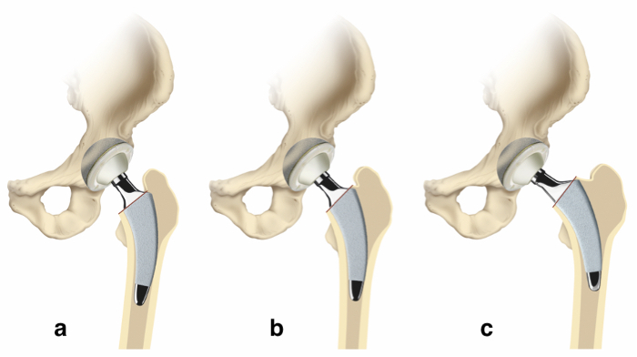
Figure 5: Individualized level of osteotomy and stem alignment. The positioning of the stem in the proximal femur is dependent on the resection level of the femoral neck. Given a varus anatomy, a high resection also results in a varus position of the implant, maintaining a large femoral offset (c). On the other hand, given a valgus anatomy, a low resection results in a valgus position, causing a small femoral offset (a).10,12 From Kutzner et al.2 Please click here to view a larger version of this figure.
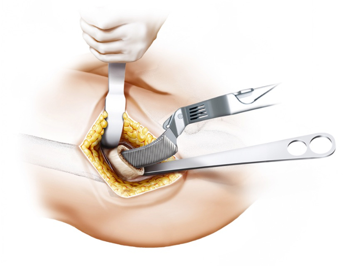
Figure 6: Round-the-corner technique. The insertion is done alongside the medial calcar, not affecting lateral structures. Especially the greater trochanter and the gluteal muscles can be spared. From Kutzner et al.2 Please click here to view a larger version of this figure.
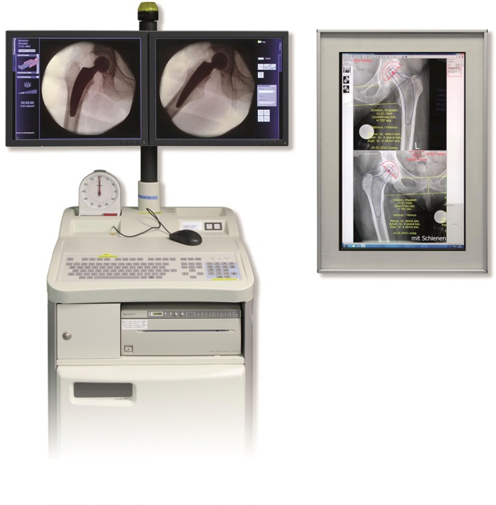
Figure 7: Intraoperative fluoroscopy. After completing trial reduction, the assessment of intraoperative radiography in two planes using a digital image intensifier should be considered mandatory to be able to compare the positioning of the rasp (trial implant) to the preoperative planning. Potential adjustments can be performed subsequently. From Kutzner et al.2 Please click here to view a larger version of this figure.
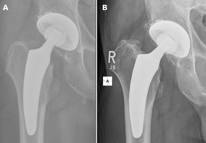
Figure 8: Example of undersizing. Insufficient cortical contact and undersizing (a) results in subsequent subsidence in the 2 year follow-up (b). Please click here to view a larger version of this figure.
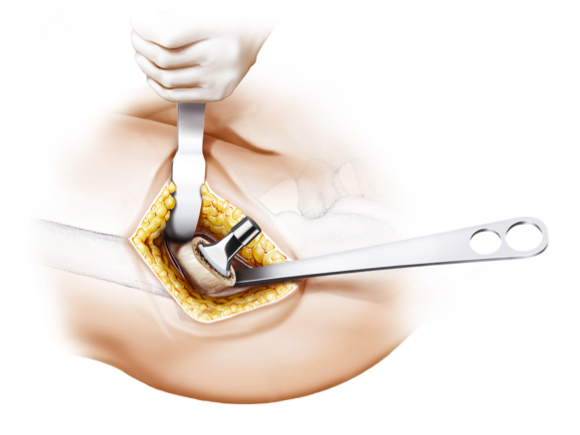
Figure 9: Implantation of the original calcar-guided short stem. The original implant positions itself exactly like the trial rasp. From Kutzner et al.2
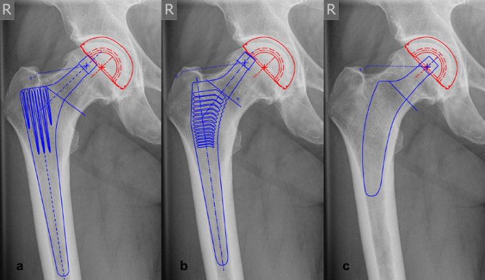
Figure 10: Examples of offset-reconstruction in varus-hips with different stem types. (a, b) Offset-reconstruction cannot be achieved due to diaphyseal anchorage and design of conventional straight stems. (c) Varus-alignment with a calcar-guided short stem accomplishes an exact reconstruction of offset. Please click here to view a larger version of this figure.
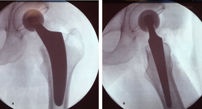
Figure 11: Example of the intraoperative fluoroscopy in two planes. (a) a.p. view; (b) axial view. Short stems will position themselves almost automatically along the anteversion and anterior tilt of the preexisting proximal femoral bone. Thus, anterior offset can also be restored. Please click here to view a larger version of this figure.
Discussion
Calcar-guided short stems provide many advantages in modern THA compared to conventional straight-stem designs in short-term follow-up. However, only a few results are published regarding mid- and long-term follow-up.
Due to the short and curved design of calcar-guided short stems, the soft-tissue sparing implantation appears to be technically easy. However, the individualized implantation technique requires distinct knowledge about the characteristics of different varus- and valgus positioning. A severe learning curve must be taken into account.
Modifications and Troubleshooting:
Given the individualized positioning of calcar-guided short stems, the preparation of a preoperative planning is absolutely mandatory8 (Figure 4). Besides the detection of the correct implant sizes, especially the alignment of the stem along with the desired level of the osteotomy can be determined. Intraoperatively, using the inserted trial rasp, after reduction of the hip a comparison to the preoperative planning can be done by performing intraoperative fluoroscopy19 (Figure 7). The lateral shoulder of the implant serves as orientation regarding leg length.
Limitations of the Technique:
Investigations suggest a broad bandwidth of different hip anatomies to be adequately reconstructed using calcar-guided short stems5,8 (Figure 2; Figure 3). Previous studies have investigated the outcome of extensive varus- and valgus stem alignment10. After 2 years no revision surgery was needed and the rate of radiographic alterations indicating abnormal stress distribution in total was low. However, especially for valgus-hips a pronounced initial subsidence was observed10.
Particularly for young and unexperienced surgeons the presented technique may come with undesired pitfalls.
Significance with Respect to Existing Methods:
The implantation technique of calcar-guided short stems with individualized neck resection differs from conventional straight stems and certain neck resecting and neck retaining short-stems. Conventional straight stems provide diaphyseal anchorage along with a mostly standardized level of osteotomy of the femoral neck. The preexisting hip anatomy can only be reconstructed by using different offset-versions of the implant5. In extensive varus anatomies for example, this often cannot be achieved properly (Figure 10). Valgization has been found to be the limiting factor in the successful reconstruction of hip geometry also in many previous short-stem designs, causing reduced offset and increased leg length9.
Critical Steps Within the Protocol:
The most critical step consists of choosing the right level of the osteotomy. Furthermore, in order to intraoperatively correctly realize the preoperative planning, a verification using fluoroscopy is needed.
Given a shortening of stem length in calcar-guided short-stem THA, primary stability potentially causes concerns18. The predominant type of fixation is metaphyseal anchoring, based on the fit-and-fill principle. However, due to the option of individualized positioning of these stem-designs, the type of anchoring might differ distinctly. In varus alignment three-point anchoring is common with cortical contact to the lateral cortex of the partially resected neck, the medial calcar, and the lateral cortex at the tip of the stem. However, depending on positioning and sizing, especially in extensive valgus alignment, pronounced diaphyseal anchorage is possible10. In those cases, a securely achieved cortical contact to the distal lateral cortex, as well as to the distal medial cortex is crucial. In valgus position a missing cortical contact of the tip has been frequently observed in the early collective including the learning curve, particularly in cases of undersizing. Therefore, surgeons should take into account, that particularly in valgus hips, undersizing accompanied with a lack of contact to the lateral cortex, might cause initial instability and subsequent implant micromovement10. The usage of intraoperative fluoroscopy to identify undersizing of the stem is therefore highly recommended19.
The present description of the technique, regarding reconstruction of offset, refers only to a two-dimensional analysis. However, given a partially retained femoral neck, short stems will position themselves almost automatically along the anteversion and anterior tilt of the preexisting proximal femoral bone. In particular in varus-hips, this leads to distinct anterior tilting in the axial plane with the stem's tip being positioned anteriorly (Figure 11). Thus, anterior offset can be reconstructed as well. Further monitoring of this new generation of short stems and the implantation technique in mid- and long-term follow-up is mandatory.
Disclosures
J. Pfeil is a medical advisor for Mathys Ltd., Switzerland.
Acknowledgements
Mathys Ltd., Switzerland supports the funding of the clinical and radiological multicenter study.
Materials
| Name | Company | Catalog Number | Comments |
|---|---|---|---|
| optimys short stem | Mathys Ltd., Switzerland | Implant |
References
- Jerosch, J. Kurzschaftendoprothesen an der Hüfte. , Springer-Verlag. (2017).
- Kutzner, K. P., Donner, S., Schneider, M., Pfeil, J., Rehbein, P. One-stage bilateral implantation of a calcar-guided short-stem in total hip arthroplasty. Oper Orthop Traumatol. , (2017).
- Anderl, C. 2-Jahres-Ergebnisse mit dem Optimys-Kurzschaft über den direkten anterolateralen Zugang. JATROS. Orthopädie & Rheumatologie 05/2015. 05, (2015).
- Pfeil, J. Minimally Invasive Surgery in Total Hip Arthroplasty [Englisch]. , Springer; Auflage. Available from: http://www.amazon.de/Minimally-Invasive-Surgery-Total-Arthroplasty/dp/3642008968/ref=sr_1_1?s=books-intl-de&ie=UTF8&qid=1399910678&sr=1-1&keywords=Siebert%5Cc+Werner (2010).
- Kutzner, K. P., Kovacevic, M. P., Roeder, C., Rehbein, P., Pfeil, J. Reconstruction of femoro-acetabular offsets using a short-stem. Int Orthop. 39 (7), 1269-1275 (2015).
- Matsushita, A., Nakashima, Y., Jingushi, S., Yamamoto, T., Kuraoka, A., Iwamoto, Y. Effects of the Femoral Offset and the Head Size on the Safe Range of Motion in Total Hip Arthroplasty. J Arthroplasty. 24, 646-651 (2009).
- Asayama, I., Chamnongkich, S., Simpson, K. J., Kinsey, T. L., Mahoney, O. M. Reconstructed hip joint position and abductor muscle strength after total hip arthroplasty. J Arthroplasty. 20, 414-420 (2005).
- Kutzner, K. P., Pfeil, J., Kovacevic, M. P. Preoperative digital planning versus postoperative outcomes in total hip arthroplasty using a calcar-guided short stem: frequent valgization can be avoided. Eur J Orthop Surg Traumatol. 27 (5), 643-651 (2017).
- Höhle, P., Schröder, S. M., Pfeil, J. Comparison between preoperative digital planning and postoperative outcomes in 197 hip endoprosthesis cases using short stem prostheses. Clin Biomech. 30 (1), 46-52 (2015).
- Kutzner, K. P., Freitag, T., Donner, S., Kovacevic, M. P., Bieger, R. Outcome of extensive varus and valgus stem alignment in short-stem THA: clinical and radiological analysis using EBRA-FCA. Arch Orthop Trauma Surg. (3), 431-439 (2016).
- Kutzner, K. P., et al. Incidence of heterotopic ossification in minimally invasive short-stem THA using the modified anterolateral approach. Hip Int. , 0-0 (2017).
- Kutzner, K. P., Kovacevic, M. P., Roeder, C., Rehbein, P., Pfeil, J. Reconstruction of femoro-acetabular offsets using a short-stem. Int Orthop. , (2014).
- Kutzner, K. P., et al. One-stage bilateral versus unilateral short-stem total hip arthroplasty: comparison of migration patterns using "Ein-Bild-Roentgen-Analysis Femoral-Component-Analysis". Int Orthop. 41 (1), 61-66 (2017).
- Kutzner, K. P., et al. Radiographic alterations in short-stem total hip arthroplasty: a 2-year follow-up study of 216 cases. Hip Int. 26 (3), 278-283 (2016).
- Kutzner, K. P., Kovacevic, M. P., Freitag, T., Fuchs, A., Reichel, H., Bieger, R. Influence of patient-related characteristics on early migration in calcar-guided short-stem total hip arthroplasty: a 2-year migration analysis using EBRA-FCA. J Orthop Surg Res. 11 (1), 29(2016).
- Kovacevic, M. P., Pfeil, J., Kutzner, K. P. Implantation of a new short stem in simultaneous bilateral hip arthroplasty - a prospective study on clinical and radiographic data of 54 consecutive patients. OUP. 10, 456-461 (2014).
- Hochreiter, J., Hejkrlik, W., Emmanuel, K., Hitzl, W., Ortmaier, R. Blood loss and transfusion rate in short stem hip arthroplasty. A comparative study. Int Orthop. , 1-7 (2016).
- Bieger, R., Ignatius, A., Decking, R., Claes, L., Reichel, H., Dürselen, L. Primary stability and strain distribution of cementless hip stems as a function of implant design. Clin Biomech. 27 (2), Bristol, Avon. 158-164 (2012).
- Loweg, L., Kutzner, K. P., Rehbein, P., Stephan, H., Pfeil, J., Schneider, M. Wertigkeit der intraoperativen Röntgenkontrolle in der primären Hüftendoprothetik. OUP. 5 (6), 334-338 (2016).
Reprints and Permissions
