The production of specialized retinal cells from pluripotent stem cells is a turning point in the development of stem cell-based therapy for retinal diseases. The present paper describes a simple method for an efficient generation of retinal organoids and retinal pigmented epithelium for basic, translational, and clinical research.
Method Article
Defined Xeno-free and Feeder-free Culture Conditions for the Generation of Human iPSC-derived Retinal Cell Models
* These authors contributed equally
In This Article
Summary
Abstract
The production of specialized cells from pluripotent stem cells provides a powerful tool to develop new approaches for regenerative medicine. The use of human-induced pluripotent stem cells (iPSCs) is particularly attractive for neurodegenerative disease studies, including retinal dystrophies, where iPSC-derived retinal cell models mark a major step forward to understand and fight blindness. In this paper, we describe a simple and scalable protocol to generate, mature, and cryopreserve retinal organoids. Based on medium changing, the main advantage of this method is to avoid multiple and time-consuming steps commonly required in a guided differentiation of iPSCs. Mimicking the early phases of retinal development by successive changes of defined media on adherent human iPSC cultures, this protocol allows the simultaneous generation of self-forming neuroretinal structures and retinal pigmented epithelial (RPE) cells in a reproducible and efficient manner in 4 weeks. These structures containing retinal progenitor cells (RPCs) can be easily isolated for further maturation in a floating culture condition enabling the differentiation of RPCs into the seven retinal cell types present in the adult human retina. Additionally, we describe quick methods for the cryopreservation of retinal organoids and RPE cells for long-term storage. Combined together, the methods described here will be useful to produce and bank human iPSC-derived retinal cells or tissues for both basic and clinical research.
Introduction
The retina is an integral part of the central nervous system (CNS) and has a limited capacity to spontaneously regenerate following a traumatic injury or diseases. Therefore, degenerative pathologies causing definitive retinal cell loss, such as age-related macular degeneration (AMD), retinitis pigmentosa (RP), glaucoma, and diabetic retinopathy, typically lead to irreversible blindness. Rescuing the degenerated retina is a major challenge for which stem cell-based therapies aiming to replace the damaged or lost cells are one of the most promising approaches1,2,3. Pluripotent stem cells as human embryonic stem cells (ESCs) cells or human-induced pluripotent stem cells (iPSCs) have the capacity to be expanded indefinitely in culture, and they have the potential to produce any cell types. Advances in our understanding of retinal development and the improvement of in vitro protocols for human iPSC differentiation have resulted in the generation of retinal organoids7,8,9,10,11,12. All of the major retinal cells, including retinal ganglion cells (RGCs), photoreceptors, and retinal pigmented epithelial (RPE) cells, have been successfully differentiated from human ESCs and iPSCs4,5,6. Based on the SFEB (serum-free culture of embryoid body-like aggregates) method developed by Eiraku et al.13, self-formation of retinal organoids can be obtained from ESC- or iPSC-derived embryoid body-like aggregates in defined extracellular matrix components7,10,14. But these protocols are intricate, requiring a large number of steps not always compatible with the large production of cells for therapeutic approaches or drug screening. Thus, the choice of the method to produce human retinal cells is critical and the method needs to be robust, scalable, and efficient.
Here, based on our previous publication15, we describe each step for a simple and efficient generation of retinal cells through retinal organoid self-formation from adherent human iPSCs cultivated in a feeder-free and xeno-free condition. Starting from routine cultures of adherent human iPSCs, this protocol requires only a simple successive medium changing to allow the generation of both iPS-derived RPE (hiRPE) cells and neuroretinal structures in 4 weeks. After a manual isolation, hiRPE can be expanded and the retinal structures can be cultured as floating organoids where the retinal progenitor cells are able to differentiate into all retinal cell types in a sequential order consistent with the in vivo human retinogenesis. Finally, for research advancement or clinical translation, we describe a cryopreservation method allowing the long-term storage of whole retinal organoids and hiRPE cells without affecting their phenotypic characteristics and functionality.
Protocol
The protocol described in this paper follows the guidelines of the Institut de la Vision's research ethics committee. The Institut de la Vision has been allowed the manipulation of human specimen according to the current French regulation. Specimen handling follows patient data protection in accordance with the Tenets of Helsinki, and national regulations after the ethical approval of the "Comité de Protection des Personnes (CPP) Ile-de-France V".
1. Preparation of Culture Media and Dishes
- Culture media
- Use iPSC medium, a chemically defined medium dedicated to pluripotent stem cell culture in feeder-free conditions16. Prepare 500 mL of medium according to the manufacturer's protocol.
- Use Basal iPSC (Bi) medium, the iPSC chemically defined medium without fibroblast growth factor 2 (FGF2) or transforming growth factor Beta (TGFß).
- Prepare 500 mL of Bi medium supplemented with 1% N2 supplement (BiN2 medium), 10 units/mL of penicillin, and 10 mg/mL of streptomycin.
- Prepare 500 mL of proneural N2-based medium (ProN2 medium) composed of DMEM:Nutrient Mixture F-12 (DMEM/F12, 1:1, L-Glutamine), 1% MEM nonessential amino acids, 1% N2 supplement, 10 units/mL of penicillin, and 10 mg/mL of streptomycin.
- Prepare proneural B27-based medium (ProB27 medium) composed of DMEM:Nutrient Mixture F-12 (DMEM/F12, 1:1, L-Glutamine), 1% MEM nonessential amino acids, 2% B27 supplement, 10 units/mL of penicillin and 10 mg/mL of streptomycin.
- Culture vessel preparation
- For human iPSC culture
- Prepare 10 mL of a vitronectin solution containing 5 μg/mL of vitronectin in 1x PBS. In 10 mL of 1x PBS, add 100 µL of the thawed vitronectin stock solution (100x).
- Distribute 2 mL of the vitronectin solution per 6-cm culture dish corresponding to 0.5 µg/cm2. Incubate it for 1 h at room temperature (RT). Remove the vitronectin solution by aspiration using a vacuum-aspiration system or a 5 mL pipette.
- Add 4 mL of iPSC medium per 6-cm dish.
- Incubate the dishes in a cell culture incubator at 37 °C and 5% CO2 for a minimum of 30 min before use.
- For human iPSC-derived RPE (hiRPE) cell culture
- Use a pluripotent stem cell-qualified matrix or substrate to coat wells according to the manufacturer's protocol. Add sufficient matrix or substrate to cover the entire growth surface area. For example, put 300 µL of matrix per well in 24-well plate and 3 mL of matrix in a T-25 cm2 flask.
- Incubate the coated culture vessels for a minimum of 1 h at 37 °C. Remove the matrix using vacuum-aspiration systems or a 5 mL pipette.
- Add 1 mL of ProN2 medium per well on 24-well plates and return the culture vessel to the incubator for a minimum of 30 min to warm and equilibrate the medium. For T-25 cm2 flasks, see step 5.2.
- For human iPSC culture
2. Maintenance and Expansion of Human iPSCs
- Maintenance of human iPSCs
- Prepare 3 mL of iPSC medium in a 15 mL tube and keep it at RT for a minimum of 15 min before use. Prepare a coated 6-cm dish as described in 1.2.1.
- Thaw a cryogenic sample of human iPSCs from a liquid nitrogen tank or a -150 °C freezer by incubation in a water bath at 37 °C for 30 s.
- Carefully disinfect the cryogenic vial using a disinfectant solution spray. Transfer the thawed human iPSCs from the cryotube to the 15 mL tube containing 3 mL of iPSC medium pre-warmed at RT.
- Centrifuge the tube at 110 x g for 3 min. Remove the supernatant by aspiration using a vacuum-aspiration system or a 5 mL pipette.
- Resuspend the cell pellet with 1 mL of iPSC medium from a vitronectin-coated 6-cm culture dish using a 2 mL pipette and transfer the cells back to the dish. Return the dish to the incubator at 37 °C and 5% CO2 for a minimum of 24 h before changing the medium.
NOTE: A ROCK inhibitor, such as Y-27632 at 10 µM, can be added to the iPSC medium in the 6-cm culture dishes to reduce apoptosis. - Change the medium daily and pass the human iPSCs every week.
- Passaging of human iPSCs
- Pass the human iPSC at a 70 - 80% confluence, classically after 7 days in culture.
- Prepare 6-cm dishes for the passaging as described in step 1.2.1. Remove the iPSC medium from the dishes with confluent iPSCs and add 2 mL of a dissociation solution for 6 min at RT.
NOTE: The incubation time depends on the iPSC clones. Try a 6 min incubation as a start. - Remove the dissociation solution using vacuum-aspiration systems or a 5 mL pipette and add 2 mL of iPSC medium pre-warmed at RT. Resuspend the iPSC colonies by pipetting them up and down 5 - 10x with a 1,000 µL pipette.
CAUTION: Avoid single cell dissociation from excessive pipetting. - Transfer 30 to 200 µL of resuspended cell clumps in a new 6-cm culture dish. Return the dishes to the cell culture incubator at 37 °C and 5% CO2 for a minimum of 24 h before changing the medium.
NOTE: The volume of resuspended cell clumps needed for the passaging depends on the iPSC clones. - Change the medium daily and pass the human iPSCs every week.
3. Generation of Retinal Organoids
- Start the iPSC differentiation following the protocol schematized in Figure 1A when the colonies reach a 60 - 70% confluence (Figure 1B).
- Prepare 4 mL of Bi medium per 6-cm dish. Warm the Bi medium to 37 °C. Change the iPSC medium to Bi medium. Note this time as day 0 (D0).
- At D2, switch the cultures in the Bi medium to a BiN2 medium, previously warmed at 37 °C. Change the medium every 2 - 3 days.
- Identify emergent self-forming retinal organoids by neuroepithelium buds, as shown in Figure 1C - 1E.
NOTE: These early retinal organoids, corresponding in their developmental stage to the optic cup, can be manually isolated for downstream experimentation.
4. Maturation of Retinal Organoids
- At D28, prepare 6-well plates containing 4 mL/well of ProB27 medium initially supplemented with 10 ng/mL of FGF2 (Figure 1A).
NOTE: Add FGF2 immediately before the media is added to the plates.
Caution: Do not filter FGF2. - Recover the retinal structures from the 6-cm dishes manually. To do this, isolate the structures making perpendicular striations with the needle around the neuroepithelial bud, as shown in Figure 1E, and detach the organoids by gently scratching them with the needle.
- Aspirate 10 - 15 organoids using a 1,000 µL pipette and transfer them in a single well of the 6-well plate containing ProB27 medium.
- Keep the retinal organoids in floating culture conditions in ProB27 medium in a cell culture incubator at 37 °C and 5% CO2. Change half of the medium every 2 - 3 days.
NOTE: Treat the organoids with FGF2 until D35. - At D35, remove half of the medium and add fresh prewarmed ProB27 medium at 37 °C without FGF2.
- Change half of the medium every 2 - 3 days during the time required to obtain the desired retinal cell types according to the emergence of retinal cell types, as depicted in Figure 2.
5. Generation and Amplification of Human iPSC-derived RPE (hiRPE) Cells
- Generation of human hiRPE cells
- Engage the iPSCs in differentiation when the colonies reach a 60 - 70% confluence, following the protocol schematized in Figure 3A.
- Prepare 4 mL per 6-cm dish of Bi medium and warm the Bi medium at 37 °C. Change the iPSC medium to Bi medium. Note this time as day 0 (D0).
- At D2, switch the cultures in the Bi medium to a BiN2 medium previously warmed at 37 °C. Change the medium every 2 - 3 days.
- At D28, change the medium to a fresh ProN2 medium previously warmed at 37 °C as depicted in Figure 3A.
NOTE: This step can be done after the organoid picking described in step 4. - Change the ProN2 medium every 2 - 3 days.
- At D42, identify emergent hiRPE cells by the observation of pigmented patches as shown in Figure 3B.
NOTE: The pigmentation of hiRPE cells appears between D35 to D56 depending on the iPSC clones.
- Amplification of hiRPE Cells
- At D42, prepare a 24-well plate previously coated with the matrix as described in step 1.2.2 and containing 1 mL of ProN2 medium per well. Place the plate for 15 min in the incubator at 37 °C and 5% CO2 before use.
- Recover the hiRPE patches from the 6-cm dish manually. To isolate patches, make a perpendicular striation with the needle around the pigmented epithelium and detach the sheet by gently scratching it with the needle. Aspirate 10 pigmented patches using a 1,000 µL pipette and transfer them in a single well of the 24-well plate.
- Keep the hiRPE patches in ProN2 medium in a cell culture incubator at 37 °C and 5% CO2 for 48 h before changing the medium. Note this hiRPE cell passage as 0 (hiRPEp0). Change the medium every 2 - 3 days with ProN2 medium.
- Pass the hiRPEp0 cells when the cells are confluent.
- Remove the medium using vacuum-aspiration systems and wash each well 1x with 1 mL of PBS using a 5 mL pipette.
- Discard the PBS using vacuum-aspiration systems, add 200 µL per well of 0.25% trypsin, and incubate it for a minimum of 15 min in an incubator at 37 °C. Add 800 µL of ProB27 medium to inactivate the trypsin.
NOTE: The use of ProN2 medium to stop the trypsin activity is not recommended. - Dissociate the sheet of hiRPE cells by pipetting it up and down. Place the cell suspension in a 15 mL tube and centrifuge it for 5 min at 110 x g.
NOTE: An additional wash can be done with ProN2 medium to remove the excess of ProB27 medium. - Remove the supernatant and gently resuspend the cell pellet in 2 mL of ProN2 medium pre-warmed at 37 °C. Count the cells with a cell counter.
- Take 1.25 million cells and place them in a matrix-coated T-25 cm2 flask (see step 2.2.2) containing 5 mL of ProN2 medium pre-warmed at 37 °C. Note this hiRPE cell passage as 1 (hiRPEp1).
- Keep the hiRPE cells in the ProN2 medium in a cell culture incubator at 37 °C and 5% CO2 for 48 h before changing the medium.
- Change the media every 2 - 3 days. When the hiRPEp1 cells reach confluence, perform the next passage or cryopreservation.
6. Cryopreservation of Retinal Organoids and hiRPE Cells
- For whole retinal organoids
- Select 5 to 20 retinal organoids using a transfer pipet and place them in a cryogenic vial. Remove any excess medium with a 1,000 µL pipette without touching the organoids at the bottom of the tube and add 250 µL of cold cryopreservation medium (see Table of Materials).
- Freeze the vials in an isopropanol-based freezing container at -80 °C for a minimum of 4 h. Transfer the frozen vials to a -150 °C freezer or liquid nitrogen tank for long-term storage.
- For hiRPE cells
- At passage 1, dissociate the hiRPE cells when the cells are confluent.
NOTE: A cryopreservation of hiRPEp0 cells is not recommended. - Aspirate the medium from the T-25 cm2 flask using vacuum-aspiration systems and wash it 1x with 3 mL of PBS using a 5 mL pipette.
- Remove the PBS using vacuum-aspiration systems, add 1 mL of 0.25% trypsin, and incubate it for a minimum of 15 min in an incubator at 37 °C. Add 5 mL of ProB27 medium to stop the trypsin activity.
NOTE: The use of ProN2 medium to inactivate trypsin is not recommended. - Dissociate the sheet of hiRPE cells by pipetting it up and down using a 10 mL pipette. Count the cells with cell counter. Place the cell suspension in a 15 mL tube and centrifuge it for 5 min at 110 x g.
NOTE: An additional wash can be performed with ProN2 medium to remove the excess of ProB27 medium. - Aspirate the supernatant using vacuum-aspiration and gently resuspend cells to obtain 2 million cells in 250 µL of cryopreservation medium.
- Transfer 250 µL of cell suspension per cryogenic vial. Freeze the vials in an isopropanol-based freezing container at -80 °C for a minimum of 4 h.
- Transfer the frozen vials to a -150 °C freezer or a liquid nitrogen tank for long-term storage.
- At passage 1, dissociate the hiRPE cells when the cells are confluent.
7. Thawing of Retinal Organoids and hiRPE Cells
- Thawing of whole retinal organoids
- Warm ProB27 medium at 37 °C (2 mL will be required for each cryogenic vial).
- From the liquid nitrogen tank or -150 °C freezer, thaw a cryogenic vial containing retinal organoids in a water bath at 37 °C for 30 s. Disinfect the cryogenic vial carefully using a disinfectant solution spray.
- Open the vial and add 1 mL of pre-warmed ProB27 medium. Transfer the organoids to a 1.5 mL tube with a transfer pipette.
- Remove the medium gently by pipetting it without touching the retinal structures at the bottom of the tube. Wash the organoids 1 more time with 1 mL of the pre-warmed ProB27 medium.
- Aspirate the organoids with a transfer pipet and place them in 1 well of a 6-well plate containing ProB27 medium pre-warmed and equilibrated in an incubator at 37 °C and 5% CO2.
NOTE: Place 10 - 15 retinal organoids per well of a 6-well plate. - Change half of the medium every 2 - 3 days during the time required to obtain the desired retinal cell types according to their emergence, as described in Figure 2.
- Thawing of hiRPE cells
- Warm ProN2 medium at 37 °C (8 mL will be required for each cryogenic vial).
- From the liquid nitrogen tank or -150 °C freezer, thaw a cryogenic sample of hiRPEp1 cells by an incubation into a water bath at 37 °C for 30 s. Disinfect the cryogenic vial carefully using disinfectant solution spray.
- Open the tube and add 1 mL of pre-warmed ProN2 medium and transfer the cell suspension to a 15 mL tube containing 2 mL of pre-warmed ProN2 medium. Centrifuge it for 5 min at 110 x g.
- Remove the supernatant and gently resuspend the cell pellet in 2 mL of ProN2 medium pre-warmed at 37 °C.
- Count the cells with a cell counter.
- Take 1.25 million cells and place them in the matrix-coated T-25 cm2 flask (see step 2.2.2) containing 5 mL of ProN2 medium pre-warmed at 37 °C. At this stage, note the passage as 2 (hiRPEp2).
- Keep the hiRPEp2 cells in ProN2 medium in the cell culture incubator at 37 °C and 5% CO2 for 48 h before changing the medium.
- Change the media every 2 - 3 days. When the hiRPE cells reach confluence, perform the next passage or cryopreservation.
NOTE: After 2 weeks in culture, 8 - 10 million hiRPE cells can be collected for further experimentations.
Results
The first step for human iPSC differentiation cultivated in feeder-free conditions16 is to shut down self-renewal machinery using Bi medium to encourage a spontaneous differentiation (Figure 1A). Then, at D2, the Bi medium is complemented with an N2 supplement to guide differentiating iPSCs cells towards the neural and retinal lineages. This process leads to the appearance of neuroretinal buds at around D28 (Figure 1C - 1E). Self-forming neuroretinal structures can be isolated using a needle as illustrated in Figure 1E and transferred to culture plates to allow the maturation of the retinal organoids in floating culture conditions using ProB27 medium (Figure 1F). To favor growth and development of the neural retina, FGF2 is added to the medium for 1 week (Figure 1A).
At D28, the emerging retinal structures contain mainly retinal progenitor cells which co-express the key transcription factor as PAX6, RAX, and VSX215. These progenitors give rise to the seven major classes of retinal cell types in floating culture conditions in an evolutionarily conserved birth order consistent with human retinal development. Based on qRT-PCR and immunohistochemistry previously described in Reichman et al.15, the broad curves in Figure 2 show waves of early- and late-born retinal cell generations during an in vitro maturation process. Thus, the culture time defines the cell types present in the organoids.
The isolation of hiRPE cells for any further amplification can be performed only when pigmentation is perceptible because this cell coloration allows their visualization. In this way, the ProN2 medium is used from D28 to D42 to favor the pigmentation of RPE cells15.
Depending on the human iPSC clone, the pigmented patches can be detected before or after the self-formation of retinal structures; but mostly 1 or 2 weeks after the use of ProN2 medium at D28 (Figure 3A, B). A representative bright field image of hiRPE cells expanded from the isolated patches at passage 0 is shown in Figure 3C and 3D. Then, the hiRPE cells can be expanded until passage 4, retaining their RPE phenotype without an epithelial-mesenchymal transition (EMT). Nevertheless, EMT can be prevented by the use of ROCK inhibitors such as Y-27632, allowing also an increase of the cell number passages17. Long-term cultures of hiRPE cells after thawing can be easily performed in the conditions described here to obtain a mature and functional epithelium15. An example of mature hiRPEp2 cells at week 52 with a classical cuboidal cobblestone morphology is illustrated in Figure 3E.
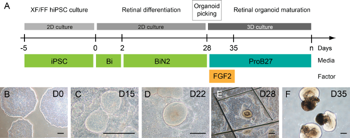
Figure 1: Generation and maturation of retinal organoids from adherent human iPSCs. (A) Differentiation protocol allowing the generation of retinal organoids. (B) Human iPSCs at D0. (C) Emerging neuroretinal epithelium at D15. (D) Self-forming neural retina-like structures at D22. (E) Here, the neuroretinal bud is isolated using a needle. (F) Representative images of retinal organoids in floating culture condition at D35. Scale bars = 200 µm. Please click here to view a larger version of this figure.
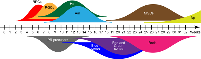
Figure 2: Waves of human iPSC-derived retinal cell generation. Abbreviations: Retinal progenitor cells (RPCs), retinal ganglion cells (RGCs), horizontal cells (Ho), amacrine cells (Am), Müller glial cells (MGCs), bipolar cells (Bp), and photoreceptors (PR). Please click here to view a larger version of this figure.
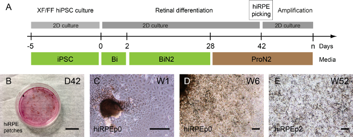
Figure 3: Generation and amplification of human iPSC-derived RPE. (A) An illustration of the differentiation protocol allowing the generation of human hiRPE cells. (B) Phase-contrast images showing pigmented patches emerging from differentiating adherent human iPSCs at Week 6 (W6). (C) hiRPEp0 cells cultivated one week (W1) after the pigmented patch picking. (D) hiRPEp0 cells at week 6 (W6) after the pigmented patch picking. (E) A representative image of hiRPEp2 cells after thawing, cultivated for 52 weeks (W52) in ProN2 medium. Scale bars = 200 µm. Please click here to view a larger version of this figure.
Discussion
This protocol describes how to produce RPE cells and retinal organoids, containing retinal RGCs and photoreceptors, from human pluripotent stem cells in xeno-free and feeder-free conditions. Compatible with the Good Manufacturing Practice (GMP) process, the method cultivated presented here allows a large production of iPSC-derived retinal cells as RPE cells, RGCs, and photoreceptors for the development of stem cell-based therapies and drug discovery approaches for the future treatment of retinal degenerative diseases. The cryopreservation of whole retinal organoids or hiRPE cells also provides a major advantage in establishing intermediate cell banks, an important step for future use in stem cell-based therapies.
The production of large stocks of specific retinal cell types at a specific stage of differentiation will be required for future clinical translation. In this regard, the generation in three months of CD73-positive photoreceptor precursors described as a transplantation-compatible cell population18, and the possibility to generate these immature photoreceptors from freeze-thawed retinal organoids15 reinforce the hope to use these cells for therapeutic purposes. Concerning retinal pigmented epithelium, the ability of hiRPE cells to proliferate in vitro allows large cell productions to bank them. Importantly, thawed human iPSC-derived RPE cells retain their RPE phenotype and function, therefore, as trophic factor secretion or photoreceptor outer segment phagocytosis15, validating their ease of use for screening strategies as well as for future therapeutic approaches.
There are a variety of protocols for the generation of iPSC-derived retinal organoids7,8,9,10,11,12,15 that vary in culture methods (embryoid body-like aggregates vs. adherent cells) as well as efficiency and robustness. The method described here starts from adherent human iPSCs and shows a reproducible efficacy, adaptability, and applicability to a wide range of human iPSC lines15. This process, based on the successive change of serum-free media, recapitulates the main steps of retinal development by exploiting the intrinsic cues of the system to guide differentiation. An important advantage of this protocol is the absence of embryonic body formation and the addition of matrix for future GMP-compliant retinal cell manufacturing protocols to produce cell therapy derivatives. In this way, no difference in the efficiency of the retinal organoid generation and maturation were found between xenogeneic and no-xenogeneic culture conditions using Cell Therapy System (CTS) supplements or not, formulated exclusively with recombinant or humanized components.
The success of the retinal differentiation method largely depends on the quality of the human iPSC cultures. The reprogramming method does not influence the differentiation efficiency of human iPSCs to retinal cells8, but their stemness status need to be optimal. Briefly, routinely cultivated human iPSCs should not show any signs of differentiation. The colonies should not overlap and must display their characteristic circular morphology. Although the efficiency of the retinal differentiation is clone-dependent, a minimum of two retinal structures per cm2 can be picked at D28, corresponding to 50 - 60 neuroretinal structures for one 6-cm dish. For the retinal organoid maturation in floating culture conditions, limiting the number of structures per well avoids structure fusion and medium overconsumption. In these culture conditions, retinal organoids can be maturated for an extensive time, required to obtain late retinal cell types.
Looking forward, retinal organoids generated in vitro by this method constitute powerful tools to model retinal diseases. Patient-specific iPSC-derived retinal cell models will be used to better understand complex or genetic diseases by the exploration of their molecular and cellular mechanisms. These models will be particularly suitable for drug discovery through high-throughput screening, cell and gene therapies, or genome editing approaches, to develop innovative treatments for retinal dystrophies.
Disclosures
Sacha Reichman, Olivier Goureau, and José-Alain Sahel are inventors on pending patents related to the generation of retinal cells from human pluripotent stem cells.
Acknowledgements
The authors would like to thank the members of Goureau's team for their input during the set-up of the methods described here, and G. Gagliardi and M. Garita for their critical reading. This work was supported by grants from the ANR (GPiPS: ANR-2010-RFCS005; SightREPAIR: ANR-16-CE17-008-02), the Retina France Association and the technology transfer company SATT Lutech. It was also performed in the frame of the LABEX LIFESENSES (ANR-10-LABX-65) supported by the ANR within the Investissements d'Avenir program (ANR-11-IDEX-0004-02).
Materials
| Name | Company | Catalog Number | Comments |
|---|---|---|---|
| Vitronectin (VTN-N) Recombinant Human Protein, Truncated | ThermoFisher Scientific | A14700 | Coating |
| CTS Vitronectin (VTN-N) Recombinant Human Protein, Truncated | ThermoFisher Scientific | A27940 | Coating |
| Essential 8 Medium | ThermoFisher Scientific | A1517001 | medium |
| Essential 6 Medium | ThermoFisher Scientific | A1516401 | medium |
| CTS (Cell Therapy Systems) N-2 Supplement | ThermoFisher Scientific | A1370701 | supplement CTS |
| N-2 Supplement (100X) | ThermoFisher Scientific | 17502048 | supplement |
| B-27 Supplement (50X), serum free | ThermoFisher Scientific | 17504044 | supplement |
| CTS B-27 Supplement, XenoFree | ThermoFisher Scientific | A1486701 | supplement CTS |
| DMEM/F-12 | ThermoFisher Scientific | 11320074 | medium |
| MEM Non-Essential Amino Acids Solution (100X) | ThermoFisher Scientific | 11140035 | supplement |
| Penicillin-Streptomycin (10,000 U/mL) | ThermoFisher Scientific | 15140122 | antibiotic |
| CellStart CTS | ThermoFisher Scientific | A1014201 | Matrix CTS |
| Geltrex hESC-Qualified, Ready-To-Use, Reduced Growth Factor Basement Membrane Matrix | ThermoFisher Scientific | A1569601 | Matrix |
| Gentle Cell Dissociation Reagent | Stemcell Technologies | 7174 | dissociation solution |
| Cryostem Freezing Media | clinisciences | 05-710-1D | Cryopreservation medium |
| Fibroblast growth factor 2 (FGF2) | Preprotech | 100-18B | FGF2 |
| Fibroblast growth factor 2 (FGF2) animal free | Preprotech | AF-100-18B | FGF2 Xeno free |
| AGANI needle 23G | Terumo | AN*2332R1 | Needle |
| Flask 25 cm² Tissue Culture Treated | Falcon | 353109 | T-25 cm² |
| 24 well plate Tissue Culture Treated | Costar | 3526 | 24-well plate |
| 6 well plate Tissue Culture Treated | Costar | 3516 | 6-well plate |
References
- Zhao, C., Wang, Q., Temple, S. Stem cell therapies for retinal diseases: recapitulating development to replace degenerated cells. Development. 144, 1368-1381 (2017).
- Dalkara, D., Goureau, O., Marazova, K., Sahel, J. -A. Let There Be Light: Gene and Cell Therapy for Blindness. Human Gene Therapy. 27, 134-147 (2016).
- Wright, L. S., Phillips, M. J., Pinilla, I., Hei, D., Gamm, D. M. Induced pluripotent stem cells as custom therapeutics for retinal repair: Progress and rationale. Experimental Eye Research. 123, 161-172 (2014).
- Leach, L. L., Clegg, D. O. Making Stem Cells Retinal: Methods for Deriving Retinal Pigment Epithelium and Implications for Patients with Ocular Disease. Stem Cells. 33, 2363-2373 (2015).
- Gill, K. P., Hewitt, A. W., Davidson, K. C., Pébay, A., Wong, R. C. B. Methods of Retinal Ganglion Cell Differentiation From Pluripotent Stem Cells. Translational Vision Science and Technology. 3, 2(2014).
- Giacalone, J. C., Wiley, L. A., et al. Concise review: Patient-specific stem cells to interrogate inherited eye disease. STEM CELLS Translational Medicine. 5, 132-140 (2016).
- Nakano, T., Ando, S., et al. Self-formation of optic cups and storable stratified neural retina from human ESCs. Cell Stem Cell. 10, 771-785 (2012).
- Reichman, S., Terray, A., et al. From confluent human iPS cells to self-forming neural retina and retinal pigmented epithelium. Proceedings of the National Academy of Sciences of the United States of America. 111, 8518-8523 (2014).
- Meyer, J. S., Howden, S. E., et al. Optic vesicle-like structures derived from human pluripotent stem cells facilitate a customized approach to retinal disease treatment. Stem Cells. 29, 1206-1218 (2011).
- Zhong, X., Gutierrez, C., et al. Generation of three-dimensional retinal tissue with functional photoreceptors from human iPSCs. Nature Communications. 5, 4047(2014).
- Mellough, C. B., Collin, J., et al. IGF-1 Signalling plays an important role in the formation of three dimensional laminated neural retina and other ocular structures from human embryonic stem cells. Stem Cells. 33, 2416-2430 (2015).
- Gonzalez-Cordero, A., Kruczek, K., et al. Recapitulation of human retinal development from human pluripotent stem cells generates transplantable populations of cone photoreceptors. Stem Cell Reports. 9, 820-837 (2017).
- Eiraku, M., Takata, N., et al. Self-organizing optic-cup morphogenesis in three-dimensional culture. Nature. 472, 51-56 (2011).
- Meyer, J. S., Shearer, R. L., et al. Modeling early retinal development with human embryonic and induced pluripotent stem cells. Proceedings of the National Academy of Sciences of the United States of America. , 16698-16703 (2009).
- Reichman, S., Slembrouck, A., et al. Generation of storable retinal organoids and retinal pigmented epithelium from adherent human ips cells in xeno-free and feeder-free conditions. Stem Cells. 35, 1176-1188 (2017).
- Chen, G., Gulbranson, D. R., et al. Chemically defined conditions for human iPSC derivation and culture. Nature Methods. 8, 424-429 (2011).
- Croze, R. H., Buchholz, D. E., et al. ROCK inhibition extends passage of pluripotent stem cell-derived retinal pigmented epithelium. Stem Cells Translational Medicine. 3, 1066-1078 (2014).
- Eberle, D., Schubert, S., Postel, K., Corbeil, D., Ader, M. Increased integration of transplanted CD73-positive photoreceptor precursors into adult mouse retina. Investigative Ophthalmology & Visual Science. 52, 6462-6471 (2011).
Reprints and Permissions
