We describe a protocol to measure vortex formation time, an index of left ventricular filling efficiency, using standard transesophageal echocardiography techniques in patients undergoing cardiac surgery. We apply this technique to analyze vortex formation time in several groups of patients with differing cardiac pathologies.
A subscription to JoVE is required to view this content. Sign in or start your free trial.
Method Article
Noninvasive Determination of Vortex Formation Time Using Transesophageal Echocardiography During Cardiac Surgery
In This Article
Summary
Abstract
Trans-mitral blood flow produces a three-dimensional rotational body of fluid, known as a vortex ring, that enhances the efficiency of left ventricular (LV) filling compared with a continuous linear jet. Vortex ring development is most often quantified with vortex formation time (VFT), a dimensionless parameter based on fluid ejection from a rigid tube. Our group is interested in factors that affect LV filling efficiency during cardiac surgery. In this report, we describe how to use standard two-dimensional (2D) and Doppler transesophageal echocardiography (TEE) to noninvasively derive the variables needed to calculate VFT. We calculate atrial filling fraction (β) from velocity-time integrals of trans-mitral early LV filling and atrial systole blood flow velocity waveforms measured in the mid-esophageal four-chamber TEE view. Stroke volume (SV) is calculated as the product of the diameter of the LV outflow track measured in the mid-esophageal long axis TEE view and the velocity-time integral of blood flow through the outflow track determined in the deep transgastric view using pulse-wave Doppler. Finally, mitral valve diameter (D) is determined as the average of major and minor axis lengths measured in orthogonal mid-esophageal bicommissural and long axis imaging planes, respectively. VFT is then calculated as 4 × (1-β) × SV/(πD3). We have used this technique to analyze VFT in several groups of patients with differing cardiac abnormalities. We discuss our application of this technique and its potential limitations and also review our results to date. Noninvasive measurement of VFT using TEE is straightforward in anesthetized patients undergoing cardiac surgery. The technique may allow cardiac anesthesiologists and surgeons to assess the impact of pathological conditions and surgical interventions on LV filling efficiency in real time.
Introduction
Fluid mechanics is a critical yet often underappreciated determinant of left ventricular (LV) filling. A three-dimensional rotational body of fluid, known as a vortex ring, is generated whenever a fluid traverses an orifice1,2,3. This vortex ring improves the efficiency of fluid transport compared with a continuous linear jet4. Movement of blood through the mitral valve during early LV filling causes a vortex ring to form5,6,7,8 and facilitates its propagation into the chamber by preserving fluid momentum and kinetic energy9. These actions enhance LV filling efficiency4,10,11,12,13. The ring not only inhibits blood flow stasis in the LV apex14,15,16,17 but also directs flow preferentially beneath the anterior mitral leaflet7,18, effects that decrease the risk of apical thrombus formation and facilitate filling of the LV outflow track19, respectively. Contrast echocardiography17, Doppler vector flow mapping6,20,21, magnetic resonance imaging7, and particle imaging velocimetry9,22,23,24 have been used to demonstrate the appearance and behavior of trans-mitral vortex rings under normal and pathological conditions. The left atrial-LV pressure gradient, the degree of diastolic mitral annular excursion, the minimum LV pressure achieved during diastole, and the rate and extent of LV relaxation are the four major determinants of the duration, size, flow intensity, and position of the trans-mitral ring2,12,25,26,27,28,29.
Vortex ring development is most often quantified with a dimensionless parameter (vortex formation time; VFT) based on fluid ejection from a rigid tube3, where VFT is defined as the product of the time-averaged fluid velocity and the duration of ejection divided by the orifice diameter. The optimal size of a vortex ring is achieved when VFT is 4 in vitro because trailing jets and energetic limitations prevent it from attaining a larger size3,4. Mitral valve VFT has been approximated clinically using transthoracic echocardiography8,30,31. Based on analysis of trans-mitral blood flow velocity and mitral valve diameter (D), it can be easily shown8 that VFT = 4 × (1-β) × EF ×α3, where β = atrial filling fraction, EF = LV ejection fraction, and α = EDV1/3/D, where EDV = end-diastolic volume. Ejection fraction is the ratio of stroke volume (SV) and EDV, allowing this equation to be simplified to VFT = 4 × (1-β) × SV/(πD3). Because VFT is dimensionless (volume/volume), this index allows direct comparison between patients of varying size without adjustment for weight or body surface area8. Optimal VFT ranges between 3.3 and 5.5 in healthy subjects8, and results are consistent with those obtained in fluid dynamics models3,32. VFT was shown to be ≤ 2.0 in patients with depressed LV systolic function, findings that are also supported by theoretical predictions8. Reductions in VFT independently predicted morbidity and mortality in patients with heart failure30. Elevated LV afterload33, Alzheimer's disease34, abnormal diastolic function19, and replacement of the native mitral valve with a prosthesis35 have also been shown to decrease VFT. Measurement of VFT may also be useful to identify blood flow stasis or thrombosis in patients with acute myocardial infarction36,37.
Our group is interested in factors that affect LV filling efficiency during cardiac surgery38,39,40,41. We use standard two-dimensional and Doppler transesophageal echocardiography (TEE) to noninvasively derive the variables required to calculate VFT. In this report, we describe this methodology in detail and review our findings to date.
Access restricted. Please log in or start a trial to view this content.
Protocol
The Institutional Review Board of the Clement J. Zablocki Veterans Affairs Medical Center approved the protocols. Written informed consent was waived because invasive cardiac monitoring and TEE are routinely used in all patients undergoing cardiac surgery in our institution. Patients with relative or absolute contraindications for TEE, those undergoing repeat median sternotomy or emergency surgery, and those with atrial or ventricular tachyarrhythmias were excluded from participation.
1. Anesthesia
- Provide each patient with intravenous midazolam (1 to 3 mg) and fentanyl (50 to 150 µg) for conscious sedation before surgery.
- Use local anesthesia (subcutaneous 1% lidocaine) for insertion of intravenous and radial artery catheters. Test the quality of the local anesthesia with a pinprick.
- Ensure that the patient receives supplemental oxygen using a nasal cannula (2 to 4 L/min).
- Place a central venous or pulmonary artery catheter using local anesthesia (subcutaneous 1% lidocaine) under sterile conditions through the right or left internal jugular vein with ultrasound guidance based on appropriate clinical indications.
- Induce anesthesia using intravenous fentanyl (5 mcg/kg), propofol (1 to 2 mg/kg), and rocuronium (0.1 mg/kg). Maintain anesthesia using inhaled isoflurane (end-tidal concentration of 1%) in an air-oxygen mixture, fentanyl (1 to 2 µg/kg/h), and rocuronium (0.05 mg/kg) titrated to effect using neuromuscular monitoring.
- Suction the stomach using an oral-gastric tube.
- Place ultrasound jelly in the patient’s hypopharynx. Lift the jaw anteriorly and advance a TEE probe into the esophagus with gentle pressure to overcome resistance of the hypopharygeus muscle.
2. Transesophageal Echocardiography
- Perform a comprehensive TEE examination following American Society of Echocardiography/Society of Cardiovascular Anesthesiologists guidelines42 in each patient.
- Place a pulse-wave Doppler sample volume between the tips of the mitral leaflets to record trans-mitral blood flow velocity in the mid-esophageal four-chamber TEE imaging plane (Figure 1).
- Identify the early LV filling and atrial systole blood flow waveforms of trans-mitral blood flow velocity, and measure their corresponding peak velocities and velocity-time integrals (VTIE and VTIA, respectively) using the echocardiography equipment’s integrated software package (Figure 1).
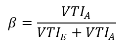
- Calculate the atrial filling fraction (β) as the ratio of atrial to total LV filling:
- Measure the maximum diameter of the LV outflow tract immediately below the aortic valve in the mid-esophageal aortic valve long axis TEE view during mid-systole (Figure 2A).
- Calculate the area of the LV outflow tract assuming circular geometry as the product of π/4 and the square of the diameter (see step 2.5 above).
- Obtain a deep transgastric long axis TEE view, and place a pulse-wave Doppler sample volume in the distal LV outflow tract to record a blood flow velocity envelope (Figure 2B) at the same level where the diameter was measured (see step 2.5 above); integrate the area of this waveform using the echocardiography equipment’s software package to obtain VTI.
- Multiply the resulting velocity-time integral (VTI) of the LV outflow track blood flow velocity waveform (Figure 2B) by the area of the outflow track (see step 2.6) to obtain stroke volume (SV).
- Record video clips of the mid-esophageal bicommissural and LV long axis TEE imaging planes, respectively42. Be sure to include several cardiac cycles in each recording.
- Visually inspect slow-motion images of the video clips (see step 2.9 above) after the ECG T-wave to choose the maximum opening of the mitral valve leaflets.
- Measure the distance between the mitral leaflets (Figures 3A and 3B) using the echocardiography equipment’s “caliper” function.
- Calculate the mitral valve diameter (D) as the average of the major (transcommissural anterior-lateral-posterior-medial) and minor (anterior-posterior) lengths.
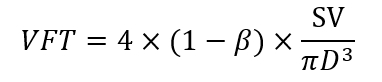
- Calculate VFT using the formula:
- Perform all quantitative echocardiographic measurements in triplicate at end-expiration.
3. Experimental Design
- Determine VFT, indices of LV diastolic function, and hemodynamics during steady-state conditions 30 minutes before and 15, 30, and 60 minutes after cardiopulmonary bypass (CPB) in 10 patients with normal preoperative LV ejection fraction under coronary artery surgery to test the hypothesis that CPB transiently decreases VFT39.
- Test the hypothesis that LV pressure-overload hypertrophy produced by aortic valve stenosis reduces VFT by examining (in one group of 8 patients undergoing aortic valve replacement) for severe aortic stenosis and comparing observations to another group of 8 patients with normal LV wall thickness undergoing coronary artery surgery40. Measure VFT, LV diastolic function, hemodynamics, and end-diastolic posterior wall thickness during steady-state conditions 30 minutes before CPB.
- Test the hypothesis that abnormal diastolic blood flow entering the LV affects trans-mitral LV filling efficiency in 8 patients with aortic valve stenosis and moderate aortic insufficiency versus 8 patients with aortic stenosis who do not have regurgitant valves38. Measure VFT and other parameters as described above (step 3.2).
- Test the hypothesis that advanced age is associated with a reduction in LV filling efficiency quantified using VFT in 7 octogenarians (82 ± 2 years) compared to 7 younger patients (55 ± 6 years)41 undergoing coronary artery surgery. Ensure that both groups have normal preoperative LV ejection fraction. Measure VFT and other parameters as described above (step 3.2).
4. Statistics
- Present the data as mean ± standard deviation.
- Evaluate data using analysis of variance (ANOVA) followed by Bonferroni’s modification of Student’s t-test.
- Use linear regression analysis to determine the relationships between VFT and end-diastolic posterior wall thickness and between VFT and age.
- Reject the null hypothesis when p < 0.05.
Access restricted. Please log in or start a trial to view this content.
Results
The current technique allowed us to reliably measure VFT during cardiac surgery under a variety of clinical conditions by obtaining each determinant from blood flow and dimensional recordings in standard TEE imaging planes. A pulse-wave Doppler sample volume was placed at the tips of the mitral leaflets in the mid-esophageal four-chamber view to obtain the trans-mitral blood flow velocity profile necessary to calculate atrial filling fraction (β; Figure 1). S...
Access restricted. Please log in or start a trial to view this content.
Discussion
The current results illustrate that VFT can be reliably measured during cardiac surgery using the TEE techniques described here. Previous descriptions of VFT used transthoracic echocardiography in conscious subjects, but this approach cannot be utilized when the chest is open. We used intraoperative TEE to determine VFT in the anesthetized patients undergoing cardiac surgery during which changes in LV filling dynamics are often encountered as a result of ischemia-reperfusion injury or surgical interventions. Our findings...
Access restricted. Please log in or start a trial to view this content.
Disclosures
The authors have no competing financial interests or other conflicts of interest pursuant to this work.
Acknowledgements
This material is the result of work supported with resources and the use of the facilities at the Clement J. Zablocki Veterans Affairs Medical Center in Milwaukee, Wisconsin.
Access restricted. Please log in or start a trial to view this content.
Materials
| Name | Company | Catalog Number | Comments |
|---|---|---|---|
| Echocardiography Machine | Philips Ultrasound, Bothall, WA | iE33 | |
| Transesophageal Echocardiography Probe | Philips Ultrasound, Bothall, WA | X7-2t | |
| Statistical Software | AnalystSoft, Walnut, CA | StatPlus:mac Pro |
References
- Collier, E., Hertzberg, J., Shandas, R. Regression analysis for vortex ring characteristics during left ventricular filling. Biomedical Sciences Instrumentation. 38 (2), 307-311 (2002).
- Kheradvar, A., Gharib, M. Influence of ventricular pressure drop on mitral annulus dynamics through the process of vortex ring formation. Annals of Biomedical Engineering. 35 (12), 2050-2064 (2007).
- Gharib, M., Rambod, E., Shariff, K. A universal time scale for vortex ring formation. Journal of Fluid Mechanics. 360 (1), 121-140 (1998).
- Krueger, P. S., Gharib, M. The significance of vortex ring formation to the impulse and thrust of a starting jet. Physics of Fluids. 15 (5), 1271-1281 (2003).
- Reul, H., Talukder, N., Muller, W. Fluid mechanics of the natural mitral valve. Journal of Biomechanics. 14 (5), 361-372 (1981).
- Kim, W. Y., et al. Two-dimensional mitral flow velocity profiles in pig models using epicardial Doppler echocardiography. Journal of the American College of Cardiology. 24 (2), 532-545 (1994).
- Kilner, P. J., et al. Asymmetic redirection of flow through the heart. Nature. 404 (6779), 759-761 (2000).
- Gharib, M., Rambod, E., Kheradvar, A., Sahn, D. J., Dabiri, J. O. Optimal vortex formation as an index of cardiac health. Proceedings of the National Academy of Sciences USA. 103 (16), 6305-6308 (2006).
- Rodriguez Munoz, D., et al. Intracardiac flow visualization: current status and future directions. European Heart Journal Cardiovascular Imaging. 14 (11), 1029-1038 (2013).
- Martinez-Legazpi, P., et al. Contribution of the diastolic vortex ring to left ventricular filling. Journal of the American College of Cardiology. 64 (16), 1711-1721 (2014).
- Dabiri, J. O., Gharib, M. The role of optimal vortex formation in biological fluid transport. Proceedings of the Royal Society B. 272 (1572), 1557-1560 (2003).
- Kheradvar, A., Gharib, M. On mitral valve dynamics and its connection to early diastolic flow. Annals of Biomedical Engineering. 37 (1), 1-13 (2009).
- Linden, P. F., Turner, J. S. The formation of "optimal" vortex rings, and the efficiency of propulsion devices. Journal of Fluid Mechanics. 427 (1), 61-72 (2001).
- Domenichini, F., Pedrizzetti, G., Baccani, B. Three-dimensional filling flow into a model left ventricle. Journal of Fluid Mechanics. 539 (1), 179-198 (2005).
- Sengupta, P. P., et al. Left ventricular isovolumic flow sequence during sinus and paced rhythms: new insights from use of high-resolution Doppler and ultrasonic digital particle imaging velocimetry. Journal of the American College of Cardiology. 49 (8), 899-908 (2007).
- Rodriguez Munoz, D., et al. Flow mapping inside a left ventricular aneurysm: a potential tool to demonstrate thrombogenicity. Echocardiography. 31 (1), E10-E12 (2014).
- Son, J. W., et al. Abnormal left ventricular vortex flow patterns in association with left ventricular apical thrombus formation in patients with anterior myocardial infarction: a quantitative analysis by contrast echocardiography. Circulation Journal. 76 (11), 2640-2646 (2012).
- Kheradvar, A., Falahatpisheh, A. The effects of dynamic saddle annulus and leaflet length on trans-mitral flow pattern and leaflet stress of a bileaflet bioprosthetic mitral valve. The Journal of Heart Valve Disease. 21 (2), 225-233 (2012).
- Kheradvar, A., Assadi, R., Falahatpisheh, A., Sengupta, P. P. Assessment of trans-mitral vortex formation in patients with diastolic dysfunction. Journal of the American Society of Echocardiography. 25 (2), 220-227 (2012).
- Chen, R., et al. Assessment of left ventricular hemodynamics and function of patients with uremia by vortex formation using vector flow mapping. Echocardiography. 29 (9), 1081-1090 (2012).
- Hendabadi, S., et al. Topology of blood transport in the human left ventricle by novel processing of Doppler echocardiography. Annals of Biomedical Engineering. 41 (12), 2603-2616 (2013).
- Sengupta, P. P., Pedrizetti, G., Narula, J. Multiplaner visualization of blood flow using echocardiographic particle imaging velocimetry. Journal of the American College of Cardiology Cardiovascular Imaging. 5 (5), 566-569 (2012).
- Sengupta, P. P., et al. Emerging trends in CV flow visualization. Journal of the American College of Cardiology Cardiovascular Imaging. 5 (3), 305-316 (2012).
- Hong, G. R., Kim, M., Pedrizzetti, G., Vannan, M. A. Current clinical application of intracardiac flow analysis using echocardiography. Journal of Cardiovascular Ultrasound. 21 (4), 155-162 (2013).
- Kheradvar, A., Milano, M., Gharib, M. Correlation between vortex ring formation and mitral annulus dynamics during ventricular rapid filling. American Society for Artificial Internal Organs Journal. 53 (1), 8-16 (2007).
- Hong, G. R., et al. Characterization and quantification of vortex flow in the human left ventricle by contrast echocardiography using vector particle image velocimetry. Journal of the American College of Cardiology Cardiovascular Imaging. 1 (6), 705-717 (2008).
- Zhang, H., et al. The evolution of intraventricular vortex during ejection studied by using vector flow mapping. Echocardiography. 30 (1), 27-36 (2013).
- Nogami, Y., et al. Abnormal early diastolic intraventricular flow 'kinetic energy index' assessed by vector flow mapping in patients with elevated filling pressure. European Heart Journal Cardiovascular Imaging. 14 (3), 253-260 (2013).
- Zhang, H., et al. The left ventricular intracavity vortex during the isovolumic contraction period as detected by vector flow mapping. Echocardiography. 29 (5), 579-587 (2012).
- Poh, K. K., et al. Left ventricular filling dynamics in heart failure: echocardiographic measurement and utilities of vortex formation time. European Heart Journal Cardiovascular Imaging. 13 (5), 385-393 (2012).
- Belohlavek, M. Vortex formation time: an emerging echocardiographic index of left ventricular filling efficiency? European Heart Journal Cardiovascular Imaging. 13 (5), 367-369 (2012).
- Dabiri, J. O., Gharib, M. Starting flow through nozzles with temporally variable exit diameter. Journal of Fluid Mechanics. 538 (1), 111-136 (2005).
- Jiamsripong, P., et al. Impact of acute moderate elevation in left ventricular afterload on diastolic trans-mitral flow efficiency: analysis by vortex formation time. Journal of the American Society of Echocardiography. 22 (4), 427-431 (2009).
- Belohlavek, M., et al. Patients with Alzheimer disease have altered trans-mitral flow: echocardiographic analysis of the vortex formation time. Journal of Ultrasound in Medicine. 28 (11), 1493-1500 (2009).
- Pedrizzetti, G., Domenichini, F., Tonti, G. On the left ventricular vortex reversal after mitral valve replacement. Annals of Biomedical Engineering. 38 (3), 769-773 (2010).
- Martinez-Legazpi, P., et al. Stasis mapping using ultrasound: a prospective study in acute myocardial infarction. Journal of the American College of Cardiology Cardiovascular Imaging. 11 (3), 514-515 (2018).
- Harfi, T. T., et al. The E-wave propagation index (EPI): a novel echocardiographic parameter for prediction of left ventricular thrombus. Derivation from computational fluid dynamic modeling and validation on human subjects. International Journal of Cardiology. 227 (1), 662-667 (2017).
- Pagel, P. S., Boettcher, B. T., De Vry, D. J., Freed, J. K., Iqbal, Z. Moderate aortic valvular insufficiency invalidates vortex formation time as an index of left ventricular filling efficiency in patients with severe degenerative calcific aortic stenosis undergoing aortic valve replacement. Journal of Cardiothoracic and Vascular Anesthesia. 30 (5), 1260-1265 (2016).
- Pagel, P. S., Gandhi, S. D., Iqbal, Z., Hudetz, J. A. Cardiopulmonary bypass transiently inhibits intraventricular vortex ring formation in patients undergoing coronary artery bypass graft surgery. Journal of Cardiothoracic and Vascular Anesthesia. 26 (3), 376-380 (2012).
- Pagel, P. S., Hudetz, J. A. Chronic pressure-overload hypertrophy attenuates vortex formation time in patients with severe aortic stenosis and preserved left ventricular systolic function undergoing aortic valve replacement. Journal of Cardiothoracic and Vascular Anesthesia. 27 (4), 660-664 (2013).
- Pagel, P. S., Dye, L., Boettcher, B. T., Freed, J. K. Advanced age attenuates left ventricular filling efficiency quantified using vortex formation time: a study of octogenarians with normal left ventricular systolic function undergoing coronary artery surgery. Journal of Cardiothoracic and Vascular Anesthesia. 32 (4), 1775-1779 (2018).
- Shanewise, J. S., et al. ASE/SCA guidelines for performing a comprehensive intraoperative multiplane transesophageal echocardiography examination: recommendations of the American Society of Echocardiography Council for Intraoperative Echocardiography and the Society of Cardiovascular Anesthesiologists Task Force for Certification in Perioperative Transesophageal Echocardiography. Journal of the American Society of Echocardiography. 12 (10), 884-900 (1999).
- Gaspar, T., et al. Three-dimensional imaging of the left ventricular outflow tract: impact on aortic valve area estimation by the continuity equation. Journal of the American Society of Echocardiography. 25 (7), 749-757 (2012).
- Karamnov, S., Burbano-Vera, N., Huang, C. C., Fox, J. A., Shernan, S. A. Echocardiographic assessment of mitral stenosis orifice area: a comparison of a novel three-dimensional method versus conventional techniques. Anesthesia and Analgesia. 125 (3), 774-780 (2017).
- Pagel, P. S., Kampine, J. P., Schmeling, W. T., Warltier, D. C. Comparison of end-systolic pressure-length relations and preload recruitable stroke work as indices of myocardial contractility in the conscious and anesthetized, chronically instrumented dog. Anesthesiology. 73 (2), 278-290 (1990).
- Pagel, P. S., Kampine, J. P., Schmeling, W. T., Warltier, D. C. Alteration of left ventricular diastolic function by desflurane, isoflurane, and halothane in the chronically instrumented dog with autonomic nervous system blockade. Anesthesiology. 74 (6), 1103-1114 (1991).
- De Hert, S. G., Rodrigus, I. E., Haenen, L. R., De Mulder, P. A., Gillebert, T. C. Recovery of systolic and diastolic left ventricular function early after cardiopulmonary bypass. Anesthesiology. 85 (5), 1063-1075 (1996).
- Gorcsan, J., Diana, P., Lee, J., Katz, W. E., Hattler, B. G. Reversible diastolic dysfunction after successful coronary artery bypass surgery. Assessment by transesophageal Doppler echocardiography. Chest. 106 (5), 1364-1369 (1994).
- Djaiani, G. N., et al. Mitral flow propagation velocity identifies patients with abnormal diastolic function during coronary artery bypass graft surgery. Anesthesia and Analgesia. 95 (3), 524-530 (2002).
- Casthely, P. A., et al. Left ventricular diastolic function after coronary artery bypass grafting: a correlative study with three different myocardial protection techniques. Journal of Thoracic and Cardiovascular Surgery. 114 (2), 254-260 (1997).
- Tulner, S. A., et al. Perioperative assessment of left ventricular function by pressure-volume loops using the conductance catheter method. Anesthesia and Analgesia. 97 (4), 950-957 (2003).
- Firstenberg, M. S., et al. Relationship between early diastolic intraventricular pressure gradients, an index of elastic recoil, and improvements in systolic and diastolic function. Circulation. 104 (12 Suppl 1), I330-I335 (2001).
- Cooke, J., Hertzberg, J., Boardman, M., Shandas, R. Characterizing vortex ring behavior during ventricular filling with Doppler echocardiography: an in vitro study. Annals of Biomedical Engineering. 32 (2), 245-256 (2004).
- Grossman, W., Jones, D., McLaurin, L. P. Wall stress and patterns of hypertrophy in the human left ventricle. Journal of Clinical Investigation. 56 (1), 56-64 (1975).
- Hess, O. M., et al. Diastolic function and myocardial structure in patients with myocardial hypertrophy. Special reference to normalized viscoelastic data. Circulation. 63 (2), 360-371 (1981).
- Hess, O. M., et al. Diastolic stiffness and myocardial structure in aortic valve disease before and after valve replacement. Circulation. 69 (5), 855-865 (1984).
- Sandstede, J. J. W., et al. Cardiac systolic rotation and contraction before and after valve replacement for aortic stenosis: a myocardial tagging study using MR imaging. American Journal of Roentgenology. 178 (4), 953-958 (2002).
- Stuber, M., et al. Alterations in the local myocardial motion pattern in patients suffering from pressure overload due to aortic stenosis. Circulation. 100 (4), 361-368 (1999).
- Nagel, E., et al. Cardiac rotation and relaxation in patients with aortic valve stenosis. European Heart Journal. 21 (7), 582-589 (2000).
- Rakowski, H., et al. Canadian consensus recommendations for the measurement and reporting of diastolic dysfunction by echocardiography: from the Investigators of Consensus on Diastolic Dysfunction by Echocardiography. Journal of the American Society of Echocardiography. 9 (5), 736-760 (1996).
- Homeyer, P., Oxorn, D. C. Aortic regurgitation: echocardiographic diagnosis. Anesthesia and Analgesia. 122 (1), 37-42 (2016).
- Landzberg, J. S., et al. Etiology of the Austin Flint murmur. Journal of the American College of Cardiology. 20 (2), 408-413 (1992).
- Flint, A. On cardiac murmurs. American Journal of Medical Sciences. 91 (1), 27(1886).
- Botvinick, E. H., Schiller, N. B., Wickramasekaran, R., Klausner, S. C., Gertz, E. Echocardiographic demonstration of early mitral valve closure in severe aortic insufficiency. Its clinical implications. Circulation. 51 (5), 836-847 (1975).
- Mann, T., McLaurin, L., Grossman, W., Craige, E. Assessing the hemodynamic severity of acute aortic regurgitation due to infective endocarditis. New England Journal of Medicine. 293 (3), 108-113 (1975).
- Borlaug, B. A., et al. Longitudinal changes in left ventricular stiffness: a community-based study. Circulation Heart Failure. 6 (5), 944-952 (2013).
- Wong, J., et al. Age-related changes in intraventricular kinetic energy: a physiological or pathological adaptation? American Journal of Physiology Heart Circulatory Physiology. 310 (6), H747-H755 (2016).
- Carrick-Ranson, G., et al. Effect of healthy aging on left ventricular relaxation and diastolic suction. American Journal of Physiology Heart Circulatory Physiology. 303 (3), H315-H322 (2012).
- Iskandrian, A. S., Hakki, A. H. Age-related changes in left ventricular diastolic performance. American Heart Journal. 112 (1), 75-78 (1986).
- Schulman, S. P., et al. Age-related decline in left ventricular filling at rest and exercise. American Journal of Physiology. 263 (6 Pt 2), H1932-H1938 (1992).
- Stork, M., et al. Age-related hemodynamic changes during diastole: a combined M-mode and Doppler echo study. Internal Journal of Cardiovascular Imaging. 6 (1), 23-30 (1991).
- Sanders, D., Dudley, M., Groban, L. Diastolic dysfunction, cardiovascular aging, and the anesthesiologist. Anesthesiology Clinics. 27 (3), 497-517 (2009).
Access restricted. Please log in or start a trial to view this content.
Reprints and Permissions
