This article presents a bovine mammary gland biopsy using core and needle biopsy tools. Harvested tissue can be used for cell culture or to assess mammary physiology and metabolism including gene expression, protein expression, protein modifications, immunohistochemistry, and metabolite concentrations.
Method Article
Bovine Mammary Gland Biopsy Techniques
In This Article
Summary
Abstract
Bovine mammary gland biopsies allow researchers to collect tissue samples to study cell biology including gene expression, histological analysis, signaling pathways, and protein translation. This article describes two techniques for biopsy of the bovine mammary gland (MG). Three healthy Holstein dairy cows were the subjects. Before biopsies, cows were milked and subsequently restrained in a cattle chute. An analgesic (flunixin meglumine, 1.1 to 2.2 mg/kg of body weight) was administered via jugular intravenous [IV] injection 15-20 min prior to biopsy. For standing sedation, xylazine hydrochloride (0.01-0.05 mg/kg of body weight) was injected via the coccygeal vessels 5-10 min before the procedure. Once adequately sedated, the biopsy site was aseptically prepared and locally anaesthetized with 6 mL of 2% lidocaine hydrochloride via subcutaneous injection. Using aseptic technique, a 2 to 3 cm vertical incision was made using a number 10 scalpel. Core and needle biopsy tools were used. The core biopsy tool was attached to a cordless drill and inserted into the MG tissue through the incision using a clock-wise drill action. The needle biopsy tool was manually inserted into the incision site. Immediately after the procedure, an assistant applied pressure on the incision site for 20 to 25 min using a sterile towel to achieve hemostasis. Stainless steel surgical staples were used to oppose the skin incision. The staples were removed 10 days post-procedure. The main advantages of core and needle biopsies is that both approaches are minimally invasive procedures that can be safely performed in healthy cows. Milk yield following the biopsy was unaffected. These procedures require a short recovery time and result in fewer risks of complications. Specific limitations may include bleeding after the biopsy and infection on the biopsy site. Applications of these techniques include tissue collection for clinical diagnosis and research purposes, such as primary cell culture.
Introduction
A biopsy is a procedure to harvest a core section of tissue from subjects for medical or research purposes. It is a minimally invasive and widely used technique to collect tissue as an alternative to euthanasia1 to allow analysis of tissue responses to treatment or other factors of interest. Samples of bovine mammary gland (MG) tissues are essential for dairy research to study gene and protein expression, histology, the organization of cellular organelles, signaling pathways, and metabolic processes in response to changes in management or the environment. Additionally, MG tissue is needed to diagnose and research certain infectious diseases, such as bovine mastitis caused by bacterial infections2.
The udder of cattle contains four separate glands and each gland contains an independent milk secretory system collectively called parenchyma. The alveoli, ducts, and connective tissue are present in the mammary parenchyma. The alveoli are microscopic spherical hollow structures that are composed of epithelial cells in the internal surface (luminal) and specialized myoepithelial cells on the external surface (basal). The alveoli are responsible for synthesis and secretion of milk. The fibrous connective tissue present in the parenchyma can separate a group of alveoli from other groups of alveoli, and each group is called a lobule3. Several factors can affect the development and function of the MG including animal physiology, nutrition, management, genetics, and the environment4. Mastitis also is a critical factor that negatively affects MG function and milk quality5. The toxins released by mastitic pathogens can cause damage to the alveoli and induce necrosis resulting in biochemical and histological changes in the MG tissue6. Thus, the histology and metabolic processes of each alveoli or lobule in the MG may be significantly different from others. As such, biopsies that are representative samples of the entire mammary gland are desirable. Small biopsies may only capture a single lobule or individual alveolus, limiting the resulting scientific or diagnostic information. For research purposes, it is necessary to be aware that a mammary biopsy provides a 'snapshot' of tissue characteristics but a biopsy cannot adequately characterize total mammary function in the absence of estimates of total mammary gland mass.
Several biopsy tools have been developed over the last 30 years for human use. Currently, adaptations of those instruments are available for use with animals. For dairy cattle, samples of MG tissue have been obtained using different techniques including surgical excision (blunt dissection)7, biopsy needles1,8, and core biopsy instruments9,10. Thus, MG biopsy techniques in lactating dairy cows have transitioned from procedures using recumbent sedation with surgical dissection using electrocautery hemostasis in 19927 to collection of core biopsies under standing sedation9,10,11,12. Surgical biopsy is an invasive method, which can be expensive and have a higher incidence of complications such as hematoma, wound problems, and tumor spread13. Currently, core biopsy and needle biopsy (also known as a tru-cut biopsy) have been widely adopted as alternatives to surgical biopsy. The advantages of core and needle biopsies compared to surgical biopsy include: the procedure is minimally invasive; major complications are rare; general anesthesia is not required; the procedure is relativley rapid; the recovery time is short; there is minimal negative effects on udder health, and only short-term effects on milk yield and composition8,9,10; and the cost is less than surgical biopsy13.
One core biopsy procedure described in 1996 used a sterile, stainless steel cannula with a removable retractable blade to remove a representative amount of tissue from the bovine MG without general anesthesia9,10,11,12. During the procedure, the instrument was attached to a cordless drill to create a low speed, rotational motion which cleanly cut a tissue core as the tool was advanced into the tissue. The benefit was a larger tissue sample (70 mm x 4 mm in diameter, about 0.75 to 1 g) 9. A recent study10 showed that the biopsy procedure described by V. C. Farret al.9 can be used to perform repeated MG tissue collections without negative impact on performance and udder health of lactating dairy cows. Most recently, a study14 was carried out in dairy cows to evaluate repeated biopsies of the MG using a larger trocar (31 cm long, outer diameter of 9.5 mm, inner diameter of 8 mm) with vacuum applied to an internal stainless steel cannula to collect the biopsy. This method used sedation (xylazine) and local anesthesia (2% lidocaine hydrochloride).
The needle biopsy is another technique to collect mammary tissue. Several studies have adopted this technique. One study1 used sedation (detomidine)and local anesthetic (1% lidocaine) in the procedure. After the biopsy, the cows received a prophylactic antibiotic treatment. The mammary gland was manually massaged before and after the milking. Blood in the milk was observed for up to 84 h after the biopsy. The amount and composition of the milk were affected for a short period of time. Recently, a study used a biopsy needle to perform repeated MG biopsies in dairy cows8. Sedation (1% acepromazine, intramuscular) and local anesthesia (2% lidocaine hydrochloride, subcutaneous) were administered to the animals. The animals did not receive intramammary drugs or antibiotics before or after the procedure, and there were no signs of infection at the biopsy site during the post-surgical period. In this study, repeated bovine MG biopsies using a needle had a minor negative impact on milk production and udder health of dairy cows. In general, the needle biopsy seems to be a less invasive method than the core biopsy instrument. However, as noted previously, it is essential for the biopsy technique to harvest a representative sample of the MG tissue. The limitation of a needle biopsy is that a small amount of bovine MG tissue is obtained (about 20 to 25 mg)8,15.
Almost all of the studies used a combination of α-2 agonist sedation and local anesthetic8,9,10, whether the biopsy was via a needle or a larger core. In the bovine MG, most nerve endings are associated with the skin. Innervation to the parenchymal tissue is largely via stretch receptors with sparse Type A nerve fibers to detect sharp surgical pain. As a result, physiological mechanisms of pain due to surgical manipulation of the MG is via skin and subcutaneous tissues3 and not deep tissues such as parenchymal tissue. Therefore, for biopsy procedures, it is only necessary to locally anesthetize the skin and subcutaneous tissues, as infiltration of local anesthetic into the deeper tissues does not significantly reduce surgical pain. After an appropriate preparation of the biopsy area, animal discomfort is, primarily, associated with restraint.
Collection of larger core samples may increase hematoma formation and increase the risk of infection within the MG parenchyma. Therefore, peri-operative protocols often include administration of parenteral antibiotics7, although that is not universals8. Achieving hemostasis is also an important factor for reducing cow morbidity. In the aforementioned study using a large core biopsy instrument14, manual pressure was applied to the biopsy site and a cow bra was used to apply ice to the wounds for at least 2 h following the procedure. Despite the large amount of tissue harvested, only slight reductions in feed intake and milk yield were observed, and the procedure was repeated every three weeks without negative effects on cow health.
Researchers performing MG biopsy in dairy cows need to consider the diagnostic or analytical quality of the resulting biopsy, ease of technique, and cow morbidity. Accurate and pre-planned surgical techniques are imperative to achieve these goals. To date, MG biopsy studies have focused on describing biopsy outcomes, as opposed to describing the biopsy technique itself, and descriptions for lactating dairy cows lack sufficient detail to allow replication. Thus, the objective of this work was to describe both needle biopsy and larger core biopsy techniques in sufficient detail to allow safe and humane replication of MG biopsy of cattle.
Protocol
All methods described were approved by the Virginia Tech Institutional Animal Care and Use Committee (IACUC).
1. Personnel
- Have at least two assistants with surgical and cow-handling experience.
- Train all assistants at least three times, if possible using cadaveric material, prior to performing procedures associated with this protocol on live animals.
NOTE: Training should be conducted by an instructor that has been previously trained and performed the technique. - Have a large animal veterinarian on-hand for drug administration and in the event that emergency treatment becomes necessary during the procedure. Emergencies that may occur during this procedure include but are not limited to: hemorrhage, α-2 agonist overdose and pulmonary edema, regurgitation and aspiration of food material, pain, and patient resistance requiring further sedation.
2. Preparation of the surgical instruments, supplies, and facility
- Inventory and purchase all equipment and supplies (see Table of Materials).
- Clean and autoclave surgical drapes, biopsy instruments, scalpel holders, surgical towels, and forceps.
- Have a properly sized squeeze chute and head gate to restrain the cows.
- Establish a work space with a table near the squeeze chute.
- Ensure that the work area is clean and has limited cow through-traffic.
- Organize equipment and supplies in the work space for easy access.
- Have proper lighting inside of the work space.
3. Preparation of the animals
- One day before the scheduled biopsy, wash and scrub the animal, particularly the udder to remove manure and soiled material.
- Follow procedures for safe and humane restraint of cattle.
- In advance of the biopsy and again at the time of the biopsy, assess the health, physical condition and behavior of the animal.
NOTE: A minimum examination includes temperature, pulse, and respiratory rates as well as examination for dermatitis over the proposed surgical site or other areas of bacterial infection. Collect milk samples from each quarter and check for mastitis. - Use only healthy cows.
- Completely milk the cow.
- Move the animal into the squeeze chute, ideally within 2 h of milking to minimize milk presence in the glands.
- Restrain the animal with a head gate.
4. Analgesia and sedation
- Place a rope halter on the head of the cow to prevent backward and forward movement.
- Pull the animal's head to one side, and tie the rope to the squeeze chute using a quick release knot to hold the head in place.
- Clean the area of injection with a 70% isopropyl alcohol swab and administer flunixin meglumine (1.1 to 2.2 mg/kg of body weight) intravenously via the jugular vein 15-20 min prior to biopsy.
NOTE:In some protocols, nonsteroidal anti-inflammatory drugs are administered after the biopsy if anti-inflammatory drugs will not affect research results.- Locate the jugular vein.
- Raise the jugular vein by application of pressure at the base of the jugular groove.
- Check to make sure that there are no bubbles in the syringe.
- Insert the needle into the raised jugular vein, and draw 0.5 mL of blood into the syringe twice and mix with the contents. If no blood shows in the syringe, relocate the needle. If the needle is resident in the vein, inject the contents.
- Gently remove the needle.
- Apply gauze with gentle pressure to the injection site to prevent bleeding.
- Administer xylazine hydrochloride intravenously (0.01 to 0.05 mg/kg of body weight) in the coccygeal vessel approximately 5-10 min prior to biopsy to allow sufficient time for establishment of sedation.
CAUTION: Check the animal for signs of pulmonary edema. Clinical signs of pulmonary edema include respiratory distress, severe dyspnea, breathing difficulties, cough, frothy sputum, and blue tongue. If signs of pulmonary edema are observed, it is recommended to use tolazine (2 to 4 mg/kg of body weight) to reverse the xylazine effects.- Raise the tail and clean the area of injection with a 70% isopropyl alcohol swab.
- Check to make sure that there are no bubbles in the syringe.
- Insert the needle into the tail vessel, draw 0.2 mL of blood into the syringe and mix with the contents to ensure the needle is resident in the vessel. If the needle is resident in the vessel, inject the syringe contents.
- Gently remove the needle.
- Apply gauze with gentle pressure to the injection site.
5. Preparation of the biopsy site
- Have an assistant tie up the tail for the procedure.
- Select the biopsy site on the udder (typically in an upper area to minimize collection of connective tissue and to avoid penetration into the gland cistern, Figure 1), and remove any soiled material or manure from the selected biopsy site.
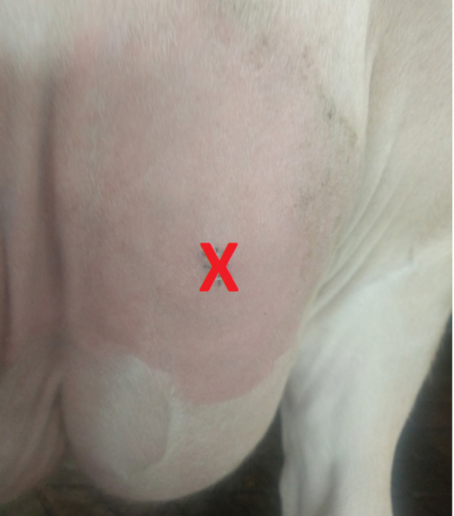
Figure 1. Image of the bovine udder illustrating the biopsy site. Please click here to view a larger version of this figure.
- Observe and palpate the skin with special attention to identify any large subcutaneous blood vessels in order to avoid these vessels during the biopsy.
- Clip the hair from a 15 cm x 15 cm area around the biopsy site.
- Prepare the biopsy area with povidone-iodine (0.75% available iodine) or chlorhexidine gluconate scrub. Alternate with 70% isopropyl alcohol at least three times to remove all visible and invisible debris. Apply the aseptic scrub solution and isopropyl alcohol in a circular motion using inside-out approach. Ensure that the antiseptic scrub solution remains in contact with the skin for at least 5 min.
- Use a butterfly infusion set with an 18 G needle to deposit 6 mL of 2% lidocaine hydrochloride subcutaneously at the incision site to create a line-block. Do not penetrate into the deeper tissues.
NOTE: Dosage of lidocaine varies between 3 and 8 mL. - Allow the local anesthetic to diffuse for 3 to 5 min. Perform another repetition of scrub solution and alcohol prior to incision. While waiting, prepare the biopsy instruments.
6. Biopsy procedure
- Use aseptic techniques when handling the biopsy tools and for the incision.
- Wash hands to remove all visible contamination and apply sterile surgical gloves.
- Arrange the surgical instruments in a sterile area in order of use. Have a number 10 scalpel, sterile gauze, an assembled biopsy instrument, and a sterile towel for hemostasis.
NOTE: Follow Procedure 1 to perform a Core biopsy or Procedure 2 to perform a Needle biopsy.
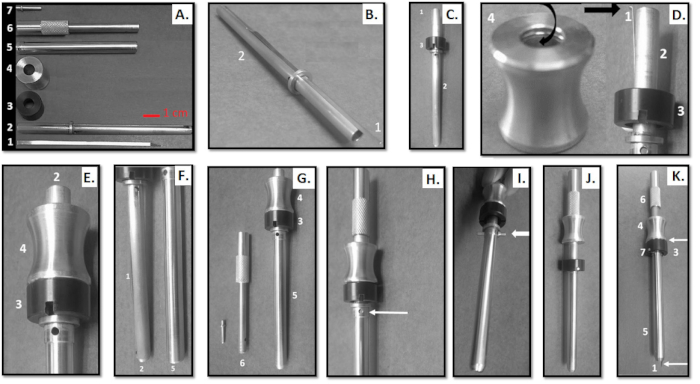
Figure 2. Schematic representation of assembly and setup of the core biopsy tool described by V. C. Farr, et al.9 Scale bar is 1 cm. Please click here to view a larger version of this figure.
- Core Biopsy Instrument (Farr et al.9)
- Assemble the core biopsy instrument using sterile gloves (Figure 2).
- Place the 7 sterile pieces on a sterile drape (Figure 2A).
- Insert the blade (piece 1) into the docking system on piece 2 (Figure 2B ).
CAUTION: Do not place fingers directly in the blade line . - Insert piece 3 on top of piece 2 (Figure 2C) ensuring that the docking station will align.
NOTE: Observe a docking system in the interior wall of piece 4 (Figure 2D). - Engage the final edge of the blade (piece 1) into the docking system of piece 4 (Figure 2D).
- Push forward piece 4 and observe if piece 4 is next to piece 3 (Figure 2E).
- Insert piece 5 on the device (blade + piece 2) (Figure 2F).
- Ensure that the docking station is aligned (Figure 2G).
- Insert piece 6 into the top of piece 2 (opposite side of the blade) (Figure 2H)
- Ensure that the docking station is aligned (Figure 2H).
- Insert the locking screw (piece 7) into the docking station (Figure 2I).
- Push forward piece 3 (black) to cover the locking screw.
CAUTION: Do not place fingers on the blade exit. - Activate the tool pushing forward piece 4 and observe the blade outside of the tool.
- Pull piece 4 back to retract the blade into the tool (ready to use).
NOTE: The blade should be activated only when the tool has penetrated the desired distance inside the tissue to be biopsied.
- Ensure that the cow is sufficiently sedated and the biopsy site is sufficiently anesthetized. Pinch the skin to ensure no reaction.
- Make a 2 to 3 cm vertical incision through the skin and subcutaneous tissues from proximal to distal using a number 10 scalpel.
- Attach the biopsy instrument to a cordless drill using sterile technique.
- Place the drill against the biopsy tool and check if the tool is firmly attached to the drill.
- Ensure adequate restraint by having an individual elevate the tail during all procedure.
- Turn on the drill using clockwise rotation and a low speed.
NOTE: The drill is not sterile, and the operator does not remain sterile when using the drill. - Advance the entire biopsy tool (around 7.5 cm) into the udder through the incision while the drill is rotating the toll.
- Turn off the drill and manually extend piece 4 of the tool.
- Turn on the drill using clockwise rotation and low speed.
- Remove the instrument containing the tissue core from the udder.
- Apply strong pressure immediately to the wound using a sterile towel for at least 20 min.
NOTE: Have an assistant perform this using their fist to ensure adequate pressure is applied. - Remove the tissue from biopsy tool using tweezers.
- Keep the sample in 1x phosphate buffered saline and evaluate the amount of tissue.
- Take the cow's vital signs every 10 min after the biopsy for at least 30 min.
- Check for bleeding after 20 min of pressure on the biopsy site. If there are drips of blood continue to apply pressure for an additional 5-10 min.
- Close the incisions using stainless steel staples at 5 mm intervals after all bleeding has stopped. Use between 5 and 8 staples.
- Apply an aerosol bandage to the biopsy area.
- Observe the animal for 50 min after the procedure.
- Assemble the core biopsy instrument using sterile gloves (Figure 2).
- Needle Biopsy Instrument
- Follow the manufacturer's instructions for the device.
- Remove the biopsy needle from the package using sterile techniques.
- Discard the needle if any damage is observed.
- Attach the needle to the device.
- Close the cover and cock the device.
- Ensure that the cow is sufficiently sedated and the biopsy site is sufficiently anesthetized. Pinch the skin to ensure no reaction.
- Make a 1 to 2 cm vertical incision through the skin and subcutaneous tissues from proximal to distal using a number 10 scalpel.
NOTE: The incision for the biopsy needle instrument can be smaller (1-2 cm) than that for the core biopsy instrument. - Insert the biopsy needle into the incision site (around 10 to 13 cm from skin).
- Activate the biopsy needle device to collect the tissue.
- Remove the needle from the udder.
- Apply immediate, strong pressure to the wound using a sterile towel for at least 20 min.
- Remove the tissue from the biopsy needle using tweezers.
- Keep the sample in 1x phosphate buffered saline and evaluate the amount of tissue.
- Take vital signs every 10 min after the biopsy for at least 30 min.
- Check for bleeding after 20 min of pressure on the biopsy site.
- Close the incisions using stainless steel staples after all bleeding has stopped.
- Apply an aerosol bandage to the biopsy area.
- Observe the animal for 50 min after the procedure.
7. Post-biopsy animal care
- Document all drugs administered to animals.
NOTE: Milk was discarded during a period of 36 h after the procedure; meat withdrawal was 4 d. Withdrawal periods for milk and meat may vary depending on the country or jurisdiction and the drugs used. Please check local rules and regulations. - Check for the presence of blood in the milk for 7 to 10 d after the biopsy.
- Hand strip blood clots from the biopsied quarter at subsequent milkings and ensure that complete milk removal occurs.
NOTE: Presence of blood clots from the biopsied quarter for 1-3 milkings following the procedure is expected. Blood in the milk may be observed for 1 to 6 d after the biopsy. - Observe milk yield, and if possible, the individual daily feed intake until surgical staples have been removed.
- Monitor the animal twice a day for body temperature, respiration and heart rates, and demeanor until surgical staples have been removed.
- Check the biopsy site twice daily for swelling, tenderness, and any signs of drainage until the surgical staples have been removed. If these are observed, consult a veterinarian.
NOTE: Keep the biopsy site clean and reapply the aerosol bandage every one to three days as needed. - Remove the staples from the incision site 10 to 14 d after the biopsy, depending on the healing rate.
- Consult a veterinarian if any signs of local or systemic infection are observed.
Results
In the present protocol, the core biopsy technique produced tissue sample of 200 to 600 mg while the needle biopsy produced samples of 10 to 30 mg per biopsy. The animals were observed twice a day for 10 d after the procedure. No complications occurred during the procedure or in the post-operative period, with vital parameters of the cows remaining within normal limits (average respiratory rate = 31.4 ± 7.04 (SD) breaths per min, average heart rate = 75.9 ± 8.9 beats per min, and average rectal temperature = 38.2 ± 0.68 °C, Figure 3).
Following manual pressure on the biopsy area to achieve hemostasis (about 25 min), the open wound showed minimal bleeding (Figure 4). The cows were fed the same diet ad libitum, and the animals were kept in the same housing facility during the post-operative period. The animals did not show signs of infection at the biopsy site; there was no pain, bleeding, swelling, drainage, or elevated temperature (Figure 5). The wound healed within 8 to 10 d of the biopsy (Figure 5C).
One animal developed minor skin irritation due to wound rubbing on day 8 after the procedure. This cow was treated by cleaning the biopsy site using povidone-iodine solution and 70% isopropyl alcohol. The stainless steel staples were removed to allow drainage and povidone iodine ointment (1%) was applied twice a day on the biopsy site for 2 to 3 d. The skin irritation disappeared 2 d after applying povidone-iodine ointment and the wound healed with no further complications.
Blood clots were removed by hand stripping of milk from the biopsied quarter prior to machine milking. In the present protocol, the presence of blood clots in the milk was observed for up to three milkings (about 36 h) after the biopsy. Blood contamination of milk was observed for up to 48 h after the biopsy. There were no visual differences observed between core and needle biopsies relative to the amount of blood in the milk. The animals did not show a significant decrease in milk yield after the procedure (Figure 6). Average milk composition was 4.37% fat, 3.34% protein, and 4.64% lactose. The average milk somatic cell count (SCC) was less than 200,000 cells per mL.
The MG tissue obtained can be used for different research purposes such as primary cell cultures (Figure 7).
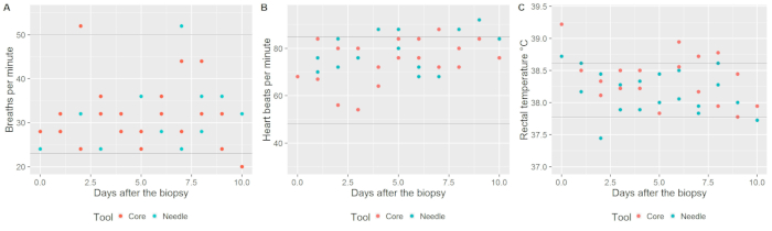
Figure 3. Evaluation of animal health indicators after the bovine mammary gland biopsy. Please click here to view a larger version of this figure.
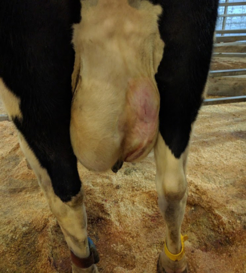
Figure 4. Biopsy site appearance after manual pressure was applied to the biopsy area to achieve hemostasis. Please click here to view a larger version of this figure.
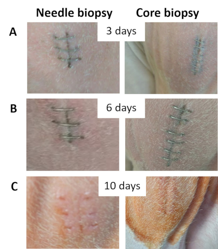
Figure 5. Biopsy site photographs. (A) Biopsy site 3 d after the procedure. (B) Biopsy site 6 d after the procedure. (C) Biopsy site 10 d after the procedure illustrating that the wound was adequately healed. Please click here to view a larger version of this figure.
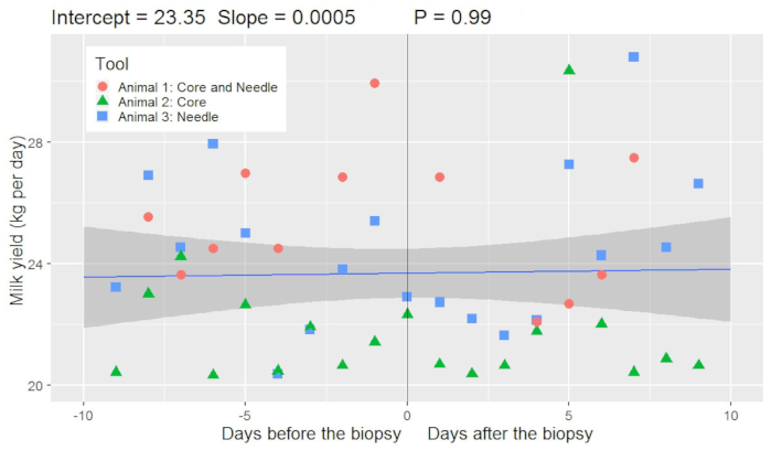
Figure 6. Daily milk production before and after the bovine mammary gland biopsy (n = 3 cows). A linear model estimated that 10 days before the biopsy the animals produced 23.35 kg of milk per day. The milk yield did not change significantly (P > 0.05) from 10 days before to 10 days after the biopsy although a slight increase in milk yield was observed (increase of 0.0005 kg/d of milk for each day from 10 days before the biopsy). Three animals were used in this protocol; one animal was biopsied twice (Animal 1: core biopsy on the left side of the udder and needle biopsy on the right side of the udder) and two animals were biopsied once (Animal 2 and 3: only on the right side of the udder using the core or needle procedure). Please click here to view a larger version of this figure.
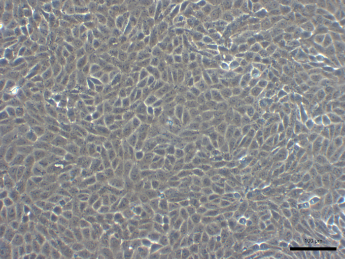
Figure 7. Representative images of primary bovine mammary epithelial cells culture. Scale bar is 100 µm. Please click here to view a larger version of this figure.
Discussion
Core and needle biopsy methods were described in this protocol9,16. A detailed evaluation of the animal health and incidence of mastitis14 before the biopsy is required for both procedures. For research purposes, performing the technique in animals with obvious signs of inflammation or infectious diseases should be avoided. This will reduce the risk of complications during and after a biopsy. All biopsy instruments and devices should be clean, disinfected, and sterilized to avoid contamination of the biopsy site. Before the procedure, it is necessary to minimize surgical site infections (SSI). In general, SSI is associated with animal morbidity, lost performance, and higher production costs in dairy cows. Studies previously reported methods to prevent SSI due to biopsy including clipping the hair around the incision site, washing the biopsy area to remove contamination9,14, and using antiseptic agents (70% alcohol, iodine surgical scrub9, 2% chlorhexidine acetate solution14, 10% povidone-iodine8) for skin preparation. In some studies, administration of antibiotics was adopted during7 or immediately after the biopsy9; however, antibiotic prophylaxis was not used in the present work, and no infections of the biopsy site were observed. To prevent contamination of the biopsy wound, the tail should be secured to prevent contact with the biopsy site until the aerosol bandage has been applied. In this protocol, staples are removed about 10 to 14 d after the biopsy.
Prior to a core biopsy, it is important to properly set up the tool and attach it to the drill; choose a slow rotation speed, select the forward rotation, and do not use the reverse mode of the drill. During the procedure, it is important to use digital pressure on both sides of the incision to keep the skin edges apart and have a long enough incision to allow tool entry without contacting the skin or connective tissue. If this procedure is not done, a drag on the incision edges during rotation of the core biopsy instrument may occur and cause additional skin tissue trauma which will increase the risk of infection and may delay wound healing. The present biopsy procedures described in this protocol were performed by a board-certified large animal surgeon. The procedures were performed successfully (Figure 5). Both techniques are relatively easy and fast to perform as compared to the surgical excision procedure.
Bleeding from the biopsy site are common after MG biopsy in dairy cows8,9,10,14. In the present protocol, bleeding observed was minimal (Figure 4), which may be due to application of adequate (strong) pressure on the wound immediately after the procedure. Strong pressure for at least 20 min is required, and it may be required for more than 30 min in some cases. If moderate to severe bleeding is observed after application of pressure to the biopsy site, it is recommended to continue pressure, and immediately contact a veterinarian.
As hemostasis is an important factor for reducing cow morbidity, one study packed hemostatic pads into the biopsy site to control bleeding9. However, use of hemostatic pads in such a manner has a high potential risk for microbiological contamination in the dairy farm environment. Another study14 applied manual pressure to the biopsy site between repeated biopsies and after biopsy and skin closure, and applied ice to the site for at least 2 h following the biopsy. In the present protocol, strong pressure applied to the biopsy site for 20 to 25 min was adequate to control bleeding.
A successful biopsy technique should result in minimum blood in the milk which persists for a short period of time after the procedure. To avoid an interruption of milk secretion during milk letdown and mastitis infections, intramammary blood clots should be removed by hand stripping from the biopsied quarter10,14. In the present protocol, blood was observed in the milk up to 48 h after the biopsy. However, in a study that used a larger number of animals, blood in the milk was observed in the majority of cows for less than 6 days. Few animals showed blood in the milk after 6 days8. For this reason, daily observation of milk appearance is necessary for 6 d after the procedure. Animals did not exhibit any signs of mastitis infection when a large tissue sample was obtained14. However, previous research which performed repeated biopsies on the same animal using a needle found that mastitis infection incidence was approximately 12% following the procedure8. In the present protocol, neither animal had visual signs of clinical mastitis infection after the procedure. There is also a slight chance that the incision site may become infected after the biopsy.
Tolazoline hydrochloride, a drug that reverses the effects of sedation, should be available in the event of an overdose from xylazine. An excessive dose of xylazine may cause pulmonary edema. Clinical signs of pulmonary edema include respiratory distress, severe dyspnea, breathing difficulties, cough, and frothy sputum. If signs of pulmonary edema are observed, it is recommended to use tolazine (2 to 4 mg/kg of body weight) to reverse the xylazine effects.
The present protocol describes the technique to perform both a needle biopsy and core biopsy. In general, the advantage of a core biopsy as compared to needle biopsy is the larger tissue sample (0.75 to 1 g)9 with minimum negative effect on udder health. The needle biopsy is a less invasive method than the core biopsy instrument. However, multiple biopsy attempts to obtain a larger amount of tissue using a needle biopsy procedure may increase the risk of major bleeding after the procedure, as well as blood clots in the milk. Both techniques caused minimal cow morbidity and were easily achieved with minimal training of surgical personnel. A short-term change in daily milk yield (8 to 10 % decrease) and its composition9 and feed intake reduction14 is expected after the core and needle biopsies, respectively. Limitations of the needle biopsy procedure are the small volume of tissue removed with the needle. The instrument needs to be activated only when the needle is within the tissue to be biopsied and multiple attempts often are necessary to obtain adequate amounts of tissue, which increases the risk of blood in the milk and mastitis after the procedure. Limitations of the core biopsy include a higher risk of bleeding after the procedure if a large amount of tissue (>200 mg) is obtained. In addition, the biopsy tool is more difficult to assemble requiring adequate training before use.
Disclosures
The authors have nothing to disclose.
Acknowledgements
This research was supported by the National Animal Nutrition Program and Virginia Tech. The technical assistance of Ms. Tara Pilonero, Dr. Julie Settlage, and Dr. Izabelle Teixeira are gratefully acknowledged.
Materials
| Name | Company | Catalog Number | Comments |
|---|---|---|---|
| 70% Isopropyl alcohol | Walmart | 565106257 | 1 L |
| Aerosol spray to kill and repel flies | MWI | 14339 | 1 bottle (if necessary) |
| Biopsy needles, 12 G × 16 cm | C. R. Bard | MN1216 | 2 |
| Butterfly infusion set (18 G needle) | Fisher | 22-258087 | 1 |
| Cell culture dishes | Fisher | 08-772E | 4 |
| Cordless drill (low speed) | Hitachi | DS10DFL | 1 |
| Core biopsy instrument, Farr et al. (1996) | 2, custom metal fabrication. To request the tool contact the authors. | ||
| Cows | cattle healthy | ||
| Flunixin meglumine | MWI/VetOne | 501018 | 1.1 to 2.2 mg/kg of body weight |
| Folding table | Amazon | 1 | |
| Forceps | Fisher | 09-753-50 | 1 |
| Gloves non-sterile | Fisher | 17-200-845 | 9, size dependent |
| Hard brush | Sullivan Supply | 18270 | 1, to wash the cows |
| Head gate | 1, customized for dairy cows | ||
| Lidocaine hydrochloride 2% injectable | MWI/VetOne | 510212 | 6 mL was used, from 3 to 8 mL |
| Livestock body wash | eZall Technologies | 39384 | 1, to wash the cows |
| Needle 18 G (1 to 1/5“) | Fisher | 14-821-15A | 6 |
| Needle 20 G (1 to 1/5“) | Fisher | 14-815-526 | 6 |
| Phosphate buffered saline (0.9%) | Fisher | 20-012-043 | 1 L |
| Povidone Iodine ointment | Jeffers | #VED1A | 500 g (if necessary) |
| Povidone iodine scrub solution (0.75% iodine) | MWI/VetOne | 510094 | 1 L |
| Reusable biopsy instrument | C. R. Bard | MG1522 | 1 |
| Rope halter | Nasco farm and ranch | C10852 | 2, cow size |
| Scalpel blades (#10) | MWI | 033870 | 2 |
| Scalpel holders (#3) | MWI | 602008 | 1 |
| Self seal autoclave pouch 10 x 5.5 in. | Fisher | 19-130-0038 | 1 case of 800 |
| Sterile cotton gauze sponges | Fisher | 22-037-986 | 4 |
| Sterile gauze pads | Fisher | 19-090-735 | 4 |
| Sterile surgical gloves | Surgical gloves | 19-166-679 | 3, size dependent |
| Sterile surgical towels | Fisher | 50-118-0339 | 6 |
| Stethoscope | Littmann Master Classic II | 1392 | 1 |
| Surgical clipper | Oster A5 | 078005-010-003 | 1, preparation of the animal for biopsy |
| Surgical clipper blades (#40 ) | Oster | 78919-016 | 1, preparation of the animal for biopsy |
| Surgical staple remover | MWI | 541 | 1 |
| Surgical stapler pre-loaded | MWI | 17713 | 5 |
| Syringes of 10 mL | Fisher | 14-823-224 | 3 |
| Syringes of 2 mL | Fisher | 22-028854 | 3 |
| Syringes of 20 mL | Fisher | 22-034507 | 3 |
| Syringes of 5 mL | Fisher | 22-028855 | 3 |
| Thermometer | Agri-Pro Enterprises Inc | 72000 | 1 |
| Tolazine hydrochloride | Akorn Animal Health | 61311-486-10 | 2 to 4 mg/kg of body weight (if necessary) |
| Tweezer | Fisher | 14-955-025 | 1, for handle the tissue sample |
| Water hose | Miracle-Gro, Walmart | 554990501 | 1, to wash the cows |
| Water-resistant aerosol bandage (aluminum powder 40 mg) | MWI | 010728 | 1 bottle |
| Xylazine hydrochloride | MWI/VetOne | 510650 | 0.01 to 0.05 mg/kg of body weight |
References
- Khatun, M., Sorensen, P., Ingvartsen, K. L., Bjerring, M., Rontved, C. M. Effects of combined liver and udder biopsying on the acute phase response of dairy cows with experimentally induced E. coli mastitis. Animal. 7 (10), 1721-1730 (2013).
- Buitenhuis, B., Rontved, C. M., Edwards, S. M., Ingvartsen, K. L., Sorensen, P. In depth analysis of genes and pathways of the mammary gland involved in the pathogenesis of bovine Escherichia coli-mastitis. BMC Genomics. 12, 130(2011).
- Nickerson, S. C., Akers, R. M. Mammary Gland | Anatomy. Fuquay JW, Fox PF and McSweeney PLH (eds.), Encyclopedia of Dairy Sciences, Second Edition. 3, Academic Press. San Diego. 328-337 (2011).
- Akers, R. M. A 100-Year Review: Mammary development and lactation. Journal of Dairy Science. 100 (12), 10332-10352 (2017).
- Ruegg, P. L. A 100-Year Review: Mastitis detection, management, and prevention1. Journal of Dairy Science. 100 (12), 10381-10397 (2017).
- Zhao, X., Lacasse, P. Mammary tissue damage during bovine mastitis: Causes and control. Journal of Animal Science. 86 (13), 57-65 (2008).
- Knight, C. H., Hillerton, J. E., Teverson, R. M., Winter, A. Biopsy of the bovine mammary gland. British Veterinary Journal. 148 (2), 129-132 (1992).
- Lima, J. A., et al. Effects of bovine mammary gland biopsy and increased milking frequency on post-procedure udder health, histology, and milk yield. Animal. 10 (5), 838-846 (2016).
- Farr, V. C., et al. An improved method for the routine biopsy of bovine mammary tissue. Journal of Dairy Science. 79 (4), 543-549 (1996).
- Weng, X., et al. Short communication: Repeated mammary tissue collections during lactation do not alter subsequent milk yield or composition. Journal of Dairy Science. 100 (10), 8422-8425 (2017).
- Peterson, D. G., Matitashvili, E. A., Bauman, D. E. Diet-induced milk fat depression in dairy cows results in increased trans-10, cis-12 CLA in milk fat and coordinate suppression of mRNA abundance for mammary enzymes involved in milk fat synthesis. Journal of Nutrition. 133 (10), 3098-3102 (2003).
- Baumgard, L. H., Matitashvili, E., Corl, B. A., Dwyer, D. A., Bauman, D. E. trans-10, cis-12 conjugated linoleic acid decreases lipogenic rates and expression of genes involved in milk lipid synthesis in dairy cows. Journal of Dairy Science. 85 (9), 2155-2163 (2002).
- Kasraeian, S., Allison, D. C., Ahlmann, E. R., Fedenko, A. N., Menendez, L. R. A comparison of fine-needle aspiration, core biopsy, and surgical biopsy in the diagnosis of extremity soft tissue masses. Clinical Orthopaedics and Related Research. 468 (11), 2992-3002 (2010).
- de Lima, L. S., et al. A new technique for repeated biopsies of the mammary gland in dairy cows allotted to Latin-square design studies. Canadian Journal of Veterinary Research-Revue Canadienne De Recherche Veterinaire. 80 (3), 225-229 (2016).
- Ollier, S., et al. Whole intact rapeseeds or sunflower oil in high-forage or high-concentrate diets affects milk yield, milk composition, and mammary gene expression profile in goats. Journal of Dairy Science. 92 (11), 5544-5560 (2009).
- Soberon, F., et al. Effects of increased milking frequency on metabolism and mammary cell proliferation in Holstein dairy cows. Journal of Dairy Science. 93 (2), 565-573 (2010).
Reprints and Permissions
