This manuscript describes a FACS-based protocol for isolation of papillary and reticular fibroblasts from human skin. It circumvents in vitro culture which was inevitable with the commonly used isolation protocol via explant cultures. The emanating fibroblast subsets are functionally distinct and display differential gene expression and localization within the dermis.
Method Article
Isolation of Papillary and Reticular Fibroblasts from Human Skin by Fluorescence-activated Cell Sorting
Opens in a new tab
In This Article
Summary
Loading...
$$\rightleftharpoonup{xx}$$
$$\longleftharp{xx}$$,
$$\longrightharp{xx}$$,
Abstract
Loading...
$$\rightleftharpoonup{xx}$$
$$\longleftharp{xx}$$,
$$\longrightharp{xx}$$,
Fibroblasts are a highly heterogeneous cell population implicated in the pathogenesis of many human diseases. In human skin dermis, fibroblasts have traditionally been attributed to the superficial papillary or lower reticular dermis according to their histological localization. In mouse dermis, papillary and reticular fibroblasts originate from two different lineages with diverging functions regarding physiological and pathological processes and a distinct cell surface marker expression profile by which they can be distinguished.
Importantly, evidence from explant cultures from superficial and lower dermal layers suggest that at least two functionally distinct dermal fibroblasts lineages exist in human skin dermis as well. However, unlike for mouse skin, cell surface markers enabling the discrimination of different fibroblast subsets have not yet been established for human skin. We developed a novel protocol for the isolation of human papillary and reticular fibroblast populations via fluorescence-activated cell sorting (FACS) using the two cell surface markers Fibroblast Activation Protein (FAP) and Thymocyte antigen 1 (Thy1)/CD90. This method enables the isolation of pure fibroblast subsets without in vitro manipulation, which was shown to affect gene expression, thus permitting accurate functional analysis of human dermal fibroblast subsets in regard to tissue homeostasis or disease pathology.
Introduction
Loading...
$$\rightleftharpoonup{xx}$$
$$\longleftharp{xx}$$,
$$\longrightharp{xx}$$,
As the main cellular component of connective tissue, fibroblasts are primarily responsible for the deposition of collagen and elastic fibers that ultimately form the extracellular matrix1, and thus, their role in tissue homeostasis, regeneration and disease has been underestimated for a long time. However, fibroblasts have recently come under the spotlight of researchers, not only because they represent a prominent cellular source for induced pluripotent stem cells2 but also because of their high plasticity and implication in the pathogenesis of a wide number of diseases such as organ fibrosis3,4,5 or cancer6,7.
Human skin is composed of a multi-layered epithelium, the epidermis, and its underlying connective tissue, the dermis, which can be histologically subdivided into the upper papillary and the lower reticular dermis and is mainly composed of fibroblasts and extracellular matrix8 and the hypodermis. According to their location within the tissue, dermal fibroblasts have roughly been classified into papillary and reticular fibroblasts1.
Importantly, recent data indicate that these dermal fibroblast subpopulations are not only histologically distinguishable, but also that their function is considerably diverse. In mouse skin, papillary and reticular fibroblasts arise from two distinct lineages during embryogenesis9. Several lines of evidence suggest that the two lineages exert different roles not only in tissue homeostasis, hair follicle morphogenesis, wound repair and fibrosis7,9,10, but they also respond to different signals from neoplastic epidermal stem cells11, suggesting diverging roles in cancer pathogenesis. Conveniently, both lineages express a distinct set of mutually exclusive cellular markers in adult mouse skin, thus enabling the isolation of pure dermal fibroblast populations and subsequent extensive analysis of their specific functions in vitro9,11.
Correspondingly, at least two distinct fibroblast subsets with distinct morphology and functions, including diverging proliferation rates, tissue remodeling capacities12,13, as well as the ability to support the growth of epidermal stem cells in vitro, have been described for human skin dermis14,15. However, most of the published studies on human dermal fibroblasts have been conducted using mixed fibroblast populations isolated from explant cultures from dermatomed skin, since specific cell surface marker sets enabling the isolation of pure human papillary or reticular fibroblast subpopulations in analogy to mouse dermis were yet to be established.
We have recently demonstrated, that human skin papillary and reticular fibroblasts are characterized by specific cell surface markers that enable the isolation of the respective subpopulations via fluorescence-activated cell sorting (FACS)16: FAP+CD90- fibroblasts represent papillary fibroblasts primarily located in the upper dermis, presenting higher proliferation rates, a distinct gene signature but no adipogenic potential. FAP+CD90+ and FAP-CD90+ fibroblasts belong to the reticular lineage of the lower dermal compartments, which are less proliferative but readily undergo adipogenesis—a hallmark for reticular fibroblasts. This method enables to extensively study these distinct fibroblast subpopulations not only in regard to their specific functions under physiological conditions but also in the context of the pathogenesis of cutaneous diseases including skin cancer.
However, since fibroblasts alter their cell surface marker expression in two-dimensional in vitro culture16,17,19, the application of our protocol is limited to the isolation of primary fibroblasts from human dermis and does not permit the identification of papillary or reticular fibroblasts in mixed cell culture populations. Importantly, although the expression of cell surface markers changes in vitro, we have demonstrated that the fibroblast subsets isolated according to the protocol described below retain their specific functionality when cultivated16, thus enabling in vitro studies of subset-specific properties under physiological or pathological conditions.
In conclusion, we developed a protocol for the isolation of distinct fibroblast subsets via FACS that for the first time permits the isolation of pure fibroblast populations from human skin dermis in a naïve state.
Protocol
Loading...
$$\rightleftharpoonup{xx}$$
$$\longleftharp{xx}$$,
$$\longrightharp{xx}$$,
Human skin was obtained from Caucasian men and women aged 26 to 61 years (sun-protected skin; abdominal corrections, breast reductions). Written informed patient consent was obtained before tissue collection in accordance with the Declaration of Helsinki, and with approval from the Vienna Medical University Institutional Review Board.
NOTE: We provide a protocol for the isolation of functionally distinct fibroblast subpopulations either from full thickness dermis (please refer to section 1) or after sectioning human skin dermis into its different layers (skip section 1 and directly refer to section 2).
1. Preparation of Full Thickness Dermis
- Place a 10 cm x 10 cm human skin piece (e.g., from abdominal corrections or breast reductions) with the epidermis facing downwards on thick filter paper.
- Hold the epidermis tightly on its edges with forceps and scrape off the subcutaneous fat layer with a scalpel. Then slice the tissue into 5 mm wide strips before putting them into a Petri dish.
NOTE: This facilitates penetration of Dispase II (referred to as dissociating enzyme henceforth, see section 3) and is used if the epidermis needs to be separated from the entire dermis. For this skip section 2 and directly refer to section 3. To avoid contamination, keep the tissue sterile after surgical removal or clean it thoroughly with ethanol or iodine solution. If at all, ethanol treatment of the skin surface causes only minor cell death. Work under sterile conditions in a tissue culture flow hood.
2. Sectioning of Human Skin Dermis into Papillary and Reticular Layers with a Dermatome
- Place a 10 cm x 10 cm skin piece (e.g., from abdominal corrections or breast reductions) on thick filter paper, with the epidermis facing upwards. Hold the skin tightly on its edges with forceps.
- Slice the skin with an electric dermatome, adjusted to a cutting thickness/depth of 300 µm, by sliding the head of the dermatome away from oneself, with the blade being at a 90° angle in relation to the surface of the skin.
- Take the first layer consisting of epidermis and papillary dermis (300 µm thick, “Dermal layer 1”, Figure 1 and Figure 2) and put it in a Petri dish. Proceed immediately to section 3 or add 1x PBS to keep the tissue from drying out.
NOTE: To avoid contamination, keep tissue sterile after surgical removal or clean it thoroughly with ethanol or iodine solution. Work under sterile conditions in a tissue culture flow hood.
CAUTION: Handle dermatome carefully since the blades are very sharp. - Repeat step 2.2 adjusting the dermatome to a cutting thickness of 700 µm and slice the remaining dermis. Place the upper slice which is defined as the upper reticular dermis (“Dermal layer 2”, Figure 2) into a separate Petri dish. Proceed immediately to section 4 or add 1x PBS to keep the tissue from drying out.
- Scrape away the subcutaneous fat layer with a scalpel from the residual lower reticular dermal layer (>1,000 µm) and discard it. Collect the lower reticular dermis (“Dermal layer 3”, Figure 2) in another Petri dish. Proceed immediately to section 4 or add 1x PBS to keep the tissue from drying out.
NOTE: Alternatively, instead of scraping the fat off with a scalpel, remove the fat layer with scissors. It may help to put the reticular dermis on ice prior to scraping the fat away.
3. Separation of Epidermis and Dermis
- Prepare a 3 U/mL dissociating enzyme (i.e., Dispase II) solution in sterile 1x PBS. Place the 5 mm skin stripes (from section 1), or the epidermis/papillary dermis (from step 2.2), with the epidermis facing upward in a 10 cm Petri dish with 10 mL of 3 U/mL dissociating enzyme solution. Incubate the Petri dish at 37 °C for 1 h.
- The protocol may be paused here. Incubate epidermis/papillary dermis in protease in the refrigerator at 4 °C overnight instead. As needed store the other layers (Dermal layer 1, 2 and 3) overnight immersed in 1x PBS in 50 mL tubes at 4 °C.
CAUTION: Work carefully with protease, it may irritate skin and eyes upon contact.
- The protocol may be paused here. Incubate epidermis/papillary dermis in protease in the refrigerator at 4 °C overnight instead. As needed store the other layers (Dermal layer 1, 2 and 3) overnight immersed in 1x PBS in 50 mL tubes at 4 °C.
- After incubation, transfer the epidermis/papillary dermis to the dry lid of the Petri dish and separate the epidermis from the upper dermis (Dermal layer 1) with two forceps each holding the edge of either the epidermis or dermis.
4. Enzymatic Digestion of Dermal Tissue
- Mince each dermal layer (Dermal layer 1, 2 and 3) separately with scissors/scalpel as thoroughly as possible; the smaller the pieces, the better the tissue digestion.
NOTE: The toughness of the dermis can vary depending on the body part it originates from (e.g., abdominal or upper arm skin). - Prepare the digestion mix by combining 1.25 mg/mL collagenase I, 0.5 mg/mL collagenase II, 0.5 mg/mL collagenase IV and 0.1 mg/mL hyaluronidase in Dulbecco's modified Eagle's medium (DMEM) with L-glutamine (see Table of Materials), 10% fetal calf serum (FCS) and 1% penicillin/streptomycin (P/S).
CAUTION: Work carefully with collagenases as these may irritate skin and eyes upon contact. - Put each of the minced tissues with 10 mL of the prepared digestion mix into a 50 mL tube and incubate in a 37 °C warm water bath for 1 h under permanent agitation. During digestion invert the tubes several times.
NOTE: One should consider adjusting the digestion volume according to the size of the minced skin piece. Do not overload 10 mL digestion mix with too much tissue otherwise the cell yield will be minimal. Less than one third of tissue within the total volume is recommended (1:2 w/v).
5. Preparation of Single Cell Suspension and Erythrocyte Lysis
- Stop enzymatic tissue digestion by adding 10 mL fibroblast medium (DMEM with L-glutamine, 10% FCS and 1% P/S) to the digested tissue. Work on ice from here on.
- Pour the contents of each tube through a regular sterile stainless tea strainer and collect cell suspension in a clean Petri dish. Wash the strainer with medium and mash undigested tissue pieces with the edge of a syringe-plunger.
- Afterwards, pipette collected cell suspension through a 70 µm cell strainer into a 50 mL tube. Rinse the cell strainer with medium and collect flow through into the same tube.
- Centrifuge tubes at 500 x g at 4 °C for 10 min. Remove and discard supernatant and wash cell pellet with 5 mL fibroblast medium. Repeat the centrifugation step.
- Remove and discard the supernatant and resuspend the pellet in 1 mL self-made ACK erythrocyte lysis buffer (0.15 M NH4Cl, 10 mM KHCO3, 0.1 mM Na2EDTA; pH 7.2‒7.4). Leave cells in lysis buffer for approximately 1 min at ambient temperature (20‒22 °C).
- Stop lysis by adding 9 mL of 1x PBS with 10% FCS and centrifuge the tubes again at 500 x g at 4 °C for 5 min. Discard the supernatant.
CAUTION: The incubation time in erythrocyte lysis may be adjusted if the lysis seems incomplete (red pellet). However, do not leave the cells in erythrocyte lysis buffer for too long to avoid lysis of fibroblasts and immune cells.
6. Blocking and Staining Fibroblasts for FACS
- Prepare 5 µL of human Fc-receptor blocking solution in 100 µL of 1x PBS with 10% FCS and resuspend cells in 100 µL of human Fc blocker. Incubate on ice for 10 min.
- Design a FACS staining panel to stain human dermal fibroblasts. Mix anti-human FAP APC (1:20), anti-human CD90 AF700 (1:30), anti-human E-cadherin PE (1:20), anti-human CD31 FITC (1:30), anti-human CD45 Pacific blue (1:20), anti-human ITGA6 PE (1:20), anti-human CD235ab Pacific blue (1:1,000) and anti-human CD106 Pacific blue (1:100) in 1x PBS with 10% FCS.
- After Fc receptor blocking, spin cells down at 500 x g at 4 °C for 3 min. Remove and discard the supernatant and resuspend cells in 100 µL of antibody mix. Incubate cells in the dark at 4 °C for 20 min.
- Prior to staining, remove 20 µL of a sample with a high cell number for unstained and single stain FACS controls.
- Wash the stained cells and FACS controls twice with 500 µL 1x PBS with 10% FCS and centrifuge at 500 x g at 4 °C for 3 min. In the meantime, add 4′,6-diamidino-2-phenylindole (DAPI) to 1x PBS with 10% FCS to make a final concentration of 1 µg/mL.
- Remove and discard supernatant, resuspend cells in 200 µL DAPI solution and filter each cell suspension through a 70 µm cell strainer into a 5 mL FACS tube.
NOTE: If FACS is going to be performed with delay, add DAPI and filter shortly before recording.
7. Gating Strategy for Human dermal Fibroblast Subset Isolation and FACS
- Record flow cytometry controls, set correct voltage settings and perform the appropriate compensation. Gate for single cells and live (DAPI-), Pacific blue, PE and FITC negative cells (CD45-, CD31-, CD235ab-, CD106-, ITGA6- and E-cadherin-) to get three fibroblast populations: FAP+CD90-, FAP+CD90+ and FAP-CD90+ cells. See Figure 3.
- Sort three fibroblast subpopulations into separate 1.5 mL screw cap microcentrifuge tubes filled either with 350 µL lysis buffer for RNA isolation or filled with 350 µL fibroblast growth medium (see the Table of Materials) for cell culture. Invert tubes immediately after the sort and either put lysis buffer tubes into liquid nitrogen for snap freezing or put medium tubes on ice.
CAUTION: Prepare lysis buffer with 0.1% b-Mercaptoethanol in fume hood and avoid inhalation.
8. Culturing of Fibroblasts and Adipogenesis Assay
- Following FACS, spin cells down at 500 x g for 3 min at 4 °C and plate equal cell numbers (5,000‒10,000 cells/well) in fibroblast growth medium (see the Table of Materials) in 48-well cell culture dishes. Leave cells to grow until they reach 70% confluency.
- Add 5 µg/mL insulin, 5 µM troglitazone, 0.5 mM 3-isobutyl-1-methylxanthine (IBMX) and 1 µM dexametasone to fibroblast growth medium to prepare adipocyte differentiation medium.
- Replace medium of the cultured cells with adipocyte differentiation medium and let cells differentiate for 14 days. Exchange medium after day 2 with reduced adipogenesis medium containing 5 µg/mL insulin and 5 µM troglitazone. Replenish medium every 3rd or 4th day.
- Fix cells with 4% PFA for 20 min at ambient temperature (20‒22 °C) at differentiation day 14. Wash wells with 60% isopropanol and let it evaporate completely. Stain cells with 5 mM Oil Red O (filter prior to use) for 20 min. Wash stained cells four times with distilled water. Proceed to perform microscopy.
CAUTION: PFA is toxic and harmful. Handle carefully.
NOTE: The protocol can be paused here. Fixed cells can be stored in 1x PBS at 4 °C until Oil Red O staining is performed. Cover dish with paraffin film prior to storage to avoid evaporation. - Image cells immersed in water with an inverted microscope at 10x magnification with transmitted light.
Results
Loading...
$$\rightleftharpoonup{xx}$$
$$\longleftharp{xx}$$,
$$\longrightharp{xx}$$,
An overview of the main steps for processing skin tissue to obtain a single cell suspension is shown in Figure 1, displaying the dermatoming of the skin (Figure 1A), different dermal layers (Figure 1B), removal of the subcutaneous fat layer (Figure 1C) and the separation of the epidermis and papillary dermis (Figure 1D), as well as the different steps of the manual and enzymatic tissue dissociation (Figure 1E,F). A scheme of the three dermal layers is provided in Figure 2.
Figure 3 displays the gating strategy of a flow cytometry staining panel for the analysis of different fibroblast subsets from human skin. Additional cell surface markers that are not expressed on fibroblasts permit the exclusion of various other cells present in the skin such as immune cells, epidermal cells, mesenchymal stem cells (MSCs), red blood cells or endothelial cells to attain maximal purity in the isolated populations. It is not critical to use the identical FACS panel used in Figure 2 for the identification of these fibroblast subpopulations, however this is our recommendation. One may use antibodies labelled with different fluorescent dyes or alter the combination of exclusion markers.
Of note, this protocol enables the identification of three fibroblast populations in human skin which present with different intradermal localization, gene expression profiles and also functions (Figure 4). FAP+CD90- are enriched in the papillary dermis whereas FAP+CD90+ and FAP-CD90+ are more abundant in the reticular dermis (Figure 4A). All three subpopulations exhibit the typical fibroblast morphology upon sorting in cell culture (Figure 4B). Interestingly, they differ regarding their ability to differentiate into adipocytes which is a hallmark for reticular fibroblasts. Combining these results with gene expression profiling via real-time polymerase chain reaction (RT-PCR)16 (Figure 4C), showing that FAP+CD90- cells express high levels of markers commonly attributed to papillary fibroblasts such as CD26, NTN and PDPN while CD90+ cells express the known reticular markers such as CD36, ACTA2 and PPARγ at high levels, we conclude that FAP+CD90- cells belong to the papillary and CD90+ cells belong to the reticular lineage.

Figure 1: Isolation of dermal single cell suspension from intact human skin. (A) Skin is sliced with an electric dermatome into papillary and reticular dermis. (B) Papillary dermis with adjacent epidermis is 300 µm thick (top) while upper reticular dermis is 700 µm thick (bottom). (C) Subcutaneous fat layer is scraped-off of lower reticular dermis with a scalpel. (D) After incubation of papillary dermis in dissociation enzyme solution at 37 °C for 1 h, the epidermis can easily be removed with forceps. (E) Different dermal layers are minced with scissors and transferred into an enzyme digestion mix consisting of collagenase I, II and IV and hyaluronidase. (F) After 1 h of dissociation at 37 °C, tissue digestion is stopped and the suspension is poured through a tea strainer to remove undigested skin pieces. (G) Cell suspension is filtered once more through a 70 µm cell strainer and centrifuged at 500 x g at 4 °C for 10 min. (H) After centrifugation, the cell pellet is washed with 1x PBS with 10% FCS and is ready for FACS staining. Please click here to view a larger version of this figure.

Figure 2: Sectioning of human skin dermis into papillary and reticular layers with a dermatome. Scheme showing the three dermal layers obtained by slicing full thickness skin into papillary dermis (including epidermis; 0‒300 µm; dermal layer 1), upper reticular (300‒1,000 µm; dermal layer 2) and lower reticular dermis (>1,000 µm; dermal layer 3) with a dermatome. Please click here to view a larger version of this figure.
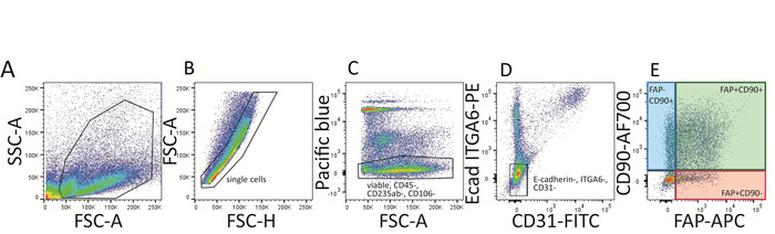
Figure 3: FACS gating strategy for human dermal fibroblast subpopulations. (A‒E) First, cells are gated on single (B) and viable (DAPI-) cells (C). Immune cells (CD45+), mesenchymal stem cells (CD106+), red blood cells (CD235ab+) are excluded (C) and Pacific blue negative cells are gated further on E-cadherin (Ecad) and ITGA6 double negative cells (PE channel, D). E-cadherin and ITGA6 are markers expressed on epidermal cells. Next, CD31-FITC positive cells (endothelial and lymphatic cells) are excluded (D) resulting in three fibroblast populations expressing either one or both of the two cell surface fibroblast markers FAP and CD90: FAP+CD90-, FAP+CD90+ and FAP-CD90+ (E). Please click here to view a larger version of this figure.

Figure 4: FAP+CD90-, FAP+CD90+ and FAP-CD90+ fibroblasts differ in dermal localization, gene expression and functionality. (A) Representative FACS plots of gated fibroblasts isolated from papillary, upper reticular and lower reticular dermis. Gating strategy is explained in Figure 3. FAP+CD90- fibroblasts are enriched in the papillary dermis (19.2%) compared to lower reticular dermis (0.83%). FAP+CD90+ cells can be found throughout the dermis but their highest abundance is in the upper reticular dermis (40.7%). FAP-CD90+ are enriched in the lower reticular dermis (64.4%). (B) Of note, all three sorted subpopulations display typical fibroblast morphology upon culture for 7 days (left). Interestingly, they behave differently in an adipogenesis assay. After 14 days in culture, FAP+CD90- do not differentiate into adipocytes while FAP+CD90+ and FAP-CD90+ readily undergo adipogenesis (right; Oil Red O stains lipid-bearing cells red). Scale bars = 1,000 µm. (C) Directly sorted FAP+CD90- fibroblasts express the papillary fibroblast markers CD26, NTN1 and PDPN, while FAP-CD90+ cells express CD36, ACTA2 and PPARγ, known to be expressed by the reticular lineage. * p ≤ 0.05; *** p ≤ 0.0005 compared to FAP+/CD90- cells; # p ≤ 0.05; ### p ≤ 0.0005 compared to FAP-/CD90+ cells. Please click here to view a larger version of this figure.
Discussion
Loading...
$$\rightleftharpoonup{xx}$$
$$\longleftharp{xx}$$,
$$\longrightharp{xx}$$,
In this article, we describe a method for the isolation of papillary and reticular fibroblasts from human skin. CD90 has been widely used for the identification or isolation of dermal fibroblasts18,20,21. However, we have demonstrated that besides CD90+ fibroblasts, human dermis also harbors a CD90- fibroblast population expressing FAP16, which has been established as a marker for activated fibroblasts and cancer-associated fibroblasts (CAFs)22,23,24,25. Importantly, we were able to identify three fibroblast subpopulations FAP+CD90-, FAP+CD90+ and FAP-CD90+ in skin biopsies from all healthy human donors. We therefore conclude that FAP is not only a marker for activated fibroblasts or CAFs but also normal tissue fibroblasts.
Of note, the FAP-CD90- cell population remaining after application of the above-described exclusion- and gating strategy does not contain fibroblasts, since these cells do not proliferate in fibroblast cultivation medium in vitro, but most likely a mixed cell population including lymphatic cells and pericytes amongst others16.
The cell yield obtained by use of the above-described protocol can vary depending on the body-part that the skin piece used for the isolation originates from. Dermis from different body parts differs regarding its structure, thickness as well as collagen composition. For example, skin from the face or the upper arm is much thinner than skin from the belly or the thigh, which also frequently display a thicker subcutaneous fat layer. Additionally, age and sex of the skin donors may further not only impact the tissue dissociation efficiency, but may also affect the distribution of the three fibroblast subpopulations (Figure 3) when isolated from full thickness skin. This results from the fact that the papillary dermis shrinks and that total fibroblast numbers decrease with age11,26,27,28. Furthermore, the cell pellet from papillary dermis will probably be larger than from reticular dermis, since the upper dermis is more densely populated by fibroblasts than the reticular dermis. Besides, the lower dermis is also tougher and more densely packed with collagen, making it harder to dissociate the tissue and to release the fibroblasts. Of note, the cell pellet may appear very red, which is why red blood cell lysis is recommended.
In addition to the identification of three subpopulations in intact human skin, we also show that in dermatomed skin, each fibroblast subset is enriched either in papillary or reticular dermis16. Precise slicing of the skin with the dermatome is critical to obtain a proper enrichment of each subpopulation from different dermal layers. Since the papillary dermis is very thin, the dermatomed slice representing it should not exceed a thickness of 300 µm. Upper reticular and lower reticular fibroblasts both represent the reticular lineage and display similar functions and gene signatures, therefore, one could also consider not separating them.
Importantly, all three fibroblasts populations are found throughout the dermis and are not exclusively present in one layer, which is why explant cultures from papillary or reticular dermis result in mixed fibroblast cultures. However, FAP+CD90- papillary fibroblast are most abundant in the papillary dermis and follow a gradient from superficial to lower dermal layers while FAP+CD90+ and FAP-CD90+ fibroblasts follow an inverse gradient from the lower to the superficial layers16. Furthermore, the majority of CD90+ fibroblasts of the papillary dermis are almost exclusively found surrounding blood vessels and express the perivascular fibroblast marker CD14629, and thus probably exhibit different functions than the remaining CD146- reticular fibroblasts16. CD146 could be used as an additional marker in the gating strategy to exclude this population.
Following the dissociation of dermal layers, the isolated cells are stained with a specially designed antibody cocktail containing various antibodies for the exclusion of immune cells, endothelial and lymphatic cells, epidermal cells, erythrocytes and MSCs to obtain pure fibroblast populations. Of note, choosing a marker for the identification and exclusion of MSCs may be tricky because of the high number of published MSC markers30,31. Since MSCs express CD90 like fibroblasts, additional MSC markers such as CD105 or CD271 could prove useful for their identification. However, MSCs only represent a very low percentage of all dermal cells and since CD90+ fibroblasts display typical morphological features of fibroblasts upon sorting, one could argue that the exclusion of MSCs by use of distinct cell surface markers might be unnecessary.
Importantly, we analyzed FAP and CD90 gene expression after keeping the cells in culture for 7-14 days after sorting (data not shown) and found that expression of both markers is upregulated in the respective sorted single positive (FAP+CD90- or FAP-CD90+) cells16. We therefore emphasize that the above-described marker sets and protocol permit the isolation of primary fibroblast subsets directly from the tissue but not from previously cultured mixed fibroblast populations.
Nevertheless, we demonstrate that the functionality of all three subpopulations is retained in cell culture regardless of the alteration of cell surface marker expression, since fibroblasts sorted as FAP+CD90- papillary fibroblasts do not acquire the ability to undergo adipogenesis after a longer period of culture, while fibroblasts sorted as FAP+CD90+ or FAP-CD90+ reticular fibroblasts maintain their ability to differentiate into adipocytes 16. Importantly, we also found that papillary and reticular-specific genes are still expressed to a higher extent in FAP+CD90- and CD90+ respectively.
In conclusion, we have established a protocol for the isolation of functionally distinct fibroblast subsets via FACS that for the first time permits the isolation and analysis of pure and naïve fibroblast subpopulations from human skin dermis. This method establishes a major advancement to the commonly used fibroblast explant culture isolation protocol from upper and lower dermis as (i) an opposing gradient of papillary and reticular fibroblasts exists from the skin surface to the hypodermis and (ii) fibroblasts change their gene signature in vitro.
Disclosures
Loading...
$$\rightleftharpoonup{xx}$$
$$\longleftharp{xx}$$,
$$\longrightharp{xx}$$,
The authors have nothing to disclose.
Acknowledgements
Loading...
$$\rightleftharpoonup{xx}$$
$$\longleftharp{xx}$$,
$$\longrightharp{xx}$$,
We gratefully acknowledge assistance in FACS-sorting by Bärbel Reininger and Wolfgang Bauer. This work was supported by grants to B. M. L. from the Austrian Science Fund (FWF: V 525-B28), and the Federation of European Biochemical Societies (FEBS, Follow-Up Research Fund). S. F. is recipient of a DOC Fellowship of the Austrian Academy of Sciences (OeAW). We gratefully acknowledge excellent support from the core facilities at the Medical University of Vienna. We thank Bernhard Gesslbauer, Christine Radtke and Martin Vierhapper for providing human skin material.
Materials
| Name | Company | Catalog Number | Comments |
|---|---|---|---|
| Material: | |||
| 3-isobutyl-1-methylxanthine | Sigma | I5879 | |
| Ammonium chloride | Sigma | 9718 | used for selfmade ACK Lysis Buffer |
| Amniomax-C100 (1x) Basal Medium | Gibco | 17001-074 | used for fibroblast growth medium |
| β-Mercaptoethanol | Scharlau | ME0095 | CAUTION |
| C100 Supplement | Thermo Scientific | 12556015 | used for fibroblast growth medium |
| Collagenase I | Thermo Scientific | 17100017 | CAUTION |
| Collagenase II | Gibco | 17101015 | CAUTION |
| Collagenase IV | Sigma | C5138 | CAUTION |
| DAPI | Thermo Scientific | 62248 | |
| Dexamethasone | Sigma | D8893 | |
| Dispase II | Roche | 4942078001 | CAUTION |
| Dulbecco‘s Modified Eagle Medium + GlutaMAX | Gibco | 31966-021 | |
| EDTA disodium salt | Sigma | E1644 | used for selfmade ACK Lysis Buffer |
| Fetal bovine serum (heat inactivated) | Gibco | 10500-064 | |
| Hyaluronidase | Sigma | H4272 | CAUTION |
| Insulin | Sigma | I5500 | |
| Isopropanol | Merck | 1,096,341,011 | |
| OilRed O | Sigma | O-0625 | |
| Paraformaldehyd | Sigma | 158127 | CAUTION Cancerogenic. Skin and eye irritant. Handle with care. |
| Penicillin/Streptomycin | Gibco | 15070-063 | |
| Phosphate-buffered saline without Ca2+ & Mg2+ | Lonza | BE17-512F | |
| Potassium bicarbonate | Sigma | 237205 | used for selfmade ACK Lysis Buffer |
| PureLink RNA MicroKit | Invitrogen | 12183-016 | |
| SuperScript III First-Strand Synthesis SuperMix for qRT-PCR | Invitrogen | 11752 | |
| Taqman 2x Universal PCR Master Mix | Applied Biosystems | 4324018 | CAUTION Skin and eye irritant. |
| Troglitazone | Sigma | T2573 | |
| Name | Company | Catalog Number | Comments |
| Flow cytometry Antibodies: | |||
| anti human CD31-AF488 | Biolegend | 303109 | Dilution: 1:30, Lot: B213986, RRID: AB_493075 |
| anti human CD45-Pacific blue | Biolegend | 304029 | Dilution: 1:20, Lot: B218608, RRID: AB_2174123 |
| anit human CD49f/ITGA6-PE | Serotec | MCA699PE | Dilution: 1:20, Lot: 0109, RRID: AB_566833 |
| anti human CD90/Thy-1-AF700 | Biolegend | 328120 | Dilution: 1:30, Lot: B217250, RRID: AB_2203302 |
| anti human CD106-AF421 | BD | 744309 | Dilution: 1:100, Lot: 7170537, RRID: x |
| anti human CD235ab-Pacific blue | Biolegend | 306611 | Dilution: 1:1000, Lot: B224563, RRID: AB_2248153 |
| anti-human E-cadherin-PE | Biolegend | 147304 | Dilution: 1:20, Lot: B197481, RRID: AB_2563040 |
| anti human FAP-APC | R&D | FAB3715A | Dilution: 1:20, Lot: AEHI0117111, RRID: x |
| Human TruStain FcX | Biolegend | 422302 | Dilution: 1:20, Lot: B235080, RRID: x |
| Name | Company | Catalog Number | Comments |
| Equipment: | |||
| 1.4 mL micronic tubes | Thermo Scientific | 4140 | |
| 1.5 mL screw cap micro tube | Sarstedt | 72,692,405 | |
| 5 mL Polystyrene Round Bottom Tube with cell strainer cap | Falcon | 352235 | |
| 48-well cell culture cluster | Costar | 3548 | |
| 50 mL polypropylene canonical tubes | Falcon | 352070 | |
| 70 µm cell strainer nylon | Falcon | 352350 | |
| Aesculap Acculan 3Ti Dermatome | VWR | AESCGA670 | |
| Aesculap Dermatome blades | VWR | AESCGB228R | |
| MicroAmp Fast Optical 96well | Applied Biosystems | 4346906 | |
| Primary cell culture dish | Corning | 353803 | |
| Scalpel blades | F.S.T | 10022-00 | |
| Tea strainer | x | x | |
| Name | Company | Catalog Number | Comments |
| Fibroblast growth medium: | |||
| AmnioMAX basal medium with AmnioMAX C-100 Supplement, 10% FCS and 1% P/S |
References
Loading...
$$\rightleftharpoonup{xx}$$
$$\longleftharp{xx}$$,
$$\longrightharp{xx}$$,
- Sorrell, J. M., Caplan, A. I. Fibroblast heterogeneity: more than skin deep. Journal of Cell Science. 117 (Pt 5), 667-675 (2004).
- Lowry, W. E., et al. Generation of human induced pluripotent stem cells from dermal fibroblasts. Proceedings of the National Academy of Sciences of the United States of America. 105 (8), 2883-2888 (2008).
- Kisseleva, T. The origin of fibrogenic myofibroblasts in fibrotic liver. Hepatology. 65 (3), 1039-1043 (2017).
- LeBleu, V. S., et al. Origin and function of myofibroblasts in kidney fibrosis. Nature Medicine. 19 (8), 1047-1053 (2013).
- Travers, J. G., Kamal, F. A., Robbins, J., Yutzey, K. E., Blaxall, B. C. Cardiac Fibrosis: The Fibroblast Awakens. Circulation Research. 118 (6), 1021-1040 (2016).
- Kalluri, R., Zeisberg, M. Fibroblasts in cancer. Nat Rev Cancer. 6 (5), 392-401 (2006).
- Rinkevich, Y., et al. Skin fibrosis. Identification and isolation of a dermal lineage with intrinsic fibrogenic potential. Science. 348 (6232), (2015).
- Abhishek, S., Palamadai Krishnan, S. Epidermal Differentiation Complex: A Review on Its Epigenetic Regulation and Potential Drug Targets. Cell Journal. 18 (1), 1-6 (2016).
- Driskell, R. R., et al. Distinct fibroblast lineages determine dermal architecture in skin development and repair. Nature. 504 (7479), 277-281 (2013).
- Mastrogiannaki, M., et al. beta-Catenin Stabilization in Skin Fibroblasts Causes Fibrotic Lesions by Preventing Adipocyte Differentiation of the Reticular Dermis. Journal of Investigative Dermatology. 136 (6), 1130-1142 (2016).
- Lichtenberger, B. M., Mastrogiannaki, M., Watt, F. M. Epidermal beta-catenin activation remodels the dermis via paracrine signalling to distinct fibroblast lineages. Nature Communications. 7, 10537(2016).
- Harper, R. A., Grove, G. Human skin fibroblasts derived from papillary and reticular dermis: differences in growth potential in vitro. Science. 204 (4392), 526-527 (1979).
- Hiraoka, C., et al. Two clonal types of human skin fibroblasts with different potentials for proliferation and tissue remodeling ability. Journal of Dermatological Science. 82 (2), 84-94 (2016).
- Higgins, C. A., et al. Multifaceted role of hair follicle dermal cells in bioengineered skins. Br Journal of Dermatology. 176 (5), 1259-1269 (2017).
- Korosec, A., Lichtenberger, B. M. Ch. 12. Skin Tissue Models. Marques, A. P., Pirraco, R. P., Cerqueira, M., Reis, R. L. , Elsevier. https://doi.org/10.1016/B978-0-12-810545-0.00012-7 279-301 (2017).
- Korosec, A., et al. Lineage identity and location within the dermis determine the function of papillary and reticular fibroblasts in human skin. Journal of Investigative Dermatology. , (2018).
- Higgins, C. A., Chen, J. C., Cerise, J. E., Jahoda, C. A., Christiano, A. M. Microenvironmental reprogramming by three-dimensional culture enables dermal papilla cells to induce de novo human hair-follicle growth. Proceedings of the National Academy of Sciences of the United States of America. 110 (49), 19679-19688 (2013).
- Philippeos, C., et al. Spatial and Single-Cell Transcriptional Profiling Identifies Functionally Distinct Human Dermal Fibroblast Subpopulations. Journal of Investigative Dermatology. 138 (4), 811-825 (2018).
- Walmsley, G. G., et al. Live fibroblast harvest reveals surface marker shift in vitro. Tissue Engineering Part C: Methods. 21 (3), 314-321 (2015).
- Jiang, D., Rinkevich, Y. Defining Skin Fibroblastic Cell Types Beyond CD90. Frontiers in Cell and Developmental Biology. 6, 133(2018).
- Sorrell, J. M., Caplan, A. I. Fibroblasts-a diverse population at the center of it all. International Review of Cell and Molecular Biology. , 161-214 (2009).
- Huang, Y., et al. Isolation of Fibroblast-Activation Protein-Specific Cancer-Associated Fibroblasts. BioMed Research International. 2017, 4825108(2017).
- Jiang, G. M., et al. The application of the fibroblast activation protein alpha-targeted immunotherapy strategy. Oncotarget. 7 (22), 33472-33482 (2016).
- Pure, E., Blomberg, R. Pro-tumorigenic roles of fibroblast activation protein in cancer: back to the basics. Oncogene. 37 (32), 4343-4357 (2018).
- Hamson, E. J., Keane, F. M., Tholen, S., Schilling, O., Gorrell, M. D. Understanding fibroblast activation protein (FAP): substrates, activities, expression and targeting for cancer therapy. PROTEOMICS – Clinical Applications. 8 (5-6), 454-463 (2014).
- Gilchrest, B. A. A review of skin ageing and its medical therapy. Br Journal of Dermatology. 135 (6), 867-875 (1996).
- Gilchrest, B. A., Krutmann, J. Skin Aging. , Springer. 198(2006).
- Makrantonaki, E., Zouboulis, C. C. Molecular mechanisms of skin aging: state of the art. Annals of the New York Academy of Sciences. 1119, 40-50 (2007).
- Crisan, M., et al. A perivascular origin for mesenchymal stem cells in multiple human organs. Cell Stem Cell. 3 (3), 301-313 (2008).
- Kundrotas, G. Surface markers distinguishing mesenchymal stem cells from fibroblasts. Acta Medica Lituanica. 19 (2), 75-79 (2012).
- Lupatov, A. Y., Vdovin, A. S., Vakhrushev, I. V., Poltavtseva, R. A., Yarygin, K. N. Comparative analysis of the expression of surface markers on fibroblasts and fibroblast-like cells isolated from different human tissues. Bulletin of Experimental Biology and Medicine. 158 (4), 537-543 (2015).
Reprints and Permissions
Request permission to reuse the text or figures of this JoVE article
Request Permission