The preparation of SNS pincer cobalt(II) model complexes of liver alcohol dehydrogenase is presented here. The complexes can be prepared by reacting the ligand precursor with CoCl2·6H2O and can then be recrystallized by allowing diethyl ether to slowly diffuse into an acetonitrile solution that contains the cobalt complex.
Method Article
Preparation of SNS Cobalt(II) Pincer Model Complexes of Liver Alcohol Dehydrogenase
In This Article
Summary
Abstract
Chemical model complexes are prepared to represent the active site of an enzyme. In this protocol, a family of tridentate pincer ligand precursors (each possessing two sulfur and one nitrogen donor atom functionalities (SNS) and based on bis-imidazole or bis-triazole compounds) are metallated with CoCl2·6H2O to afford tridentate SNS pincer cobalt(II) complexes. Preparation of the cobalt(II) model complexes for liver alcohol dehydrogenase is facile. Based on a quick color change upon adding the CoCl2·6H2O to acetonitrile solution that contains the ligand precursor, the complex forms rapidly. Formation of the metal complex is complete after allowing the solution to reflux overnight. These cobalt(II) complexes serve as models for the zinc active site in liver alcohol dehydrogenase (LADH). The complexes are characterized using single crystal X-ray diffraction, electrospray mass spectrometry, ultra-violet visible spectroscopy, and elemental analysis. To accurately determine the structure of the complex, its single crystal structure must be determined. Single crystals of the complexes that are suitable for X-ray diffraction are then grown via slow vapor diffusion of diethyl ether into an acetonitrile solution that contains the cobalt(II) complex. For high quality crystals, recrystallization typically takes place over a 1 week period, or longer. The method can be applied to the preparation of other model coordination complexes and can be used in undergraduate teaching laboratories. Finally, it is believed that others may find this recrystallization method to obtain single crystals beneficial to their research.
Introduction
The purpose of the presented method is to prepare small-molecule analogs of LADH to further understand the catalytic activity of metalloenzymes. LADH is a dimeric enzyme that contains a cofactor-binding domain and zinc(II) metal-containing catalytic domain1. LADH, in the presence of co-factor NADH, can reduce ketones and aldehydes to their respective alcohol derivatives2. In the presence of NAD+, LADH can perform reverse catalysis of oxidation of alcohols to ketones and aldehydes2. The crystal structure of LADH’s active site shows that its zinc(II) metal center is bound to one nitrogen atom, provided by a histidine side chain and two sulfur atoms and offered by two cysteine ligands3. Further research has shown that the zinc metal center is ligated with a labile water molecule, resulting in pseudo-tetrahedral geometry around the metal center4.
We have previously reported and utilized SNS pincer ligand precursors as well as metallated the ligand precursors with ZnCl2 to form Zn(II) complexes that contain the tridentate ligand precursor5,6,7. These ligand precursors are shown in Figure 1. These zinc(II) complexes exhibited activity for the stoichiometric reduction of electron-poor aldehydes and are thus model complexes for LADH. Subsequently, the synthesis and characterization of a series of copper(I) and copper(II) complexes that contain SNS ligand precursors have been reported8,9,10.
Although LADH is a zinc(II) enzyme, we are interested in preparing cobalt(II) model complexes of LADH in order to obtain more spectroscopic information about the cobalt(II) analogs of LADH. The cobalt(II) complexes are colored, whereas the zinc(II) complexes are off-white. Since the cobalt(II) complexes are colored, ultraviolet visible spectra of the complexes can be obtained, in which information about the strength of the ligand field in cobalt(II) complexes can also be gathered. By using information from Gaussian calculations and the experimentally obtained ultra-violet visible spectra, information about the strength of the ligand field can be deduced. Cobalt(II) is a good substitute for zinc(II), since both ions have similar ionic radii and similar Lewis acidities11,12.
The presented method involves synthesizing and characterizing model complexes to attempt to mimic the natural catalytic behavior of LADH5,6. We have previously metallated a family of ligand precursors with ZnCl2 to form zinc(II) model complexes of LADH, which modeled the structure and reactivity of the zinc active site in LADH4. Through multiple experiments, these pincer ligands have proven to be robust under different environmental conditions and have remained stable with a diverse collection of attached R-groups.5,6
Tridentate ligands are preferable compared to monodentate ligands, because they have been found to be more successful with metalation due to the strong chelate effects of tridentate ligands. This observation is due to a more favored entropy of tridentate pincer ligand formation in comparison to a monodentate ligand13. Furthermore, tridentate pincer ligands are likely to prevent dimerization of the metal complexes, which is favored because dimerization is likely to slow catalytic activity of a complex14. Thus, using tridentate pincer ligands has been proven successful in organometallic chemistry in the preparation of catalytic active and robust complexes. SNS pincer complexes have been less studied than other pincer systems, as pincer complexes usually contain second and third row transition metals15.
This research on metalloenzymes can help further the understanding of their enzymatic activity, which can be applied to other areas in biology. This method of synthesizing model complexes compared to the alternative method (synthesizing the entire protein of LADH) is favorable for a number of reasons. The first advantage is that model complexes are low in molecular mass and are still capable of accurately representing catalytic activity and environmental conditions of the natural enzyme’s active site. Second, model complexes are simpler to work with and produce reliable and relatable data.
This manuscript describes the synthetic preparation and characterization of two cobalt(II) pincer model complexes of LADH. Both complexes feature a pincer ligand that contains sulfur, nitrogen, and sulfur donor atoms. The first complex (4) is based on an imidazole precursor, and the second (5) is based on a triazole precursor. The complexes show reactivity for the stoichiometry reduction of electron poor aldehydes in the presence of a hydrogen donor. These reactivity results will be reported in a subsequent manuscript.
Protocol
1. Synthesis of chloro-(n3-S,S,N)-[2,6-bis(N-isopropyl-N’-methyleneimidazole-2-thione)pyridine]cobalt(II)tetrachlorocobaltate [4]
- To prepare complex 4, add 0.121 g (3.12 x 10⁻⁴ mol) of 2,6-bis(N-isopropyl-N’-methyleneimidazole-2-thione)pyridine (C19H25N5S2)6 to 15 mL of acetonitrile in a 100 mL round bottom flask. Next, to this solution, add 0.0851 g (3.58 x 10⁻⁴ mol) of cobalt chloride(II) hexahydrate (CoCl2·6H2O). The reaction solution should change color from light yellow to emerald green immediately after the cobalt(II) chloride hexahydrate is added.
- Add a stir bar to the flask. Reflux and stir the reaction for 20 h to ensure complete reaction. Remove the solvent using a rotovap under reduced pressure.
2. Recrystallization of chloro-(n3-S,S,N)-[2,6-bis(N-isopropyl-N’-methyleneimidazole-2-thione)pyridine]cobalt(II)tetrachlorocobaltate [4] by slow vapor diffusion
- Dissolve the solute in acetonitrile (7.5 mL), filter the solution, and place the solution evenly in 1 dram vials. Fill each vial with 1.5 mL of acetonitrile solution.
- Add cotton to cap the vials, which allows for slow vapor diffusion. Fit the cotton snugly in the opening at the top of the vial.
- Place the vials in a 240 mL jar containing 50 mL of diethyl ether. Close the jar with a cap.
- Allow the crystals to grow over a period of 1 week.
NOTE: Recrystallization may take longer than 1 day.
3. Synthesis of chloro-(n3-S,S,N)-[2,6-bis(N-isopropyl-N’-methylenetriazole-2-thione)pyridine]cobalt(II)tetrachlorocobaltate [5]
- To prepare complex 5, add 0.183 g (4.70 x 10⁻⁴ mol) of 2,6-bis(N-isopropyl-N’-methylenetriazole-2-thione)pyridine (C17H23N7S2)6 to 15 mL of acetonitrile in a 100 mL round bottom flask. To this solution, add 0.223 g (9.37 x 10⁻⁴ mol) of cobalt chloride hexahydrate (CoCl2·6H2O). The reaction solution should change color from light yellow to royal blue immediately after the cobalt(II) chloride hexahydrate is added.
- Add a stir bar to the flask. Reflux and stir the reaction for 20 h to ensure complete reaction. Remove the solvent using a rotovap under reduced pressure.
4. Recrystallization of chloro-(n3-S,S,N)-[2,6-bis(N-isopropyl-N’-methylenetriazole-2-thione)pyridine]cobalt(II)tetrachlorocobaltate [5] by slow vapor diffusion
- Dissolve the solute in acetonitrile (9.0 mL), filter the solution, and place the solution evenly in 1 dram vials. Fill each vial with 1.5 mL of acetonitrile solution.
- Add cotton to cap the vials, which allows for slow vapor diffusion. Fit the cotton snugly in the opening at the top of the vial.
- Place the vials in a jar containing 50 mL of diethyl ether. Close the vial with a cap.
- Allow the crystals to grow over a period of 1 week.
NOTE: Recrystallization may take longer than 1 day.
5. X-ray crystallography
- Mount a crystal of 4 on a nylon loop. Collect the data on a Rigaku Oxford Diffraction diffractometer. Here, X-ray diffraction data is collected at 173(2) K. Solve the crystal structure using Olex216 and ShelXT17 structure solution programs using direct methods. Refine the structure with the ShelXL18 refinement package using least squares minimization.
- Mount a crystal of 5 on a nylon loop. Collect the X-ray diffraction data on a Rigaku Oxford Diffraction diffractometer. Here, X-ray diffraction data is collected at 173(2) K. Solve the crystal structure using Olex216 and ShelXT17 structure solution programs using direct methods. Refine the structure with the ShelXL18 refinement package using least squares minimization.
Results
Synthesis
The syntheses of complexes 4 and 5 were successfully carried out by reacting an acetonitrile solution containing a bis-thione ligand precursor with cobalt (II) chloride hexahydrate (Figure 2). This reaction occurred at a reflux temperature in the presence of air. In general, complexes 4 and 5 were observed to be soluble in acetonitrile, dimethyl sulfoxide, dichloromethane, and methanol. Complex 4 was green in color and complex 5 was blue in color. The percent yield for complexes 4 and 5 was quantitative.
X-ray crystallography
Single crystals of complexes 4 and 5 were obtained via a slow vapor diffusion method, in which the compounds were dissolved in acetonitrile, and diethyl ether vapor was allowed to slowly diffuse into each solution. This recrystallization method is an excellent way to grow single crystals for hard-to-crystallize samples. Table 1 shows refinement data for the two complexes, and the single crystal structures are shown in Figure 3 and Figure 4. Based on the single crystal structures, each unit cell contains two cobalt(II) SNS pincer cations and one [CoCl4]2- counter-anion. The oxidation state of the cobalt ion in the cation and anion is Co2+. The crystal structures of complexes 4 and 5 have been deposited in the Cambridge Structural Database (deposition numbers 1946448 and 1946449).
Both complexes display pseudo-tetrahedral geometry about the cobalt(II) metal center with one nitrogen and two sulfur donor atoms coordinated to the metal center. Furthermore, both complexes feature a tetrachloride counter-anion. The Co-N and Co-S bond lengths for complexes 4 and 5 are nearly identical in value. The Co-N bond length is 2.084(3) Å in 4 and 2.0763(16) Å in 5. The Co-S bond lengths in 4 are 2.2927(12) Å and 2.3386(11) Å. Similarly, the Co-S bond lengths in 5 are 2.3180(6) Å and 2.3227(6) Å. For complexes 4 and 5, the bond lengths are similar to those previously reported19. The Co-Cl bond lengths are 2.2256(13) Å in 4 and 2.2116(6) Å in 5.
The carbon-sulfur bond lengths of 1.710(4) Å and 1.714(4) Å in 4 and 1.693(2) Å and 1.698(2) Å in 5 are similar for the two complexes and between what is typically observed for C-S single bonds (1.83 Å) and C=S double bonds (1.61 Å)20.
As previously mentioned, complexes 4 and 5 both contain a tetrachloride counter-anion. The counter-anion Co-Cl bond lengths for 4 are 2.2709(12) Å, 2.2709(12) Å, 2.2949(11) Å and 2.2950(11) Å. These are comparable to those of complex 5, which are 2.2737(6) Å, 2.2737(6) Å, 2.2956(6) Å, and 2.2956(6) Å. The Co-N and Co-S bond lengths in 4 and 5 are in good agreement with the Co-N(histidine) and Co-S(cysteine) bond lengths in the cobalt(II)-substituted analog of liver alcohol dehydrogenase. In this enzyme, the cobalt-N(histidine) bond length is 2.04 Å, and the cobalt-S(cysteine) bond lengths are 2.29 Å and 2.33 Å.21
In complex 4, the N-Co-S bond angles are 108.77(10)° and 114.03(10)°, whereas in complex 5 they are 112.58(5)° and 114.15(5)°. The N-Co-S bond angles are close to each other, and any differences may be due to the varying electronics of the two complexes. The N-Co-Cl bond angles in 4 and 5 are 107.91(10)° and 107.59(5)°, respectively. The S-Co-S angle was measured as 99.79(5)° for 4 and 102.78(3)° for 5. Lastly, the S-Co-Cl bond angles for 4 are 117.98(5)° and 108.43(5)° and for 5 are 111.76(3)° and 107.93(3)°.
The tau-4 parameter was also determined for complexes 4 and 5. The tau-4 parameter for complex 4 is 0.907, and the tau-4 parameter for complex 5 is 0.94522. Both tau-4 parameters are more consistent with tetrahedral geometry about the cobalt center than square planar geometry. The tau-4 parameter for a tetrahedral complex is equal to one, and the tau-4 parameter for a square planar complex is equal to zero.
Elemental analysis
To study the bulk purity of 4 and 5, the recrystallized complexes underwent elemental analyses. The results are summarized in Table 2. The data here suggests that complexes 4 and 5 are pure, because the calculated percentages of carbon, hydrogen, and nitrogen are in excellent agreement with the found percentages of carbon, hydrogen, and nitrogen.
Electrospray mass spectrometry
The preparation of complexes 4 and 5 was also confirmed using electrospray mass spectrometry. The electrospray mass spectra were collected using a direct flow injection. The injection volume was 5 µL. The data was collected on an Agilent QTOF instrument in positive and negative ion modes. The optimized conditions were as follows: capillary = 3000 kV, cone = 10 V, source temperature = 120 °C. For complex 4, in positive ion mode, the molecular ion was observed at m/z = 481.0631. In negative ion mode, the [CoCl3]- ion was observed at m/z 163.8433. For complex 5, in positive ion mode, the molecular ion was observed at m/z 483.0503. In negative ion mode, the [CoCl3]- ion was observed at m/z 163.8413.
Ultra-violet visible spectroscopy
Complexes 4 and 5 were analyzed using ultraviolet visible spectroscopy to gain further insight on the electronic environment of the complexes. Complexes 4 and 5 were dissolved in acetonitrile to form separate solutions. Complex 4 was 1.0 x 10⁻⁴ M in concentration and complex 5 was 9.2 x 10⁻⁴ M in concentration. Complex 4 exhibited three peaks in the visible region at 680 nm (ε = 1300 M-1cm-1), 632 nm (ε = 1100 M-1cm-1), and 589 nm (ε = 1200 M-1cm-1). Complex 5 exhibited four peaks in the visible region at 682 nm (ε = 1300 M-1cm-1), 613 nm (ε = 850 M-1cm-1), 588 nm (ε = 790 M-1cm-1), and 573 nm (ε = 820 M-1cm-1).
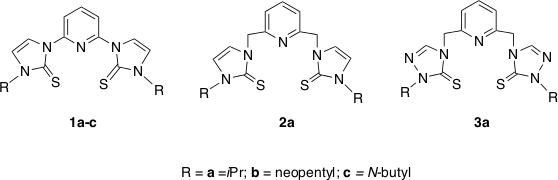
Figure 1: SNS pincer ligand precursors previously utilized. Ligand precursors based on bis-imidazole and bis-triazole moieties. (A) R = iPr, (B) R= neopentyl, (C) R = N-butyl. Please click here to view a larger version of this figure.
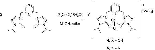
Figure 2: Synthesis of complexes 4 and 5. Synthetic scheme to prepare complexes 4 and 5. Please click here to view a larger version of this figure.
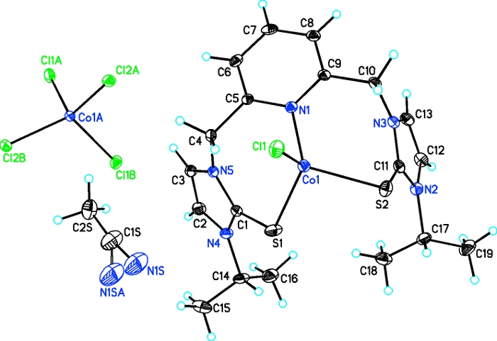
Figure 3: Solid-state structure of complex 4. Solid-state single crystal structure of complex 4. Please click here to view a larger version of this figure.
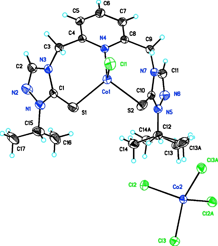
Figure 4: Solid-state structure of complex 5. Solid-state single crystal structure of complex 5. Please click here to view a larger version of this figure.
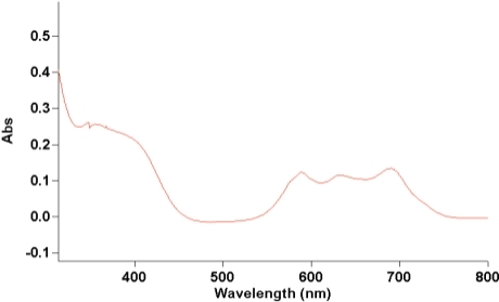
Figure 5: Ultra-violet visible spectrum of complex 4. Ultra-violet visible spectrum of complex 4 (1.0 x 10⁻⁴ M) in acetonitrile. Please click here to view a larger version of this figure.
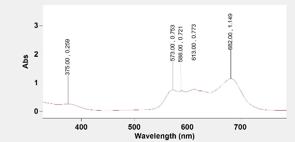
Figure 6: Ultra-violet visible spectrum of complex 5. Ultra-violet visible spectrum of complex 5 (9.15 x 10⁻⁴ M) in acetonitrile. Please click here to view a larger version of this figure.
| 4 | 5 | |
| α/° | 90 | 90 |
| β/° | 97.2252(19) | 90.770(2) |
| γ/° | 90 | 90 |
| Volume/Å3 | 5462.6(2) | 4852.0(2) |
| Z | 4 | 4 |
| ρcalcg/cm3 | 1.516 | 1.6 |
| μ/mm 1 | 11.526 | 1.56 |
| F(000) | 2556 | 2380 |
| Crystal size/mm3 | 0.24 × 0.22 × 0.06 | 0.28 × 0.08 × 0.06 |
| Radiation | CuKα (λ = 1.54184) | MoKα (λ = 0.71073) |
| 2Θ range for data collection/° | 7.39 to 142.76 | 6.596 to 65.254 |
| Index ranges | -26 ≤ h ≤ 29, -8 ≤ k ≤ 8, -39 ≤ l ≤ 31 | -27 ≤ h ≤ 28, -17 ≤ k ≤ 13, -33 ≤ l ≤ 32 |
| Reflections collected | 10233 | 21514 |
| Independent reflections | 5235 [Rint = 0.0565, Rsigma = 0.0739] | 8079 [Rint = 0.0262, Rsigma = 0.0315] |
| Data/restraints/parameters | 5235/0/312 | 8079/0/289 |
| Goodness-of-fit on F2 | 0.978 | 1.035 |
| Final R indexes [I>=2σ (I)] | R1 = 0.0529, wR2 = 0.1246 | R1 = 0.0398, wR2 = 0.0845 |
| Final R indexes [all data] | R1 = 0.0758, wR2 = 0.1361 | R1 = 0.0610, wR2 = 0.0964 |
| Largest diff. peak/hole / e Å-3 | 0.99/-0.55 | 0.59/-0.46 |
Table 1: Tabulated refinement data for complexes 4 and 5. X-ray refinement and collection data for complexes 4 and 5.
| Complex | Calc. % C | Found % C | Calc. % H | Found % H | Calc. % N | Found % N |
| 4, [C38H50Cl2Co2N10S4][CoCl4]•2[CH3CN] | 40.46 | 40.26 | 4.53 | 4.39 | 13.48 | 13.17 |
| 5, [C34H46Cl2Co2N14S4][CoCl4]•[CH3CN] | 35.75 | 36.20 | 4.08 | 4.20 | 17.37 | 17.40 |
Table 2: Elemental analysis results for complexes 4 and 5. Elemental analyses results for percent carbon, hydrogen, and nitrogen for complexes 4 and 5.
Discussion
The preparation of complexes 4 and 5 is facile. The key step is to add the solid CoCl2·6H2O to an acetonitrile solution that contains the respective ligand precursor. The solution turns dark green within seconds after the addition of CoCl2·6H2O to form complex 4. The solution turns bright blue after the addition of CoCl2·6H2O to form complex 5. To ensure complete reaction, the solution is placed on reflux overnight.
To grow single crystals of complexes 4 and 5, the acetonitrile solution that contains complexes 4 or 5 needs to be concentrated. The complexes must be dissolved in a minimal amount of acetonitrile to produce the solutions that contain the complex as concentrated as possible. Single crystals of 4 and 5 are grown by adding acetonitrile solution that contain complex 4 or 5 to 1 dram vials. These 1 dram vials that contain a solution of complex 4 or 5 are placed in a closed jar that contained diethyl ether. To slow the rate at which diethyl ether diffuses into the acetonitrile solution, a cotton ball is added to each 1 dram vial. The cotton ball must be very snug to slow the rate of diffusion. The use of cotton to slow the diffusion of diethyl ether can be utilized by others to grow single crystals for tough samples.
If the concentration of the metal complex in acetonitrile for the recrystallization is not strong enough, single crystals will not form. The product after the recrystallization attempt may be an oily residue. Researchers need to ensure that the metal complex has a high enough concentration for single crystals to form.
To the best of our knowledge, no other cobalt(II) substituted model complexes of liver alcohol dehydrogenase have been published in the literature. Future work will focus on comparing experimentally obtained UV-visible spectra to the spectra predicted by Gaussian calculations to determine the ligand field strength of pincer ligands. Current work in the Miecznikowski laboratory is focusing on preparing cobalt substituted model complexes of liver alcohol dehydrogenase that do not contain [CoCl4]2- as the counter-anion. These complexes are currently being screened for the reduction of electron poor aldehydes and ketones.
Disclosures
The authors have nothing to disclose.
Acknowledgements
John Miecznikowski received financial support from the following for this project: the Connecticut NASA Space Grant Alliance (Award Number P-1168), the Fairfield University Science Institute, College of Arts and Sciences Publication Fund, Fairfield University Faculty Summer Research Stipend, and National Science Foundation-Major Research Instrumentation Program (Grant Number CHE-1827854) for funds to acquire a 400 MHz NMR spectrometer. He also thanks Terence Wu (Yale University) for assistance in acquiring electrospray mass spectra. Jerry Jasinski acknowledges the National Science Foundation-Major Research Instrumentation Program (Grant Number CHE-1039027) for funds to purchase an X-ray diffractometer. Sheila Bonitatibus, Emilse Almanza, Rami Kharbouch, and Samantha Zygmont acknowledge the Hardiman Scholars Program for providing their summer research stipend.
Materials
| Name | Company | Catalog Number | Comments |
|---|---|---|---|
| 100 mL Round Bottomed Flask | Chem Glass | CG150691 | 100mL Single Neck Round Bottomed Flask, 19/22 Outer Joint |
| Acetonitrile | Fisher | HB9823-4 | HPLC Grade |
| Chiller for roto-vap | Lauda | L000638 | Alpha RA 8 |
| Cobalt Chloride hexahydrate | Acros Organics | AC423571000 | Acros Organics |
| Diethyl Ether | Fisher | E-138-1 | Diethyl Ether Anhydorus |
| graduated cylinder | Fisher | S63456 | 25 mL graduated cylinder |
| hotplate | Fisher | 11-100-49SH | Isotemp Basic Stirring Hotplate |
| jars | Fisher | 05-719-481 | 250 mL jars |
| Ligand | ----- | ----- | Synthezied previously by Professor Miecznikowski |
| medium cotton balls | Fisher | 22-456-80 | medium cotton balls |
| one dram vials | Fisher | 03-339 | one dram vials with TFE Lined Cap |
| pipet | Fisher | 13-678-20B | 5.75 inch pipets |
| pipet bulbs | Fisher | 03-448-21 | Fisher Brand Latex Bulb for pipet |
| recrystallizing dish for sand bath | Fisher | 08-741 D | 325 mL recrystallizing dish for sand bath |
| reflux condensor | Chem Glass | CG-1218-A-22 | Condenser with 19/22 inner joint |
| Rotovap | Heidolph Collegiate | 36000090 | Brinkmann; Heidolph Collegiate Rotary Evaporator with Heidolph WB eco bath Heidolph Rotary Evaporator |
| sea sand for sandbath | Acros Organics | 612355000 | washed sea sand for sand bath |
| Stir bar | Fisher | 07-910-23 | Egg-Shaped Magnetic Stir Bar |
| Vacum grease | Fisher | 14-635-5D | Dow Corning High Vacuum Grease |
| vacuum pump for rotovap | Heidolph Collegiate | 36302830 | Heidolph Rotovac Valve Control |
References
- Holm, R. H., Kennepohl, P., Solomon, E. I. Structural and Functional Aspects of Metal Sites in Biology. Chemical Reviews. 96 (7), 2239-2314 (1996).
- Ibers, J. A., Holm, R. H. Modeling coordination sites in metallobiomolecules. Science. 209 (4453), 223-235 (1980).
- Kannan, K. K., et al. Crystal structure of human erythrocyte carbonic anhydrase B. Three-dimensional structure at a nominal 2.2-A resolution. Proceedings of the National Academy of Sciences USA. 72 (1), 51-55 (1975).
- Eklund, H., Brändén, C. I. Structural differences between apo- and holoenzyme of horse liver alcohol dehydrogenase. Journal of Biological Chemistry. 254, 3458-3461 (1979).
- Miecznikowski, J. R., et al. Syntheses, Characterization, Density Functional Theory Calculations and Activity of Tridentate SNS Zinc Pincer Complexes. Inorganica Chimica Acta. 376, 515-524 (2011).
- Miecznikowski, J. R., et al. Syntheses, Characterization, Density Functional Theory Calculations, and Activity of Tridentate SNS Zinc Pincer Complexes Based on Bis-Imidazole or Bis-Triazole Precursors. Inorganica Chimica Acta. 387, 25-36 (2012).
- Sunderland, J. R., et al. Investigation of liver alcohol dehydrogenase catalysis using an NADH biomimetic and comparison with a synthetic zinc model complex. Polyhedron. 114, 145-151 (2016).
- Miecznikowski, J. R., et al. Synthesis and characterization of three- and five-coordinate copper(II) complexes based SNS ligand precursors. Polyhedron. 80, 157-165 (2014).
- Miecznikowski, J. R., et al. Synthesis, Characterization, and Computational Study of Three-Coordinate SNS Copper(I) Complexes based on Bis-Thione Ligand Precursors. Journal of Coordination Chemistry. 67, 29-44 (2014).
- Lynn, M. A., et al. Copper(I) SNS Pincer Complexes: Impact of Ligand Design and Solvent Coordination on Conformer Interconversion from Spectroscopic and Computational Studies. Inorganica Chimica Acta. 495, (2019).
- Web Elements. , Available from: https://www.webelements.com/zinc/atom_sizes.html (2019).
- Web Elements. , Available from: https://www.webelements.com/cobalt/atom_sizes.html (2019).
- Caballero, A., Díez-Barra, E., Jalón, F. A., Merino, S., Tejeda, J. 1,1'-(pyridine-2,6-diyl)bis(3-benzyl-2,3-dihydro-1H-imidazol-2-ylidine), a new multidentate N-heterocyclic bis-carbene and its silver(I) complex derivative. Journal of Organometallic Chemistry. 617-618, 395-398 (2001).
- Albrecht, M., van Koten, G. Platinum Group Organometallics Based on "Pincer" Complexes: Sensors, Switches, and Catalysis. Angewandte Chemie International Edition. 40 (20), 3750-3781 (2001).
- Peris, E., Crabtree, R. H. Key factors in pincer ligand design. Chemistry Society Reviews. 47, 1959-1968 (2018).
- Dolomanov, O. V., Bourhis, L. J., Gildea, R. J., Howard, J. A. K., Puschmann, H. A complete structure, solution, refinement, and analysis program. Journal of Applied Crystallography. 42, 339-341 (2009).
- Sheldrick, G. M. Integrated Space Group and Crystal Structure Determination. Acta Crystallography. 71, 3-8 (2015).
- Sheldrick, G. M. Crystal Structure Refinement with SHELXL. Acta Crystallography. 71, 3-8 (2015).
- Pauling, L. Metal-metal bond lengths in complexes of transition metals. Proceedings of the National Academies of the Sciences of the United States of America. 73, 4290-4293 (1976).
- Trzhtsinskaya, B. V., Abramova, N. D. Imidazole-2-Thiones: Synthesis, Structure, Properties. Sulfur Reports. 10 (4), 389(1991).
- Schneider, G., Eklund, H., Cedergren-Zeppezauer, E., Zeppezauer, M. Crystal structure of the active site in specifically metal-depleted and cobalt substituted horse liver alcohol dehydrogenase derivatives. Proceedings of the National Academies of the Sciences of the United States of America. 80, 5289-5293 (1983).
- Yang, L., Powell, D. R., Houser, R. P. Structural variation in copper(I) complexes with pyridylmethylamide ligands: structural analysis with a new four-coordinate geometry index, τ4. Dalton Transactions. , 955-964 (2007).
Reprints and Permissions
