A protocol for studying FcγRIIIa-driven events by therapeutic antibodies in human natural killer cells is described here. This artificial stimulation platform permits the interrogation of downstream effector functions such as degranulation, chemokine/cytokine production, and signaling pathways mediated by the FcγRIIIa and Fc portions of antibodies involved in binding.
Method Article
Assessment of Human Natural Killer Cell Events Driven by FcγRIIIa Engagement in the Presence of Therapeutic Antibodies
Opens in a new tab
In This Article
Summary
Loading...
$$\rightleftharpoonup{xx}$$
$$\longleftharp{xx}$$,
$$\longrightharp{xx}$$,
Abstract
Loading...
$$\rightleftharpoonup{xx}$$
$$\longleftharp{xx}$$,
$$\longrightharp{xx}$$,
One mechanism of action for clinical efficacy by therapeutic antibodies is the promotion of immune-related functions, such as cytokine secretion and cytotoxicity, driven by FcγRIIIa (CD16) expressed on natural killer (NK) cells. These observations have led to research focusing on methods to increase Fc receptor-mediated events, which include removal of a fucose moiety found on the Fc portion of the antibody. Further studies have elucidated the mechanistic changes in signaling, cellular processes, and cytotoxic characteristics that increase ADCC activity with afucosylated antibodies. Additionally, other studies have shown the potential benefits of these antibodies in combination with small molecule inhibitors. These experiments demonstrated the molecular and cellular mechanisms underlying the benefits of using afucosylated antibodies in combination settings. Many of these observations were based on an artificial in vitro activation assay in which the FcγRIIIa on human NK cells was activated by therapeutic antibodies. This assay provided the flexibility to study downstream effector NK cell functions, such as cytokine production and degranulation. In addition, this assay has been used to interrogate signaling pathways and identify molecules that can be modulated or used as biomarkers. Finally, other therapeutic molecules (i.e., small molecule inhibitors) have been added to the system to provide insights into the combination of these therapeutics with therapeutic antibodies, which is essential in the current clinical space. This manuscript aims to provide a technical foundation for performing this artificial human NK cell activation assay. The protocol demonstrates key steps for cell activation as well as potential downstream applications that range from functional readouts to more mechanistic observations.
Introduction
Loading...
$$\rightleftharpoonup{xx}$$
$$\longleftharp{xx}$$,
$$\longrightharp{xx}$$,
Over the last few decades, there has been tremendous focus on developing targeted cancer therapies using antibodies. Therapeutic antibodies, such as trastuzumab and rituximab, operate through multiple mechanisms, including the prevention of dimerization of signaling molecules and mobilization of the immune system1,2. The latter is accomplished through antibody-dependent cellular cytotoxicity (ADCC), in which lymphocytes called natural killer (NK) cells are brought to a target cell by the antibody1,2. By placing the cells in proximity with each other, the NK cell is activated and can lyse a tumor/target cell through the secretion of effector molecules3.
At the molecular level, the Fab portion of the antibody binds its cognate antigen expressed on the tumor cells, while its Fc portion engages the FcγRIIIa expressed on NK cells to bring the two cells together1,2. After engagement of the FcγRIIIa, signaling pathways (i.e., MAPK and PI3K pathways) drive cytoskeletal rearrangement, cytokine production, and cytotoxicity4,5,6,7,8,9. Thus, ADCC is an FcγRIIIa-driven event mediated by NK cells and antibodies.
Because ADCC was thought to be a mechanism of action for these therapeutic antibodies, researchers searched for methods to increase ADCC by modifying the antibody. One modification was the removal of fucose on the oligosaccharide chain attached to asparagine 297, which increases the binding affinity of the Fc portion of the antibody to the FcγRIIIa10,11,12. In animal studies, mice receiving afucosylated antibodies exhibited slower tumor growth compared to mice treated with its fucosylated counterpart13. More importantly, obinutuzumab (e.g., Gazyva, an approved afucosylated antibody) showed better efficacy relative to rituximab (e.g., Rituxan, its fucosylated counterpart) in patients diagnosed with chronic lymphocytic leukemia or follicular lymphoma14,15.
Until recently, the mechanisms underlying increased ADCC via afucosylated antibodies were unknown. Combined with the fact that there are numerous research programs developing therapeutic antibodies to utilize FcγRIIIa-driven mechanisms to target cancer cells, it is imperative to develop in vitro assays that examine the molecular and cellular aspects promoted by these antibodies. This provides fundamental understanding of the mechanisms of action as well as the potential to discover biomarkers. As such, an artificial activation assay was developed to study antibody-dependent FcγRIIIa-mediated functions in addition to signaling and cellular characteristics8. Through these studies, the mechanisms underlying increased efficacy of afucosylated antibodies have been elucidated in which enhanced binding affinity increases signaling to promote cellular properties and cytotoxic characteristics8.
The current trend in clinical trials is use of a combination of therapeutic molecules16. One of the most commonly mutated pathways is the PI3K pathway, which has prompted tremendous effort in developing small molecule inhibitors that target components of this pathway17,18,19,20. Yet, how these molecules act in combination with therapeutic antibodies is relatively unknown, especially in combinations where the inhibitor may affect molecules that require the PI3K pathway in order to function, such as those driven by therapeutic antibodies.
To this end, the in vitro assay employed for the afucosylated antibody studies has also been used to study the combination of PI3K inhibitors and therapeutic antibodies. These studies defined the molecular characteristics of PI3K inhibition on therapeutic antibody PI3K-driven events and described how afucosylated antibodies can offset this loss of signaling9. These findings are relevant as they lend potential guidance for designing clinical trials. In addition, this series of experiments also led to the first described observations for kinetic regulation of the PI3K signaling pathway to modulate chemokine/cytokine transcription and production, which may serve as potential biomarkers9.
The artificial in vitro activation assay used to define the signaling and cellular characteristics described above has been designed to study FcγRIIIa-driven events in NK cells mediated by antibodies in the absence of target cells. Without target cells in the system, all of the signaling events and functions observed can be attributed directly to the NK cells. In the presented assay, antibody is added to purified NK cells, at which point the Fc portion binds the FcγRIIIa. This is followed by crosslinking of the antibody using an anti-human κ light chain antibody to artificially stimulate the cells. Crosslinking of the antibody mimics binding of the target antigen to generate a signaling platform that elicits downstream events. Depending on the length of stimulation, researchers can assess signaling, cellular processes, cytotoxic characteristics, and effector functions8,9. Similarly, this assay also provides flexibility in studying these events when antibodies are combined with other molecules9.
Together, this is an ideal in vitro assay to study therapeutic antibodies that elicit NK cell responses through their FcγRIIIa as part of the mechanism of action. This protocol describes the performance of this in vitro activation assay and provides insight into the various readouts that can be performed.
Protocol
Loading...
$$\rightleftharpoonup{xx}$$
$$\longleftharp{xx}$$,
$$\longrightharp{xx}$$,
The following protocol is in accordance with the guidelines of iQ Bioscience’s human research ethics committee.
1. Isolation of PBMCs and enrichment/purification of NK cells
NOTE: Other methods for the isolation of peripheral blood mononuclear cells (PBMCs) and enrichment/purification can also be performed.
- Draw 200 mL of blood under regulated conditions into a tube containing heparin.
- Add 15 mL of the density gradient medium into a 50 mL tube with a porous barrier incorporated. Spin the tube at 1000 x g for 30 s.
- Add 12.5 mL of blood into the tube followed by another 12.5 mL of PBS, and gently invert 3x. Spin the tube at 800 x g for 15 min at room temperature (RT) with no breaks.
- Prepare a 50 mL conical tube with 40 mL of PBS for each tube with a porous barrier while the cells are spinning.
- Carefully remove the tubes and inspect after centrifugation. Check for a thin, visibly white layer of PBMCs above the porous barrier, between the 20 mL and 25 mL demarcations on the 50 mL tube.
- Carefully remove as much liquid as possible from the top using a pipette without disturbing the thin, white PBMC layer.
- With a clean 10 mL serological pipette, gently remove the PBMC layer in addition to the clear, yellowish liquid layer down to the filter of the tube.
- Expel the PBMCs and liquid into the 50 mL conical containing PBS prepared in step 1.4. Spin tubes at RT and 300 x g for 5 min.
- Aspirate the PBS wash and add an additional 40 mL of PBS. Spin tubes at RT and 300 x g for 5 min.
- After washing, count the cells and resuspend them in 40 µL of 2% BSA/PBS per 1 x 10⁷ cells.
- Proceed to the isolation of NK cells using the method of choice. Choices include manual or automated magnetic bead-based sorting9,21. Fluorescence-activated cell sorting can also be performed22.
- Remove a small aliquot of cells to determine the purity of NK cells by flow cytometry. Gating strategy includes the following: gate on live cells based on forward vs. side scatter profile, then assess the purity based on NK markers (e.g., CD3, CD56) in the specific live population. Typical purity is >95%.
- After isolation is completed, spin cells at RT and 300 x g for 5 min to pellet and aspirate.
- Resuspend the cell pellet to 1 x 10⁷ cells/mL in RPMI 1640 with nutrients, 10% heat-inactivated FBS, 1 mM sodium pyruvate, 55 mM 2-ME, and 10 mM HEPES (pH = 7.2).
2. Antibody-mediated activation of NK cells via FcγRIIIa
- Set a refrigerated microcentrifuge to 4 °C.
- Dispense 1 x 10⁶ (100 µL of) resuspended NK cells into 1.5 mL tube(s) and place on ice.
NOTE: Cells can be dispensed into a 96 well U-bottom plate if there are numerous samples. - Prepare 1 µL of 100 µg/mL rituximab (or antibody of interest) per sample and add 1 µL to the cells for a final concentration of 1 μg/mL antibody.
NOTE: Other molecules can be added to the cells at this point or earlier for the assessment of combination effects. Here, ritixumab was used for stimulation. In prior studies, small molecule inhibitors have been added to stimulations9. - Incubate the sample on ice for 30 min. During incubation, prepare 50 μL of 50 µg/mL anti-human κ light chain antibody per sample in media and warm to 37 °C on a heat block or in a water bath.
- After incubation of cells on ice, add 1 mL of ice-cold media and spin at 135 x g for 5 min at 4 °C. Wash again with 1 mL of ice cold-media.
- Aspirate the supernatant after the last wash and add 50 µL of the anti-human κ light chain mixture prepared in step 2.5.
- Immediately incubate samples for the end-user determined timepoints at 37 °C on a heat block or in a water bath.
- Stop stimulation by adding 1 mL of ice-cold media and immediately spin samples in a refrigerated centrifuge at 135 x g for 5 min. Wash once more with 1 mL of ice-cold media.
3. Downstream applications and readouts
- Interrogation of signaling molecules in FcγRIIIa-stimulated NK cells by western blot analysis
NOTE: Different protein separation and membrane transfer apparatuses may be used.- After the last washing with ice-cold media (step 2.8), lyse cells with 20 µL of RIPA buffer containing phosphatase and protease inhibitors mix for 30 min on ice.
- Spin tubes at 2100 x g for 15 min at 4 °C. Transfer the lysate to a clean 1.5 mL tube and add the reagents required for protein separation.
- Separate proteins on SDS-PAGE gel and transfer onto a nitrocellulose or PVDF membrane following standard protocols.
- Block and probe the membrane according to the manufacturer’s datasheet for the primary detection antibody (here, pAKT, pPRAS 40, and pERK1/2 were used).
- Add HRP conjugated secondary antibody and chemiluminescence reagent per the manufacturer’s recommendations on the PVDF membrane. Use X-ray film or detection instrument to visualize (here, pAKT was detected at 60 kDa, pPRAS40 at 40 kDa, and pERK1/2 at 42 kDa and 44 kDa).
- Isolation of mRNA and preparation for gene expression analysis of FcγRIIIa-stimulated NK cells (Table of Materials)
- After the timepoint for stimulation is reached (step 2.7), place the tube on ice and immediately spin in a refrigerated centrifuge at 135 x g for 5 min (similar to step 2.8).
- Remove the supernatant and wash 2x with 1 mL of ice-cold PBS.
- Lyse cells using guanidium isothiocyanate RNA extraction (Table of Materials). Use 1 µg of mRNA to perform reverse transcription using random hexamers with a commercial kit (Table of Materials).
NOTE: Any method of RNA extraction or cDNA synthesis can be used9,23,24. - Freeze cDNA at -20 ˚C until gene expression analysis.
- Cytoskeletal rearrangement assessment in FcγRIIIa-stimulated NK cells
- After the last washing with ice-cold media (step 2.8), resuspend the cell pellet with 50 µL of 3.7% paraformaldehyde for 10 min at RT.
- Add 1 mL of PBS and spin at 135 x g for 5 min at RT. Repeat this washing once more.
- Add 100 µL of 0.1% Triton X-100/PBS for 5 min to permeabilize cells, and spin cells at 135 x g for 5 min at RT to pellet.
- Add 100 µL of 5 units/mL AF488-labeled phalloidin diluted in 1% BSA/PBS and incubate for 20 min at RT.
- Add 1 mL of PBS and spin at 135 x g for 5 min at RT to wash. Resuspend the cell pellet in the desired volume for flow cytometric analysis.
- Assessing degranulation of FcγRIIIa-stimulated NK cells using CD107a surface staining
- Incubate the cells with the antibody of interest on ice as described in steps 2.1–2.4. During incubation, prepare 50 µL of antibody mixture per sample. Prepare the mixture in cell media with a final concentration of 50 µg/mL anti-human κ light chain antibody and 1 µg/mL fluorochrome-labeled CD107a.
- After incubation of the cells on ice, add 1 mL of ice-cold media. Spin at 135 x g for 5 min at 4 °C, then wash again with 1 mL of ice cold-media.
- Aspirate the supernatant after the last washing, then add 50 µL of the anti-human κ light chain mixture and incubate at 37 °C for the desired timepoints.
- At the desired timepoints, add 1 mL of PBS and spin at 135 x g for 5 min at RT. Aspirate supernatant and add 100 µL of 4% paraformaldehyde. Incubate at RT for 10 min.
- Add 1 mL of PBS and spin at 135 x g for 5 min at RT to wash. Resuspend the cell pellet in the desired volume for flow cytometric analysis.
- Assessment of chemokine/cytokine production from FcγRIIIa-stimulated NK cells
NOTE: Different methods and/or kits may be used for assessment of chemokine and cytokine production.- After the timepoint for stimulation is reached (step 2.7), immediately place the tube on ice and spin in a refrigerated centrifuge at 135 x g for 5 min at 4 °C.
- Transfer the supernatant into a clean vessel. Freeze the supernatant until chemokine or cytokine assessment using the desired assay.
NOTE: Cell pellets can now be washed with ice-cold PBS and processed for signaling, gene expression, and cytoskeletal rearrangement studies.
Results
Loading...
$$\rightleftharpoonup{xx}$$
$$\longleftharp{xx}$$,
$$\longrightharp{xx}$$,
It is essential that NK cell purity is high because the Fc portion of antibodies can bind the FcγRIIIa expressed on other cell types, such as monocytes. With high purity, the observations made can be attributed directly to FcγRIIIa-driven events in NK cells. Here, NK cells had greater than 90% purity based on CD56 and CD3 stains (Figure 1). In addition, the viability was >95%. Care should be used when using isolations with lower viability. To ensure events were driven by the FcγRIIIa, western blots for phospho-AKT (pAKT), phospho-PRAS40 (pPRAS40), and phospho-ERK1/2 (pERK1/2) were performed, in which NK cells were activated for 1–5 min. As shown, an accumulation of these molecules was observed (Figure 2). Similarly, activated NK cells expressed MIP-1α, MIP-1β, IFN-γ, and TNF-α, as shown by gene expression analysis (Figure 3).
Additionally, cytoskeletal rearrangement was observed in activated cells (Figure 4). A percentage of NK cells stimulated for 4 h expressed CD107a on the cell surface (Figure 5). Additionally, IFN-γ, TNF-α, MIP-1α, MIP-1β, and RANTES were detected in the supernatant after 3 h of stimulation (Figure 6). These readouts are expected based on published studies as activated NK cells will have these phosphorylated proteins, as well as the gene expression and production of the mentioned chemokines and cytokines8,9. In all experiments, stimulation conditions should include an anti-human κ light chain only control and antibody without anti-human κ light crosslinking control. These NK cells should not show any phospho-signaling, degranulation, chemokine/cytokine production, or chemokine/cytokine gene expression.
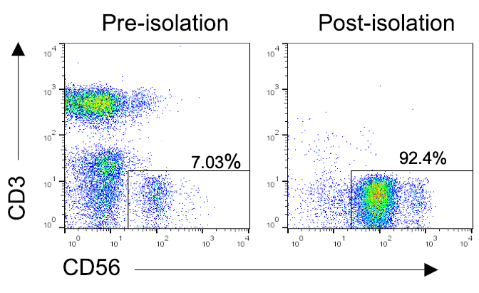
Figure 1: Representative flow profiles of NK cell purity after isolation from PBMCs. PBMCs were isolated from blood of a healthy donor, followed by enrichment of NK cells using a negative selection method for human NK cell isolation (Table of Materials). Cells were stained with CD56 and CD3 before and after enrichment to determine purity. Representative dot plots of CD56 vs. CD3 before and after isolation. Please click here to view a larger version of this figure.
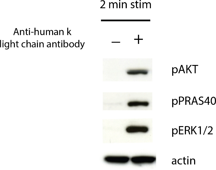
Figure 2: FcγRIIIa-activated NK cells exhibit phosphorylated signaling molecules. Cells were stimulated with rituximab for 2 min, and cell lysates were made according to the protocol. Lysates were separated on a 4%–12% gel, followed by transfer onto a PVDF membrane. The membrane was probed with antibodies against pAKT, pPRAS40, pERK1/2, and actin. Please click here to view a larger version of this figure.
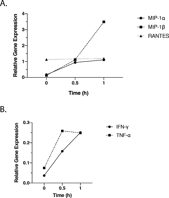
Figure 3: FcγRIIIa-activated NK cells express chemokine and cytokine genes. NK cells were stimulated with rituximab for 0 h, 0.5 h, and 1 h. mRNA was collected, reverse-transcribed, and subjected to qPCR analysis for MIP-1α, MIP-1β, RANTES, IFN-γ, and TNF-α (Table of Materials). (A) Relative expression of MIP-1α, MIP-1β, and RANTES at each timepoint. (B) Relative expression of IFN-γ and TNF-α at each timepoint. Values were normalized to the actin gene. Please click here to view a larger version of this figure.
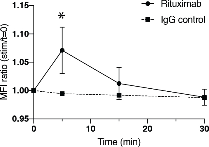
Figure 4: FcγRIIIa-activated NK cells exhibit cytoskeletal rearrangement. NK cells were stimulated with rituximab for 0 min, 5 min, 15 min, and 30 min, followed by assessment for cytoskeletal rearrangement by phalloidin staining and flow cytometry. The ratio of MFI at experimental timepoint to the MFI at time 0 of NK cells stimulated with rituximab (circle, solid line) or secondary antibody alone (square, dotted line). Bars represent the SD of four replicates. Asterisks represent statistical significance based on a two-tailed unpaired Student’s t-test (*p < 0.05). Please click here to view a larger version of this figure.
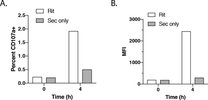
Figure 5: FcγRIIIa-activated NK cells express CD107a. NK cells were stimulated with rituximab for 4 h followed by assessment for degranulation by CD107a and flow cytometry. (A) Percentage of NK cells expressing CD107a after stimulation with rituximab (white bars) or anti-human κ light chain antibody (gray bars) for 0 h and 4 h. (B) CD107a MFI of NK cells stimulated with rituximab (white bars) or anti-human κ light chain antibody (gray bars) after 0 h and 4 h of treatment. Please click here to view a larger version of this figure.
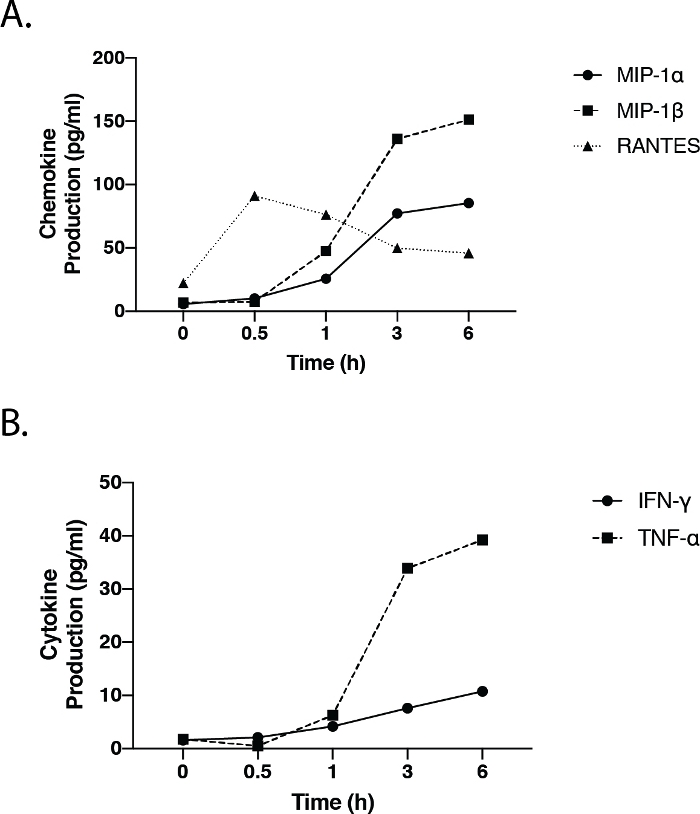
Figure 6: FcγRIIIa-activated NK cells secrete chemokines and cytokines. NK cells were stimulated for 0, 0.5, 1, 3, and 6 hr with rituximab. Supernatant was collected to measure release of MIP-1α, MIP-1β, RANTES, IFN-γ, and TNF-α by a flow- and bead-based cytokine assessment method (Table of Materials). (A) MIP-1α, MIP-1β, and RANTES production at each timepoint. (B) IFN-γ and TNF-α production at each timepoint. Please click here to view a larger version of this figure.
Discussion
Loading...
$$\rightleftharpoonup{xx}$$
$$\longleftharp{xx}$$,
$$\longrightharp{xx}$$,
This protocol describes methods for studying FcγRIIIa-driven events in NK cells mediated by antibodies. These techniques permit the evaluation of potential mechanisms of action of therapeutic antibodies, which is suggested to be ADCC1,2. Specifically, these methods provide flexibility in studying underlying molecular signaling pathways and cellular processes that are responsible for ADCC. They also allow observation of other effector functions, such as chemokine and cytokine production. In addition, these methods allow the identification of potential biomarkers and molecules that may be targeted to modulate ADCC.
The basis for this protocol is the artificial stimulation of NK cells through the FcγRIIIa with antibodies in the absence of target cells. Antibody-bound target cells typically serve to promote the crosslinking of Fc receptors to form a platform that drives signaling and downstream effects. Instead, crosslinking is accomplished using an anti-human κ light chain antibody in this assay, bypassing the need for target cells to stimulate the NK cells. Without target cells, the results and observations can be attributed directly to the NK cells, assuming that the purification process is successful.
Importantly, using anti-human κ light chain antibody to crosslink the antibody does not interfere with the binding affinity of the Fc portion for the FcγRIIIa, an interaction that dictates the strength of response10,11,12,13. Indeed, studies have shown that afucosylated antibodies increase ADCC due to their increased affinity for the FcγRIIIa10,11,12,13. Subsequent studies showed that this increased affinity is unaffected by the anti-human κ light chain secondary antibody substitute and can be used to study the basis for increased ADCC8. To ensure that the NK cell is stimulated through crosslinking of the antibody, two negative controls should be included: 1) therapeutic antibody only without the secondary anti-human κ light chain antibody, and 2) the secondary anti-human κ light chain antibody only. In both cases, no signaling or effector function should be generated.
This method also provides flexibility for studying the effects of therapeutic antibodies on small molecule inhibitors. The inhibitor can be added before crosslinking with the secondary antibody so that the inhibitor has time to engage its target. However, studies should be performed to determine the optimal time of inhibitor pretreatment; thus, the inhibitor has a maximal effect on stimulation. With that said, researchers may also choose to study the effects of an inhibitor after stimulation. In this case, the inhibitor may be added after crosslinking to study how it influences signals and processes that are already generated. Together, the method described here provides maximal flexibility in studying combinatorial effects of different small molecule inhibitors with therapeutic antibodies.
As mentioned above, a variety of readouts can be performed after stimulation. Western blotting can be performed to study signaling using SDS-PAGE and membrane transfer systems from various vendors. Similarly, gene expression can also be assessed using various RNA extraction methods, reverse transcription reagents, and gene expression instruments. Finally, staining for intracellular or extracellular protein can also be performed in which samples can be analyzed using different flow cytometers. For intracellular cytokine and CD107a staining (step 3.4, which can be assessed simultaneously), monensin and/or brefeldin A should be added to maximize signals. We have used different platforms for each experimental goal and still observed similar results. Therefore, the method can be complemented with various reagents, platforms, and instruments, depending on the study.
The crosslinking time for stimulation will depend on the goal of the study. If signaling studies are desired, typical crosslinking stimulation time is between 2 min and 10 min. pAKT, pPRAS40, and pERK1/2 accumulation peaks at 2 min and disappears after 10 min8,9. For functional studies (i.e., those involving chemokine/cytokine production), cells must be stimulated for at least 30 min, depending on the analyte9. Gene expression analysis also typically requires 30 min of stimulation9. Caution should be exercised when using RANTES gene expression as a readout, as RANTES mRNA production is independent of transcriptional activation since it is already stored in cells for prompt translation and release of protein upon stimulation25. Degranulation, in contrast, requires at least 3 h of stimulation. Despite these general observations, researchers should perform kinetic studies to determine the optimal stimulation time for the particular molecules of interest.
Similarly, researchers should titrate the antibody of interest to determine the optimal concentration because antibodies with different specificities bind to FcγRIIIa with different affinities, even if they are of the same isotype. For example, rituximab and trastuzumab are both an IgG1 isotype but trastuzumab binds more strongly to the valine polymorphism of FcγRIIIa than rituximab26,27. This difference in affinity may lead to functional differences, such as degranulation, as observed in published studies8.
Determining the optimal concentration is also important because of the low affinity the Fc portion of the antibody has for the FcγRIIIa. This may result in washing off the antibody since the protocol includes washing steps after binding of the antibody to the Fc receptor. This may then lead to a lack of sensitivity in the assays as suggested by the low percentage of CD107a positive cells after stimulation (Figure 5). However, determination of the optimal concentration should provide confidence that results are not due to a lack of sensitivity. In addition, cells are clearly activated in the biochemical and functional assays that use bulk cell as opposed to single cell readouts (Figure 2, Figure 3, Figure 6).
The protocol is also limited since it does not entirely mimic what occurs physiologically. The secondary anti-human K antibody used is to imitate the crosslinking generated by target antigen expressed on cells. Here, a saturating amount of secondary antibody is added to generate the maximum response. However, distinct target cells will express different levels of antigen, which will affect crosslinking and response. Currently, this platform is not optimized to mimic the effects of different antigen expression levels.
Another factor to consider when performing these experiments is donor-to-donor variability due to different genetic backgrounds and immunological histories among individuals. Therefore, care must be taken when comparing NK cell responses from different donors across the same assays. Similarly, only general conclusions should be made when different donors are used.
Altogether, the described method is a simple and flexible stimulation platform to study antibody driven FcγRIIIa-mediated events in NK cells. It has been used to better understand the basis for the increased ADCC and efficacy observed with afucosylated antibodies8. This method was also employed in a study combining therapeutic antibodies and PI3K small molecule inhibitors9. Additionally, a previously unknown mechanism for chemokine and cytokine production regulated by pS6 was identified9. Therefore, future studies using this artificial signaling platform can further elucidate mechanisms of regulation for effector functions driven by the FcγRIIIa. It may also potentially identify new molecules important for these mechanisms as well as new roles for known molecules.
Disclosures
Loading...
$$\rightleftharpoonup{xx}$$
$$\longleftharp{xx}$$,
$$\longrightharp{xx}$$,
All authors are or have been employees or consultants for iQ Biosciences.
Acknowledgements
Loading...
$$\rightleftharpoonup{xx}$$
$$\longleftharp{xx}$$,
$$\longrightharp{xx}$$,
The authors thank James Lee and Christopher Ng for comments and editing of this manuscript.
Materials
| Name | Company | Catalog Number | Comments |
|---|---|---|---|
| 16% paraformaldehyde | Thermo Fisher Scientific | 50-980-487 | |
| Alexa Fluor 488 phalloidin | Thermo Fisher Scientific | A12379 | |
| anti-mouse HRP antibody | Cell Signaling Technologies | 7076 | |
| AutoMACS instrument | Miltenyi | NK cell isolation method; another isolation instrument may be used | |
| B-mercaptoethanol | Thermo Fisher Scientific | 21985023 | |
| Bovine Serum Albumin Fraction V, fatty acid free | Millipore Sigma | 10775835001 | |
| CD107a APC antibody | Biolegend | 328620 | |
| Cytokine 30-Plex Human Panel | Thermo Fisher Scientific | LHC6003M | chemokine/cytokine method; another chemokine/cytokine analysis method may be used |
| FBS | Hyclone | SH30071.01 | |
| Goat anti-human κ light chain antibody | Millipore Sigma | AP502 | |
| Halt Protease and Phosphatase Inhibitor Cocktail | Thermo Fisher Scientific | 78440 | |
| HEPES | Thermo Fisher Scientific | 15630080 | |
| High Capacity cDNA Reverse Transcription Kit | Thermo Fisher Scientific | 4368814 | cDNA transcription method; used according to manufacturer's protocol |
| IFN-? primers | Thermo Fisher Scientific | Hs00989291_m1 | |
| Leukosep tube (50 ml conical with porous barrier) | Greiner | 227290 | |
| Lymphoprep (density gradient medium) | Stemcell | 7851 | |
| MIP-1? primers | Thermo Fisher Scientific | Hs00234142_m1 | |
| MIP-1β primers | Thermo Fisher Scientific | Hs99999148_m1 | |
| NK cell isolation kit | Miltenyi | 130-092-657 | NK cell isolation kit; another isolation kit may be used |
| NuPAGE 4-12% Bis-Tris Protein Gels | Thermo Fisher Scientific | another protein separation system may be used | |
| pAKT (S473) antibody | Cell Signaling Technologies | 4060 | |
| pERK1/2 antibody | Cell Signaling Technologies | 4370 | |
| pPRAS40 antibody | Cell Signaling Technologies | 13175 | |
| PVDF membrane | Thermo Fisher Scientific | nitrocellulose may be used | |
| RANTES primers | Thermo Fisher Scientific | Hs00982282_m1 | |
| RIPA buffer | Millipore Sigma | R0278 | |
| RPMI w/glutamax | Thermo Fisher Scientific | 61870 | |
| Sodium pyruvate | Thermo Fisher Scientific | 11360070 | |
| TNF-? primers | Thermo Fisher Scientific | Hs00174128_m1 | |
| Triton X-100 | Thermo Fisher Scientific | 85111 | |
| TRIzol | Thermo Fisher Scientific | 15596018 | RNA isolation method; used according to manufacturer's protocol |
| Xcell Blot II Transfer Module | Thermo Fisher Scientific | another protein separation system may be used | |
| Xcell SureLock Protein Gel Electrophoresis Chamber System | Thermo Fisher Scientific | another protein separation system may be used | |
| β-actin HRP antibody | Abcam | ab6721 | |
| β-actin primers | Thermo Fisher Scientific | Hs00982282_m1 |
References
Loading...
$$\rightleftharpoonup{xx}$$
$$\longleftharp{xx}$$,
$$\longrightharp{xx}$$,
- Hudis, C. A. Trastuzumab - Mechanism of Action and Use in Clinical Practice. New England Journal of Medicine. 357 (1), 39-51 (2007).
- Weiner, G. J. Rituximab: mechanism of action. Seminars inHhematology. 47 (2), 115-123 (2010).
- Orange, J. S. Formation and function of the lytic NK-cell immunological synapse. Nature Reviews Immunology. 8 (9), 713-725 (2008).
- Bonnema, J. D., Karnitz, L. M., Schoon, R. A., Abraham, R. T., Leibson, P. J. Fc receptor stimulation of phosphatidylinositol 3-kinase in natural killer cells is associated with protein kinase C-independent granule release and cell-mediated cytotoxicity. The Journal of Experimental Medicine. 180 (4), 1427-1435 (1994).
- Jiang, K., et al. Pivotal role of phosphoinositide-3 kinase in regulation of cytotoxicity in natural killer cells. Nature Immunology. 1 (5), 419-425 (2000).
- Kanakaraj, P., et al. Phosphatidylinositol-3 kinase activation induced upon Fc gamma RIIIA-ligand interaction. The Journal of Experimental Medicine. 179 (2), 551-558 (1994).
- Orange, J. S., Harris, K. E., Andzelm, M. M., Valter, M. M., Geha, R. S., Strominger, J. L. The mature activating natural killer cell immunologic synapse is formed in distinct stages. Proceedings of the National Academy of Sciences. 100 (24), 14151-14156 (2003).
- Liu, S. D., et al. Afucosylated Antibodies Increase Activation of FcγRIIIa-Dependent Signaling Components to Intensify Processes Promoting ADCC. Cancer Immunology Research. 3 (2), 173-183 (2015).
- Romeo, V., Gierke, S., Edgar, K. A., Liu, S. D. Effects of PI3K Inhibition on Afucosylated Antibody-Driven FcγRIIIa Events and Phospho-S6 Activity in NK Cells. The Journal of Immunology. 203 (1), 137-147 (2019).
- Mössner, E., et al. Increasing the efficacy of CD20 antibody therapy through the engineering of a new type II anti-CD20 antibody with enhanced direct and immune effector cell-mediated B-cell cytotoxicity. Blood. 115 (22), 4393-4402 (2010).
- Shields, R. L., et al. Lack of fucose on human IgG1 N-linked oligosaccharide improves binding to human Fcgamma RIII and antibody-dependent cellular toxicity. The Journal of Biological Chemistry. 277 (30), 26733-26740 (2002).
- Shinkawa, T., et al. The absence of fucose but not the presence of galactose or bisecting N-acetylglucosamine of human IgG1 complex-type oligosaccharides shows the critical role of enhancing antibody-dependent cellular cytotoxicity. The Journal of Biological Chemistry. 278 (5), 3466-3473 (2003).
- Junttila, T. T., et al. Superior In vivo Efficacy of Afucosylated Trastuzumab in the Treatment of HER2-Amplified Breast Cancer. Cancer Research. 70 (11), 4481-4489 (2010).
- Goede, V., et al. Obinutuzumab (GA101) plus chlorambucil (Clb) or rituximab (R) plus Clb versus Clb alone in patients with chronic lymphocytic leukemia (CLL) and preexisting medical conditions (comorbidities): Final stage 1 results of the CLL11 (BO21004) phase III trial. Journal of Clinical Oncology. 31, 15_suppl 7004(2013).
- Sehn, L. H., et al. Randomized Phase II Trial Comparing Obinutuzumab (GA101) With Rituximab in Patients with Relapsed CD20+ Indolent B-Cell Non-Hodgkin Lymphoma: Final Analysis of the GAUSS Study. Journal of Clinical Oncology. 33 (30), 3467-3474 (2015).
- Pons-Tostivint, E., Thibault, B., Guillermet-Guibert, J. Targeting PI3K Signaling in Combination Cancer Therapy. Trends in Cancer. 3 (6), 454-469 (2017).
- Engelman, J. A. Targeting PI3K signalling in cancer: opportunities, challenges and limitations. Nature Reviews. Cancer. 9 (8), 550-562 (2009).
- Fruman, D. A., Rommel, C. PI3K and cancer: lessons, challenges and opportunities. Nature Reviews. Drug Discovery. 13 (2), 140-156 (2014).
- Janku, F., Yap, T. A., Meric-Bernstam, F. Targeting the PI3K pathway in cancer: are we making headway. Nature Reviews. Clinical Oncology. 15 (5), 273-291 (2018).
- Samuels, Y., et al. High frequency of mutations of the PIK3CA gene in human cancers. Science. 304 (5670), New York, N.Y. 554(2004).
- Kanevskiy, L. M., Telford, W. G., Sapozhnikov, A. M., Kovalenko, E. I. Lipopolysaccharide induces IFN-γ production in human NK cells. Frontiers in Immunology. 4, (2013).
- Romagnani, C., et al. Activation of human NK cells by plasmacytoid dendritic cells and its modulation by CD4+ T helper cells and CD4+ CD25hi T regulatory cells. European Journal of Immunology. 35 (8), 2452-2458 (2005).
- Sivori, S., et al. CpG and double-stranded RNA trigger human NK cells by Toll-like receptors: Induction of cytokine release and cytotoxicity against tumors and dendritic cells. Proceedings of the National Academy of Sciences. 101 (27), 10116-10121 (2004).
- Hanna, J., et al. Novel Insights on Human NK Cells' Immunological Modalities Revealed by Gene Expression Profiling. The Journal of Immunology. 173 (11), 6547-6563 (2004).
- Swanson, B. J., Murakami, M., Mitchell, T. C., Kappler, J., Marrack, P. RANTES production by memory phenotype T cells is controlled by a posttranscriptional, TCR-dependent process. Immunity. 17 (5), 605-615 (2002).
- Jo, M., Kwon, H. S., Lee, K. H., Lee, J. C., Jung, S. T. Engineered aglycosylated full-length IgG Fc variants exhibiting improved FcγRIIIa binding and tumor cell clearance. mAbs. 10 (2), 278-289 (2018).
- Isoda, Y., et al. Importance of the Side Chain at Position 296 of Antibody Fc in Interactions with FcγRIIIa and Other Fcγ Receptors. PLoS ONE. 10 (10), 0140120(2015).
Reprints and Permissions
Request permission to reuse the text or figures of this JoVE article
Request Permission