This protocol provides an efficient collagenase digestion method for isolation of viable adipocytes and stromal vascular fraction-SVF cells from human visceral fat in a single process, including methodology to obtain high-quality RNA from adipocytes and phenotypification of SVF-macrophages through staining of multiple membrane-bound markers for analysis by flow cytometry.
Method Article
Isolation of Viable Adipocytes and Stromal Vascular Fraction from Human Visceral Adipose Tissue Suitable for RNA Analysis and Macrophage Phenotyping
Guadalupe Estrada-Gutierrez1 , Eyerahi Bravo-Flores2 , Veronica Ortega-Castillo3 , Veronica Flores-Rueda4 , Ismael Mancilla-Herrera5 , Salvador Espino Y Sosa6 , Maribel Sánchez-Martínez2 , Otilia Perichart-Perera7 , Mario Solis-Paredes8
1Research Division, Instituto Nacional de Perinatologia, 2Department of Immunobiochemistry, Instituto Nacional de Perinatologia, 3Department of Obstetrics, Instituto Nacional de Perinatologia, 4Department of Academic Programs and Continuing Education, Instituto Nacional de Perinatologia, 5Department of Infectology and Immunology, Instituto Nacional de Perinatologia, 6Clinical Research Branch, Instituto Nacional de Perinatologia, 7Department of Nutrition and Bioprogramming, Instituto Nacional de Perinatologia, 8Department of Human Genetics and Genomics, Instituto Nacional de Perinatologia, Mexico City, Mexico
Opens in a new tab
In This Article
Summary
Loading...
$$\rightleftharpoonup{xx}$$
$$\longleftharp{xx}$$,
$$\longrightharp{xx}$$,
Abstract
Loading...
$$\rightleftharpoonup{xx}$$
$$\longleftharp{xx}$$,
$$\longrightharp{xx}$$,
Visceral adipose tissue (VAT) is an active metabolic organ composed mainly of mature adipocytes and stromal vascular fraction (SVF) cells, which release different bioactive molecules that control metabolic, hormonal, and immune processes; currently, it is unclear how these processes are regulated within the adipose tissue. Therefore, the development of methods evaluating the contribution of each cell population to the pathophysiology of adipose tissue is crucial. This protocol describes the isolation steps and provides the necessary troubleshooting guidelines for efficient isolation of viable mature adipocytes and SVF from human VAT biopsies in a single process, using a collagenase enzymatic digestion technique. Moreover, the protocol is also optimized to identify macrophage subsets and perform mature adipocyte RNA isolation for gene expression studies, which allows performing studies dissecting the interaction between these cell populations. Briefly, VAT biopsies are washed, minced mechanically, and digested to generate a single-cell suspension. After centrifugation, mature adipocytes are isolated by flotation from the SVF pellet. The RNA extraction protocol ensures a high yield of total RNA (including miRNAs) from adipocytes for downstream expression assays. Simultaneously, SVF cells are used to characterize macrophage subsets (pro- and anti-inflammatory phenotype) through flow cytometry analysis.
Introduction
Loading...
$$\rightleftharpoonup{xx}$$
$$\longleftharp{xx}$$,
$$\longrightharp{xx}$$,
White adipose tissue is composed not only of fat cells or adipocytes, but also of a non-fat cell fraction known as stromal vascular fraction (SVF), which contains a heterogeneous cell population consisting in macrophages, other immune cells as regulatory T cells (Tregs), and eosinophils, preadipocytes, and fibroblasts, surrounded by vascular and connective tissue1,2. Adipose tissue (AT) is now considered an organ that regulates physiological processes related to metabolism and inflammation through adipokines, cytokines, and microRNAs produced and released by different cells into the tissue, with autocrine, paracrine, and endocrine effects3,4. In humans, white adipose tissue comprises the subcutaneous adipose tissue (SAT) and the visceral adipose tissue (VAT), with important anatomic, molecular, cellular, and physiological differences between them2,5. SAT represents up to 80% of human AT, while VAT is located within the abdominal cavity, mainly in the mesentery and omentum, being metabolically more active6. Moreover, VAT is an endocrine organ that secretes mediators with a substantial impact on body weight, insulin sensitivity, lipid metabolism, and inflammation. Consequently, VAT accumulation leads to abdominal obesity and obesity-related diseases such as type-2 diabetes, metabolic syndrome, hypertension, and cardiovascular disease risk, representing a better predictor of obesity-associated mortality6,7,8,9.
In homeostatic conditions, adipocytes, macrophages, and the other immune cells cooperate to maintain the VAT metabolism through the secretion of anti-inflammatory mediators10. However, excessive VAT expansion promotes the recruitment of activated T cells, NK cells, and macrophages. In fact, in lean VAT, the ratio of the macrophages is 5%, while this ratio rises up to 50% in obesity, with macrophage polarization from anti-inflammatory to pro-inflammatory phenotype, generating a chronic inflammatory environment10,11.
As a consequence of the obesity pandemic, an astonishing number of reports have arisen addressing different VAT research topics, including adipocyte biology, epigenetics, inflammation, endocrine properties, and emerging areas as extracellular vesicles, among others8,10,12,13. However, although the VAT environment is defined by crosstalk between adipocytes and the resident or arriving macrophages, most studies have focused on only one cell population, and there is scarce information about the interaction of these cells in VAT and their pathophysiological consequences11,14. Moreover, valuable studies addressing the adipocyte-macrophage interplay in AT were performed using cell lines, lacking the in vivo priming conditions11,14,15. A suitable strategy to dissect the interaction or the particular contribution of these cells in VAT requires the isolation of both cell types from the same fat biopsy to perform in vitro assays that mirror as similar as possible the in vivo properties that regulate VAT metabolism.
Although the non-enzymatic dissociation methods based on mechanical forces to break the AT ensure minimal manipulation, these methods cannot be used if the aim is to study SVF cells, as they have lower efficiency in cell recovery and low cell viability compared to enzymatic methods, and a larger volume of tissue is needed16,17. Enzymatic digestion using collagenase is a gentle method that allows adequate digestion of collagen and extracellular matrix proteins of fibrous tissues such as WAT18 and is frequently used when trypsin is ineffective or damaging19. The protocol provides fundamental troubleshooting guidelines for efficient isolation of viable mature adipocytes and SVF cells from human VAT biopsies in a single process, using a collagenase enzymatic digestion technique, giving information to ensure high yields (quantity, purity, and integrity) of total RNA from mature adipocytes, including microRNAs, for downstream expression applications. Simultaneously, the protocol is optimized to identify macrophage subsets from SVF cells through the staining of multiple membrane-bound markers for further analysis by flow cytometry20.
Protocol
Loading...
$$\rightleftharpoonup{xx}$$
$$\longleftharp{xx}$$,
$$\longrightharp{xx}$$,
This protocol was approved by the IRB of the Instituto Nacional de Perinatologia (212250-3210-21002-06-15). Participation was voluntary, and all the enrolled women signed the informed consent form.
1. Visceral adipose tissue collection
- Obtain VAT biopsies through partial omentectomy during cesarean section from healthy adult women with singleton pregnancies at term without labor.
- After the uterine closure and hemostasis, proceed to identify greater omentum and extend it on a wet compress. The AT exposed is VAT.
- Locate the largest blood vessel and trace an imaginary line of 7 x 5 cm to the greater omentum base.
- Identify an avascular zone and use Kelly forceps to drill the outer side of VAT.
- Use Ochsner forceps to clamp the proximal and distal side in the zone where VAT was drilled and use Metzenbaum scissors to cut the tissue.
- Tie greater omentum with black silk number 1.
- Remove Ochsner forceps and evaluate the hemostasis.
- Place the VAT biopsy into a sterile container and transport it to the lab immediately.
2. Enzymatic digestion of visceral adipose tissue and isolation of mature adipocytes and stromal vascular fraction cells
- Weigh the VAT biopsy and rinse with 1x PBS pH 7.4.
- Cut 4 g of VAT and use scissors to mince the tissue into small pieces in a dissection tray.
- Transfer minced VAT to a sterile 50 mL centrifuge tube, add 20 mL of PBS, and shake gently to remove the excess of red blood cells.
- Discard PBS and repeat the procedure twice.
- Transfer VAT to a new sterile 50 mL centrifuge tube and add 25 mL of digestion solution (0.25% collagenase type II, 5 mM glucose, 1.5% albumin in PBS).
- Incubate at 37 °C for 60 min in an orbital shaker at 125 rpm.
- Filter the digested tissue through three layers of gauze into a new sterile 50 mL centrifuge tube.
- Centrifuge at 200 x g for 5 min at 4 °C.
- Two phases are obtained, an upper phase that corresponds to mature adipocytes, while SFV cells remain in the pellet. Gently transfer the mature adipocytes into a new sterile 50 mL centrifuge tube using a transfer pipette.
- Add 20 mL of cold PBS, shake gently, and centrifuge at 200 x g for 5 min at 4 °C.
- Discard PBS and repeat the procedure twice.
- Use a transfer pipette to gently transfer the mature adipocytes into a 1.5 mL DNase-RNase free microcentrifuge tube.
- Centrifuge at 200 x g for 1 min at 4 °C and remove the excess PBS using a P200 micropipette. Repeat this step if it is necessary.
- Gently transfer 300 μL of mature adipocyte suspension into a 1.5 mL DNase-RNase free microcentrifuge tubes. Store mature adipocytes at -80 °C until RNA extraction.
NOTE: Adipocytes do not form a pellet. - Once adipocytes are separated (step 2.9), aspirate most of the digestion solution with a transfer pipette, keeping the SVF pellet at the bottom of the tube.
- Transfer the pellet with SVF cells into a new 50 mL centrifuge tube using a transfer pipette.
- Wash cell pellet gently, resuspending in 20 mL of cold 1x PBS by pipetting up and down.
- Centrifuge at 800 x g for 5 min at 4 °C and rapidly discard the supernatant by decantation.
- Perform a second wash by repeating steps 2.17 and 2.18.
- Add 5 mL of red blood cell lysis buffer equilibrated to room temperature (RT) to the SVF pellet and suspend by repeated pipetting. Do not vortex.
- Incubate for 5 min at RT and centrifuge for 5 min at 800 x g. Carefully remove the supernatant using a transfer pipette and dispose of properly.
- To neutralize the lysis buffer, add 10 mL of cold 1x PBS and gently mix until the cell pellet disaggregates.
- Centrifuge at 800 x g for 5 min to obtain SVF pellet and discard PBS. A white pellet should be visible at the bottom of the tube.
- Resuspend cells in 5 mL of 1x PBS at RT by repeated pipetting and filtered through three layers of gauze into a new 50 mL conical tube. Subsequently, these cells will be used for macrophage subpopulation characterization by flow cytometry.
3. RNA extraction from mature adipocytes
- Prepare a clean area, using RNase decontamination spray to avoid RNA degradation. Use a set of pipettes reserved for RNA procedures. All tubes and tips employed should be RNase-free.
- Thaw the mature adipocyte suspension (section 2.14) at 4 °C if it was stored to -80 °C.
NOTE: Avoid multiple freeze-thaw cycles. - Lyse cells by adding 1,000 μL of acid-guanidinium-phenol based reagent, and mix thoroughly with a P1000 micropipette to homogenize.
- Incubate for 5 min at RT to boost dissociation of nucleoprotein complexes.
- Centrifuge cell lysate for 5 min at 12,000 x g at RT. Three phases will be obtained: an upper phase (yellow) corresponding to adipocyte lipids, a middle phase (pink) corresponding to nucleic acids, and a pellet (cellular debris).
- Carefully remove the lipid layer, using a P200 micropipette.
- Transfer the middle phase into a new 2 mL tube, avoiding lipid remnant and pellet disturbing (approximately 700 μL).
4. Purification of total RNA, including microRNAs
NOTE: A column-based total RNA purification method is used to obtain high-quality total RNA.
- Add an equal volume of ethanol (95%–100%) to the sample from section 3.7, approximately 700 μL, and mix by hand inverting the tube for 10 s.
- Transfer 700 μL of the mixture into a column inserted in a collection tube, centrifuge and discard the flow-through.
- Reload the column and repeat step 4.2.
- Transfer the column into a new collection tube.
- Add 400 µL of pre-wash buffer to the column and centrifuge. Discard the flow-through and repeat this step.
- Add 700 µL of wash buffer to the column and centrifuge for 2 min.
- Transfer the column carefully into a nuclease-free tube.
- Add 50 µL of nuclease-free water directly to the column matrix and elute the RNA by centrifugation. Prepare aliquots of 10 µL using 200 µL microfuge tubes.
- Immediately put aliquots on ice and use one of them to determine RNA concentration and purity.
- Prepare an aliquot of 5 µL of total RNA adjusted to 100 ng/µL to measure RNA integrity and microRNA concentration.
- Store at -80 °C until use.
5. Determination of mature adipocyte RNA concentration, purity, and integrity; microRNA quantification assay
NOTE: RNA concentration and purity determinations are performed using a UV-Vis spectrophotometer; RNA integrity and miRNA quantification are performed using an RNA quality control analyzer.
- Wash the sample reader with molecular grade water and wipe.
- Load 2 µL of elution water (blank), change the setting to RNA, and click on the Blank button.
- Load 2 µL of sample and click on the Measure button.
- After the reading is complete, record the A260/A280 and A260/A230 ratios as well as the amount of RNA (ng/µL) (Figure 1A).
NOTE: RNA with OD260/OD280 and OD260/OD230 ratio around 2.0 is considered pure. - Measure the RNA integrity number (RIN) using an RNA integrity kit following the manufacturer´s instructions (Figure 1B).
NOTE: A RIN =10 corresponds to intact RNA, whereas a RIN ≤ 3.0 indicates a strongly degraded RNA. For expression microarrays or RT-qPCR, use RNA with RIN ≥ 6.0. - Use a small RNA kit to determine the concentration and percent of microRNAs, following the manufacturer´s instructions (Figure 1C).
NOTE: To avoid the overestimation of small and micro RNAs, use RNA samples with RIN ≥ 6.0.
6. Count and viability of stromal vascular fraction cells
- Dilute 10 μL of SVF cell suspension (section 2.24) into 90 μL of 0.4% Trypan Blue Solution (final dilution 1:10) and apply 10 μL to a standard hemocytometer.
- Count the viable cells carefully, excluding the dead cells, in four squares at the corner of the counting chamber.
- Determine the cell concentration present in the original suspension: Cell concentration = Total cell count/4 x dilution factor (10) x 10,000 = Cells/mL
- To calculate the cellular yield (cells per gram of tissue) obtained, multiply the cellular concentration by the original total sample volume (in mL) and divide by the weight of tissue digested (in grams).
- Evaluate cell viability as the percentage of the living cells as follows: % Viability = (Number of viable cells / Total number of cells) x 100.
7. Characterization of macrophage subsets from stromal vascular fraction
- Transfer a total of 1 x 10⁶ SVF cells/mL to a 5 mL round-bottom polypropylene test tube for flow cytometry and pellet cells by centrifugation at 800 x g for 5 min at 4 °C.
- Vortex carefully to loosen the pellet and resuspend the cells in 100 μL of 1x PBS at RT.
- Add pre-titrated optimal concentration of each fluorochrome-conjugated monoclonal antibody specific for a cell surface antigen; mix gently and incubate for 15 min in the dark at RT.
- Add an excess of cold 1x PBS (≈ 1 mL) and centrifuge the SVF at 400 x g for 5 min at 4 °C.
- Discard supernatants rapidly by decantation. Be careful not to disturb the pellet.
- Add 500 μL of 1x lysing solution and incubate 15 min at RT, protecting the tubes from direct light.
- Remove the solution after centrifuge at 400 x g for 5 min at 4 °C, and store at 2–8 °C in the dark until data acquisition.
- Vortex the cells thoroughly at low speed to reduce aggregation before acquiring.
- Resuspend the SVF pellet in 5 mL of sheath fluid at RT, and subsequently filter through three layers of gauze into a new 5 mL round-bottom polypropylene test tube before analysis by flow cytometry, to reduce clogging the cell sorter lines.
- Vortex each tube briefly before analysis.
- Acquire data from the samples of interest. For flow analysis, count a minimum of 10,000 events.
NOTE: If using a cell sorting flow cytometer, separate the macrophages for application in subsequent studies.
8. Gating strategy
- Plot the height or width against the area for Forward Scatter (FSC) to determine the singlet population (Figure 2A).
- Select cells plotting the area for Side Scatter (SSC) against FSC, discarding the cellular debris (Figure 2B).
- From selected cells, identify the populations expressing CD45 as hematopoietic cells (Figure 2C), and CD45/CD14 double positive cells as macrophages (Figure 2D).
- From CD45+/CD14+ macrophages, detect negative and positive HLA-DR cells (Figure 2E).
NOTE: Include the compensation controls for the antibody panel and adjust spectral overlap on a multicolor flow cytometer adequate for the panel. We perform flow cytometry analysis using a cytometer equipped with three lasers (405 nm violet laser, 488 nm blue laser, and 640 nm red laser), and detectors for the indicated fluorochromes.
Results
Loading...
$$\rightleftharpoonup{xx}$$
$$\longleftharp{xx}$$,
$$\longrightharp{xx}$$,
This protocol describes an enzymatic method using collagenase digestion followed by differential centrifugation to isolate, in a single process, viable mature adipocytes and SVF cells from VAT biopsies obtained from healthy pregnant women after partial omentectomy. In this case, we use the adipocytes for RNA extraction and the SVF for macrophage phenotyping.
The RNA extraction protocol enabled to obtain RNA with an adequate purity high integrity, and microRNAs from mature adipocytes (Figure 1). The RNA integrity assessed by an RNA quality control analyzer resulted in excellent values for the samples of adipocytes isolated with the protocol (RIN = 9.7).
The total of nucleated cells present in VAT-SVF was approximately 2.8 x 10⁶ cells/gram of VAT (2.8 x 10⁶ ± 1.7 x 10⁶), with 64% viability (63.9 ± 2.0) using the trypan blue exclusion test. SVF cells were labeled with fluorophore-conjugated primary antibodies to identify and characterize AT macrophages by fluorescence-activated cell sorting analysis. Flow cytometry analysis generate plots showing different cell populations based on cellular markers (Figure 2). Initially, by plotting the Forward Scatter-area (FSC-A) vs Forward Scatter-height (FSC-H), cell aggregates were easily eliminated from analysis (Figure 2A). Then, cellular debris was excluded by gating the cells based on correct size and complexity using Forward Scatter-area (FSC-A) and the Side Scatter-area (SSC-A) (Figure 2B). Next, to analyze macrophages, it was not necessary to exclude other immune cells. First, monocyte/macrophage lineage cells were selected by the use of CD45 and CD14 markers (Figure 2C,D). Subsequently, the CD45/CD14 double positive cells were separated based on macrophage marker HLA-DR expression, where two subsets macrophages were identified: HLA-DR- and HLA-DR+ (Figure 2E).
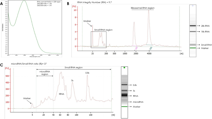
Figure 1: RNA quality control from mature adipocytes. (A) RNA concentration, 260/280, and 260/230 ratio; (B) Integrity analysis. Electropherogram, gel image, and RNA integrity number were obtained using an RNA quality control analyzer and an RNA analyzer kit; (C) microRNA quantification. Electropherogram, gel image, and microRNA percent were obtained using an RNA quality control analyzer and a small RNA kit. Please click here to view a larger version of this figure.
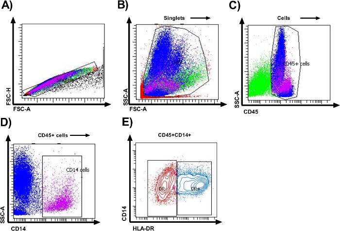
Figure 2: Identification of macrophages from visceral adipose tissue. Representative flow cytometry plots of stromal vascular fraction macrophages isolated from visceral adipose tissue collected during a cesarean section using collagenase digestion. (A) Identification of singlets using a first Forward Scatter-area (FSC-A) vs FSC-height (FSC-H) gate to remove doublets. (B) A second Forward Scatter-area (FSC-A) vs Side Scatter-area (SSC-A) gate was used to eliminate debris based on size and density. (C,D) SSC-A vs CD45 gate identified cells from hematopoietic origin, and SSC-A vs CD14 gate identified macrophages. (E) Total CD45+/CD14+ macrophages were gated based on HLA-DR marker and two subsets of macrophages were identified: HLA-DR- and HLA-DR+. Plots illustrate representative data from individual subjects. Please click here to view a larger version of this figure.
Discussion
Loading...
$$\rightleftharpoonup{xx}$$
$$\longleftharp{xx}$$,
$$\longrightharp{xx}$$,
VAT plays a crucial role in metabolic regulation and inflammation. Increasing interest in the role of adipocytes and immune cells in the chronic inflammation associated with obesity has led to the development of different techniques to separate the SVF and fat cells present in AT. However, most techniques do not allow to obtain these two different sets of cells viable for downstream applications from the same VAT biopsy in a single procedure, which could be crucial for studies regarding interactions between adipocytes and SVF cells. Therefore, we implemented this protocol that provides a detailed description to isolate viable mature adipocytes and SVF cells present in a VAT biopsy. It differs from previous reports in the time of tissue digestion and collagenase concentration, as well as time and speed of centrifugation for cell separation, enabling adequate adipocyte RNA yields and detailed macrophage subset characterization.
A large amount of enzymatic and non-enzymatic isolation techniques for AT-derived cells have been proposed, with different effects on the biological characteristics and functional properties of isolated cells17,21,22,23,24, so it is necessary to choose the most appropriate strategy according to the goal pursued. Frequently, mature adipocytes and SVF isolation from adipose tissue is achieved using tissue dissociation enzymes25,26,27. Although different enzymes can be used to dissociate AT, the enzymatic digestion with collagenase remains as the gold standard to digest this tissue27,28. Since VAT is composed of a soft matrix, it can be easily digested with Collagenase Type II. This procedure avoids improper tissue dissociation because this enzyme disrupts the extracellular matrix native collagen, releasing many more cells from the fibrous stroma, with a negligible impact on cell viability, cell yield, and cluster differentiation expression19,27,28,29.
In terms of enzymatic digestion, it is also essential to consider the enzyme concentration, the digestion period as well as the centrifugation parameters to design a proper protocol for mature adipocyte and SVF cell isolation from VAT because these factors may impact cell phenotype, recovery, and viability in the final cell suspension27,29. The use of collagenase as a proteolytic enzyme is ideal at low concentration [range 0.075% a 0.3% (w/v)], with an incubation period of less than 2 h, so the phenotypic and functional SVF characteristics such as proliferation rate, differentiation capacity, and frequency of specific cellular lineages, remain unaltered30,31. We use a maximum digestion period of 60 min and 0.25% collagenase, reducing the concentration and length of collagenase exposure to obtain a negligible impact on cellular viability and SVF cells surface markers expression, avoiding skewed results in the flow cytometry analysis. We also include gentle centrifugation steps and shorter centrifugation times of 200 g/5 min to improve cell recovery during fat cell separation, representing an advantage for the protocol, since long and intense centrifugation periods [above 400 g/1 min] increase the death of adipocytes, limiting the cellular yield32,33,34. The SVF cellular recovery achieved through the collagenase-based digestion protocol is similar to the typical yields of 2–6 x 10⁶ cells/g AT reported in the literature, whereas the SVF viability of 63.9 ± 2.0 is moderately lower than the minimum proposed threshold of 70%–80% for these type of cells35,36,37,38. This is likely because we do not use culture media to resuspend the SVF cells as standard protocols, since we characterize the macrophage population through their surface markers, and it has been reported that these cells exhibit remarkable plasticity in response to environmental conditions, even changing their phenotype39,40,41.
Additionally, it is also important to mention that the pregnant women included in this protocol as VAT donors were healthy, with no clinical evidence of metabolic comorbidities, normal weight gain during pregnancy, average age 20–25 years, and normal pregestational Body Mass Index (BMI 18.5–24.9 kg/m2; n = 7), because some authors have described that different donors, age, BMI and gender or ethnicity have an influence on the cell number, and the expression of SFV surface markers42,43,44.
On the other hand, gene expression profile analysis requires reasonable amounts of intact RNA, but its isolation from mature adipocytes is particularly difficult because these cells have a higher lipid content and in some instances, the cell number is low45,46,47,48. We optimized an RNA isolation procedure from mature adipocytes based on a standard guanidine isothiocyanate/phenol method49,50, implementing easy steps that enable eliminate lipids and cellular debris. After sample preparation, column-based RNA extraction allows us to obtain a high-quality DNA-free RNA, including microRNAs, which is quite unusual for AT samples. The purity and integrity verification of these RNAs through a microfluidics-based RNA quality control analyzer ensures that isolated RNA quality is appropriate for downstream applications such as RT-qPCR assays and expression microarrays.
Besides the advantages mentioned before, AT digestion using collagenase allows liberating adipocytes and resident immune cells simultaneously, maintaining the integrity of cell surface receptors; this is an important fact during SVF cell isolation to get reliable and interpretable flow cytometry data27. Additionally, cells isolated from lipid-laden tissues such as VAT tend to be more prone to aggregation, reducing the sorted cell yield, and increasing autofluorescence51. In the protocol, the elimination of these cell aggregates by filtration through three layers of gauze before sorting and their exclusion with the described gating strategy, minimize background fluorescence, improving sample quality for cell sorting.
Previous studies have been conducted to identify the cells present in the SVF using a single set of CD markers characteristic for these cells52,53,54,55,56,57,58,59. For more in-deep characterization of macrophage subtypes in VAT SVF, we categorized these cells by expression of specific cell surface markers. According to CD45 and CD14 markers expression used to identify monocyte/macrophage linage cells in VAT60, we found that this tissue is composed of a typical CD45+CD14+ macrophage population from hematopoietic origin. Using the same protocol, in a previous study we were able to characterize M1 (CD11c) and M2 (CD163 and CD206) macrophage populations20.
The macrophage phenotyping within AT has been categorized as a spectrum from pro-inflammatory macrophages (classically activated-M1) to anti-inflammatory macrophages (alternatively activated-M2) according to the presence of different activation markers on its surface61,62. HLA-DR macrophage marker reflects the macrophage activation degree63,64, and was incorporated in the protocol to establish the M1 or M2 phenotype: macrophage populations expressing HLA-DR+ are inflammatory and HLA-DR- represent non-inflammatory macrophages. So, based on HLA-DR marker expression, two subsets of macrophages were defined: CD45+CD14+HLA-DR- and CD45+CD14+HLA-DR+, which originate from circulating monocytes and better known as monocyte-derived macrophages within the adipose tissue65,66.
Additionally, we quantified CD11c (M1 marker), as well as CD163 and CD206 (M2 marker) on each macrophages subtype, determining that AT macrophages display all evaluated activation markers on their membrane surface, demonstrating that macrophages present in VAT from pregnant women have more complex characteristics than those described for the overly simplified classifications of M1 and M2 macrophage populations. So, applying basic flow cytometry with multicolor antibody panel and stringent cell gating makes it possible to identify and characterize the macrophages from the heterogeneous pool of cells in the SVF. The refined macrophage phenotyping obtained with the protocol can be useful in studies conducted to elucidate the dynamic changes of macrophage populations in the AT that lead to disturbance in AT homeostasis.
Although this protocol was designed to characterize VAT macrophages, adjustments made in the antibody panel could expand its applications to facilitate sorting of other immune cells residing in the AT, since numerous fluorescent antibodies are available to identify specific markers of different lineage. Moreover, the method allows that cell populations purified by cell sorting can be used for post-sort analyses such as DNA/RNA/protein extraction or ex vivo treatments, obtaining the cells directly into an appropriate media with sterility techniques.
In summary, we consider that the protocol detailed herein represents the most efficient tradeoff between time, resources, cellular yield, and cell viability, besides being highly efficient for RNA and miRNA extraction from fat cells, and for characterization of macrophage population in AT.
Disclosures
Loading...
$$\rightleftharpoonup{xx}$$
$$\longleftharp{xx}$$,
$$\longrightharp{xx}$$,
The authors have no conflicts of interest to disclose.
Acknowledgements
Loading...
$$\rightleftharpoonup{xx}$$
$$\longleftharp{xx}$$,
$$\longrightharp{xx}$$,
This study was supported from the Instituto Nacional de Perinatologia (grant numbers: 3300-11402-01-575-17 and 212250-3210-21002-06-15) and CONACyT, Fondo Sectorial de Investigacion en Salud y Seguridad Social (FOSISS) (grant number 2015-3-2-61661).
Materials
| Name | Company | Catalog Number | Comments |
|---|---|---|---|
| 0.2 mL PCR tubes | Axygen | PCR-02-C | RNase, DNase free and nonpyrogenic |
| 1.5 mL microcentrifuge tubes | Axygen | MCT-150-C | RNase, DNase free and nonpyrogenic |
| 10 mL serological pipettes | Corning | CLS4101-50EA | Individually plastic wrapped |
| 10 µL universal pipet tip | Axygen | T-300-L-R | RNase, DNase free and nonpyrogenic |
| 10 µL universal pipet tip | Axygen | T-300-R-S | RNase, DNase free and nonpyrogenic |
| 1000 µL universal pipet tip | Axygen | T-1000-B-R | RNase, DNase free and nonpyrogenic |
| 2.0 mL microcentrifuge tube | Axygen | MCT-200-C | RNase, DNase free and nonpyrogenic |
| 200 µL universal pipet tip | Axygen | T-200-Y-R | RNase, DNase free and nonpyrogenic |
| 2100 Bioanalyzer Instrument | Agilent | G2939BA | - |
| 2101 Bioanalyzer PC | Agilent | G2953CA | 2100 Expert Software pre-installed in PC |
| 5 ml Round Bottom Polystyrene Test Tube | Corning | 352003 | Snap cap, sterile |
| 50 mL centrifuge tubes | Corning | CLS430828-100EA | Polipropilene, conical bottom and sterile |
| Acid-guanidinium-phenol based reagent | Zymo Research | R2050-1-200 | TRI Reagent or similar |
| Agilent RNA 6000 Nano Kit | Agilent | 5067-1511 | - |
| Agilent Small RNA Kit | Agilent | 5067-1548 | - |
| APC/Cy7 anti-human CD14 Antibody | BioLegend | 325620 | 0.4 mg/106 cells, present on monocytes/macrophages, clone HCD14 |
| Baker | - | - | 250 ml, non sterile |
| Bovine serum albumin | Sigma-Aldrich | A3912-100G | Heat shock fraction, pH 5.2, ≥96% |
| Chip priming station | Agilent | 5065-9951 | - |
| Collagenase type II | Gibco | 17101-015 | Powder |
| D-(+)-Glucose | Sigma-Aldrich | G8270-100G | Powder |
| Direct-zol RNA Miniprep | Zymo Research | R2051 | Supplied with 50 mL TRI reagen |
| Dissecting forceps | - | - | Steel, serrated jaws and round ends |
| Dissection tray | - | - | Stainless steel |
| Ethyl alcohol | Sigma-Aldrich | E7023-500ML | 200 proof, for molecular biology |
| FACS Flow Sheath Fluid | BD Biosciences | 342003 | - |
| FACS Lysing Solution | BD Biosciences | 349202 | - |
| FACSAria III Flow Cytometer/Cell Sorter | BD Biosciences | 648282 | - |
| FASCDiva Software | BD Biosciences | 642868 | Software v6.0 pre-installed |
| Hemacytometer | Sigma | Z359629-1EA | - |
| Manual cell counter | - | - | - |
| Mayo dissecting scisors | - | - | Stainless steel |
| Microcentrifuge | - | - | Adjustable temperature |
| Nanodrop spectrophotometer | Thermo Scientific | ND2000LAPTOP | - |
| Orbital shaker | - | - | Adjustable temperature and speed |
| P10 variable volume micropipette | Thermo Scientific-Finnpipette | 4642040 | 1 to 10 μL |
| P1000 variable volume micropipette | Thermo Scientific-Finnpipette | 4642090 | 100 to 1000 μL |
| P2 variable volume micropipette | Thermo Scientific-Finnpipette | 4642010 | 0.2 to 2 μL |
| P200 variable volume micropipette | Thermo Scientific-Finnpipette | 4642080 | 20 to 200 μL |
| PCR tube storage rack | Axygen | R96PCRFSP | - |
| PE/Cy5 anti-human HLA-DR Antibody | BioLegend | 307608 | 0.0625 mg/106 cells, present on macrophages, clone L243 |
| PE/Cy7 anti-human CD45 Antibody | BioLegend | 304016 | 0.1 mg/106 cells, present on leukocytes, clone H130 |
| Phosphate buffered saline | Sigma-Aldrich | P3813-10PAK | Powder, pH 7.4, for preparing 1 L solutions |
| Pipette controller | - | - | - |
| Red Blood Cells Lysis Buffer | Roche | 11 814 389 001 | For preferential lysis of red blood cells from human whole blood |
| Refrigerated centrifuge | - | - | Whit adapter for 50 mL conical tubes |
| Sterile Specimen container | - | - | - |
| Transfer pipette | Thermo Scientific-Samco | 204-1S | Sterile |
| Trypan Blue | Gibco | 15250-061 | 0.4% Solution |
| Tube racks | - | - | For different tube sizes |
| Vortex Mini Shaker | Cientifica SENNA | BV101 | - |
References
Loading...
$$\rightleftharpoonup{xx}$$
$$\longleftharp{xx}$$,
$$\longrightharp{xx}$$,
- Adipose-derived stromal vascular fraction cells: update on clinical utility and efficacy. Critical Reviews in Eukaryotic Gene Expression. 25 (2), 145-152 (2015).">Han, S., Sun, H. M., Hwang, K. C., Kim, S. W. Adipose-derived stromal vascular fraction cells: update on clinical utility and efficacy. Critical Reviews in Eukaryotic Gene Expression. 25 (2), 145-152 (2015).
- Subcutaneous and visceral adipose tissue: structural and functional differences. Obesity Reviews. 11 (1), 11-18 (2010).">Ibrahim, M. M. Subcutaneous and visceral adipose tissue: structural and functional differences. Obesity Reviews. 11 (1), 11-18 (2010).
- Sick fat: the good and the bad of old and new circulating markers of adipose tissue inflammation. Journal of Endocrinological Investigation. 42 (11), 1257-1272 (2019).">Barchetta, I., Cimini, F. A., Ciccarelli, G., Baroni, M. G., Cavallo, M. G. Sick fat: the good and the bad of old and new circulating markers of adipose tissue inflammation. Journal of Endocrinological Investigation. 42 (11), 1257-1272 (2019).
- The endocrine function of adipose tissues in health and cardiometabolic disease. Nature Reviews Endocrinology. 15 (9), 507-524 (2019).">Scheja, L., Heeren, J. The endocrine function of adipose tissues in health and cardiometabolic disease. Nature Reviews Endocrinology. 15 (9), 507-524 (2019).
- Different gene expression profiles in subcutaneous and visceral adipose tissues from Mexican patients with obesity. Indian Journal of Medical Research. 149 (5), 616-626 (2019).">Ronquillo, M. D., et al. Different gene expression profiles in subcutaneous and visceral adipose tissues from Mexican patients with obesity. Indian Journal of Medical Research. 149 (5), 616-626 (2019).
- Subcutaneous adipose tissue and visceral adipose tissue. Indian Journal of Medical Research. 149 (5), 571-573 (2019).">Mittal, B. Subcutaneous adipose tissue and visceral adipose tissue. Indian Journal of Medical Research. 149 (5), 571-573 (2019).
- Nutrition, the visceral immune system, and the evolutionary origins of pathogenic obesity. Proceedings of the National Academy of Sciences of the United States of America. 116 (3), 723-731 (2019).">West-Eberhard, M. J. Nutrition, the visceral immune system, and the evolutionary origins of pathogenic obesity. Proceedings of the National Academy of Sciences of the United States of America. 116 (3), 723-731 (2019).
- Adaptive immunity and adipose tissue biology. Trends in Immunology. 31 (10), 384-390 (2010).">Kaminski, D. A., Randall, T. D. Adaptive immunity and adipose tissue biology. Trends in Immunology. 31 (10), 384-390 (2010).
- Body fat distribution, in particular visceral fat, is associated with cardiometabolic risk factors in obese women. PLoS One. 12 (9), 0185403(2017).">Elffers, T. W., et al. Body fat distribution, in particular visceral fat, is associated with cardiometabolic risk factors in obese women. PLoS One. 12 (9), 0185403(2017).
- Visceral adipose tissue immune homeostasis is regulated by the crosstalk between adipocytes and dendritic cell subsets. Cell Metabolism. 27 (3), 588-601 (2018).">Macdougall, C. E., et al. Visceral adipose tissue immune homeostasis is regulated by the crosstalk between adipocytes and dendritic cell subsets. Cell Metabolism. 27 (3), 588-601 (2018).
- Interaction of differentiated human adipocytes with macrophages leads to trogocytosis and selective IL-6 secretion. Cell Death & Disease. 6 (1), 1613(2015).">Sarvari, A. K., et al. Interaction of differentiated human adipocytes with macrophages leads to trogocytosis and selective IL-6 secretion. Cell Death & Disease. 6 (1), 1613(2015).
- Extracellular vesicles from adipose tissue-A potential role in obesity and type 2 diabetes. Frontiers in Endocrinology. 8 (1), Lausanne. 202(2017).">Gao, X., Salomon, C., Freeman, D. J. Extracellular vesicles from adipose tissue-A potential role in obesity and type 2 diabetes. Frontiers in Endocrinology. 8 (1), Lausanne. 202(2017).
- Genome-wide DNA methylation pattern in visceral adipose tissue differentiates insulin-resistant from insulin-sensitive obese subjects. Translational Research. 178 (1), 13-24 (2016).">Crujeiras, A. B., et al. Genome-wide DNA methylation pattern in visceral adipose tissue differentiates insulin-resistant from insulin-sensitive obese subjects. Translational Research. 178 (1), 13-24 (2016).
- Macrophage infiltration into adipose tissue: initiation, propagation and remodeling. Future Lipidology. 3 (5), 545-556 (2008).">Surmi, B. K., Hasty, A. H. Macrophage infiltration into adipose tissue: initiation, propagation and remodeling. Future Lipidology. 3 (5), 545-556 (2008).
- A paracrine loop between adipocytes and macrophages aggravates inflammatory changes: role of free fatty acids and tumor necrosis factor alpha. Arteriosclerosis, Thrombosis, and Vascular Biology. 25 (10), 2062-2068 (2005).">Suganami, T., Nishida, J., Ogawa, Y. A paracrine loop between adipocytes and macrophages aggravates inflammatory changes: role of free fatty acids and tumor necrosis factor alpha. Arteriosclerosis, Thrombosis, and Vascular Biology. 25 (10), 2062-2068 (2005).
- Maximizing non-enzymatic methods for harvesting adipose-derived stem from lipoaspirate: technical considerations and clinical implications for regenerative surgery. Scientific Reports. 7 (1), 10015(2017).">Bellei, B., Migliano, E., Tedesco, M., Caputo, S., Picardo, M. Maximizing non-enzymatic methods for harvesting adipose-derived stem from lipoaspirate: technical considerations and clinical implications for regenerative surgery. Scientific Reports. 7 (1), 10015(2017).
- Mechanical and enzymatic procedures to isolate the stromal vascular fraction from adipose tissue: preliminary results. Frontiers in Cell and Developmental Biology. 7 (1), 88(2019).">Senesi, L., et al. Mechanical and enzymatic procedures to isolate the stromal vascular fraction from adipose tissue: preliminary results. Frontiers in Cell and Developmental Biology. 7 (1), 88(2019).
- Differential effects of processing time and duration of collagenase digestion on human and murine fat grafts. Plastic and Reconstructive Surgery. 136 (2), 189-199 (2015).">Seaman, S. A., Tannan, S. C., Cao, Y., Peirce, S. M., Lin, K. Y. Differential effects of processing time and duration of collagenase digestion on human and murine fat grafts. Plastic and Reconstructive Surgery. 136 (2), 189-199 (2015).
- Bacterial collagenases - A review. Critical Reviews in Microbiology. 42 (1), 106-126 (2016).">Duarte, A. S., Correia, A., Esteves, A. C. Bacterial collagenases - A review. Critical Reviews in Microbiology. 42 (1), 106-126 (2016).
- Macrophage populations in visceral adipose tissue from pregnant women: potential role of obesity in maternal inflammation. International Journal of Molecular Sciences. 19 (4), 1074(2018).">Bravo-Flores, E., et al. Macrophage populations in visceral adipose tissue from pregnant women: potential role of obesity in maternal inflammation. International Journal of Molecular Sciences. 19 (4), 1074(2018).
- Shift toward mechanical isolation of adipose-derived stromal vascular fraction: review of upcoming techniques. Plastic and Reconstructive Surgery - Global Open. 4 (9), 1017(2016).">Conde-Green, A., et al. Shift toward mechanical isolation of adipose-derived stromal vascular fraction: review of upcoming techniques. Plastic and Reconstructive Surgery - Global Open. 4 (9), 1017(2016).
- Enzymatic and non-enzymatic isolation systems for adipose tissue-derived cells: current state of the art. Cell Regeneration. 4 (7), London, England. 1-14 (2015).">Oberbauer, E., et al. Enzymatic and non-enzymatic isolation systems for adipose tissue-derived cells: current state of the art. Cell Regeneration. 4 (7), London, England. 1-14 (2015).
- Comparison of intraoperative procedures for isolation of clinical grade stromal vascular fraction for regenerative purposes: a systematic review. Journal of Tissue Engineering and Regenerative Medicine. 12 (1), 261-274 (2018).">van Dongen, J. A., et al. Comparison of intraoperative procedures for isolation of clinical grade stromal vascular fraction for regenerative purposes: a systematic review. Journal of Tissue Engineering and Regenerative Medicine. 12 (1), 261-274 (2018).
- Isolation of adipose tissue derived regenerative cells from human subcutaneous tissue with or without the use of an enzymatic reagent. PLoS One. 14 (9), 0221457(2019).">Winnier, G. E., et al. Isolation of adipose tissue derived regenerative cells from human subcutaneous tissue with or without the use of an enzymatic reagent. PLoS One. 14 (9), 0221457(2019).
- Characteristics and potentiality of human adipose-derived stem cells (hASCs) obtained from enzymatic digestion of fat graft. Cells. 8 (3), 282(2019).">Gentile, P., Piccinno, M. S., Calabrese, C. Characteristics and potentiality of human adipose-derived stem cells (hASCs) obtained from enzymatic digestion of fat graft. Cells. 8 (3), 282(2019).
- Characterizing and quantifying leukocyte populations in human adipose tissue: impact of enzymatic tissue processing. Journal of Immunological Methods. 386 (1-2), 50-59 (2012).">Hagman, D. K., et al. Characterizing and quantifying leukocyte populations in human adipose tissue: impact of enzymatic tissue processing. Journal of Immunological Methods. 386 (1-2), 50-59 (2012).
- Tissue dissociation enzymes for adipose stromal vascular fraction cell isolation: a review. Journal of Stem Cell Research & Therapy. 5 (12), 1000321(2015).">Lockhart, R., Hakakian, C., Aronowitz, J. Tissue dissociation enzymes for adipose stromal vascular fraction cell isolation: a review. Journal of Stem Cell Research & Therapy. 5 (12), 1000321(2015).
- Comparison between stromal vascular cells' isolation with enzymatic digestion and mechanical processing of aspirated adipose tissue. Plastic and Reconstructive Surgery. 134 (4), 54(2014).">Condé-Green, A., et al. Comparison between stromal vascular cells' isolation with enzymatic digestion and mechanical processing of aspirated adipose tissue. Plastic and Reconstructive Surgery. 134 (4), 54(2014).
- Isolation of adipose-derived stem cells: a comparison among different methods. Biotechnology Letters. 36 (4), 693-702 (2014).">Markarian, C. F., et al. Isolation of adipose-derived stem cells: a comparison among different methods. Biotechnology Letters. 36 (4), 693-702 (2014).
- Optimization of parameters for a more efficient use of adipose-derived stem cells in regenerative medicine therapies. Stem cells international. 2012 (1), 303610(2012).">Aguena, M., et al. Optimization of parameters for a more efficient use of adipose-derived stem cells in regenerative medicine therapies. Stem cells international. 2012 (1), 303610(2012).
- High efficient isolation and systematic identification of human adipose-derived mesenchymal stem cells. Journal of Biomedical Science. 18 (1), 59(2011).">Yang, X. F., et al. High efficient isolation and systematic identification of human adipose-derived mesenchymal stem cells. Journal of Biomedical Science. 18 (1), 59(2011).
- Effects of centrifugation on cell composition and viability of aspirated adipose tissue processed for transplantation. Aesthetic Surgery Journal. 30 (2), 249-255 (2010).">Conde-Green, A., et al. Effects of centrifugation on cell composition and viability of aspirated adipose tissue processed for transplantation. Aesthetic Surgery Journal. 30 (2), 249-255 (2010).
- Effect of centrifugation and washing on adipose graft viability: a new method to improve graft efficiency. Journal of Plastic, Reconstructive & Aesthetic Surgery. 66 (5), 712-719 (2013).">Hoareau, L., et al. Effect of centrifugation and washing on adipose graft viability: a new method to improve graft efficiency. Journal of Plastic, Reconstructive & Aesthetic Surgery. 66 (5), 712-719 (2013).
- The effect of centrifugation on viability of fat grafts: an evaluation with the glucose transport test. Journal of Plastic, Reconstructive & Aesthetic Surgery. 63 (3), 482-487 (2010).">Xie, Y., et al. The effect of centrifugation on viability of fat grafts: an evaluation with the glucose transport test. Journal of Plastic, Reconstructive & Aesthetic Surgery. 63 (3), 482-487 (2010).
- Stromal cells from the adipose tissue-derived stromal vascular fraction and culture expanded adipose tissue-derived stromal/stem cells: a joint statement of the International Federation for Adipose Therapeutics and Science (IFATS) and the International Society for Cellular Therapy (ISCT). Cytotherapy. 15 (6), 641-648 (2013).">Bourin, P., et al. Stromal cells from the adipose tissue-derived stromal vascular fraction and culture expanded adipose tissue-derived stromal/stem cells: a joint statement of the International Federation for Adipose Therapeutics and Science (IFATS) and the International Society for Cellular Therapy (ISCT). Cytotherapy. 15 (6), 641-648 (2013).
- Immunological complications of obesity. Nature Immunology. 13 (8), 707-712 (2012).">Kanneganti, T. D., Dixit, V. D. Immunological complications of obesity. Nature Immunology. 13 (8), 707-712 (2012).
- Adipose tissue heterogeneity: implication of depot differences in adipose tissue for obesity complications. Molecular Aspects of Medicine. 34 (1), 1-11 (2013).">Lee, M. J., Wu, Y., Fried, S. K. Adipose tissue heterogeneity: implication of depot differences in adipose tissue for obesity complications. Molecular Aspects of Medicine. 34 (1), 1-11 (2013).
- Adipose tissue development and the molecular regulation of lipid metabolism. Essays in Biochemistry. 60 (5), 437-450 (2016).">Raajendiran, A., Tsiloulis, T., Watt, M. J. Adipose tissue development and the molecular regulation of lipid metabolism. Essays in Biochemistry. 60 (5), 437-450 (2016).
- The impact of surface chemistry modification on macrophage polarisation. Immunobiology. 221 (11), 1237-1246 (2016).">Rostam, H. M., et al. The impact of surface chemistry modification on macrophage polarisation. Immunobiology. 221 (11), 1237-1246 (2016).
- Extended culture of macrophages from different sources and maturation results in a common M2 phenotype. Journal of Biomedical Materials Research Part A. 103 (9), 2864-2874 (2015).">Chamberlain, L. M., Holt-Casper, D., Gonzalez-Juarrero, M., Grainger, D. W. Extended culture of macrophages from different sources and maturation results in a common M2 phenotype. Journal of Biomedical Materials Research Part A. 103 (9), 2864-2874 (2015).
- Changes in macrophage function modulated by the lipid environment. Innate Immunity. 22 (3), 141-151 (2016).">Williams, M. R., Cauvi, D. M., Rivera, I., Hawisher, D., De Maio, A. Changes in macrophage function modulated by the lipid environment. Innate Immunity. 22 (3), 141-151 (2016).
- Adipose-derived mesenchymal stromal/stem cells: tissue localization, characterization, and heterogeneity. Stem Cells International. 2012 (1), 812693(2012).">Baer, P. C., Geiger, H. Adipose-derived mesenchymal stromal/stem cells: tissue localization, characterization, and heterogeneity. Stem Cells International. 2012 (1), 812693(2012).
- Does the liposuction method influence the phenotypic characteristic of human adipose-derived stem cells. Bioscience Reports. 35 (3), 00212(2015).">Bajek, A., et al. Does the liposuction method influence the phenotypic characteristic of human adipose-derived stem cells. Bioscience Reports. 35 (3), 00212(2015).
- Effect of BMI and age on adipose tissue cellularity and differentiation capacity in women. International Journal of Obesity and Related Metabolic Disorders. 27 (8), 889-895 (2003).">van Harmelen, V., et al. Effect of BMI and age on adipose tissue cellularity and differentiation capacity in women. International Journal of Obesity and Related Metabolic Disorders. 27 (8), 889-895 (2003).
- RNA isolation from adipose tissue: an optimized procedure for high RNA yield and integrity. Laboratory Medicine. 41 (2), 104-106 (2010).">Hemmrich, K., Denecke, B., Paul, N. E., Hoffmeister, D., Pallua, N. RNA isolation from adipose tissue: an optimized procedure for high RNA yield and integrity. Laboratory Medicine. 41 (2), 104-106 (2010).
- A rapid protocol for purification of total RNA for tissues collected from pigs at a slaughterhouse. Genetics and Molecular Research. 10 (4), 3251-3255 (2011).">Mendez, V., et al. A rapid protocol for purification of total RNA for tissues collected from pigs at a slaughterhouse. Genetics and Molecular Research. 10 (4), 3251-3255 (2011).
- Technical note: Efficient protocol for isolation of total ribonucleic acid from lyophilized fat and muscle pig samples. Journal of Animal Science. 88 (2), 442-445 (2010).">Pena, R. N., Canovas, A., Estany, J. Technical note: Efficient protocol for isolation of total ribonucleic acid from lyophilized fat and muscle pig samples. Journal of Animal Science. 88 (2), 442-445 (2010).
- Isolation of total RNA and detection procedures for miRNA present in bovine-cultured adipocytes and adipose tissues. Methods in Molecular Biology. 936 (1), 181-194 (2013).">Pratt, S. L., Burns, T. A., Owens, M. D., Duckett, S. K. Isolation of total RNA and detection procedures for miRNA present in bovine-cultured adipocytes and adipose tissues. Methods in Molecular Biology. 936 (1), 181-194 (2013).
- Highly efficient method for isolation of total RNA from adipose tissue. BMC Research Notes. 6 (1), 472(2013).">Cirera, S. Highly efficient method for isolation of total RNA from adipose tissue. BMC Research Notes. 6 (1), 472(2013).
- Purification of RNA using TRIzol (TRI reagent). Cold Spring Harbor Protocols. 2010 (6), (2010).">Rio, D. C., Ares, M., Hannon, G. J., Nilsen, T. W. Purification of RNA using TRIzol (TRI reagent). Cold Spring Harbor Protocols. 2010 (6), (2010).
- Flow cytometry analyses of adipose tissue macrophages. Methods in Enzymology. 537 (1), 297-314 (2014).">Cho, K. W., Morris, D. L., Lumeng, C. N. Flow cytometry analyses of adipose tissue macrophages. Methods in Enzymology. 537 (1), 297-314 (2014).
- Characterization of the adipocyte cellular lineage in vivo. Nature Cell Biology. 15 (3), 302-308 (2013).">Berry, R., Rodeheffer, M. S. Characterization of the adipocyte cellular lineage in vivo. Nature Cell Biology. 15 (3), 302-308 (2013).
- Characterization of adipose tissue-derived stromal vascular fraction for clinical application to cartilage regeneration. In Vitro Cellular & Developmental Biology - Animal. 51 (2), 142-150 (2015).">Jang, Y., et al. Characterization of adipose tissue-derived stromal vascular fraction for clinical application to cartilage regeneration. In Vitro Cellular & Developmental Biology - Animal. 51 (2), 142-150 (2015).
- Methods and procedures in adipose stem cells: state of the art and perspective for translation medicine. Journal of Cellular Physiology. 230 (3), 489-495 (2015).">Nicoletti, G. F., De Francesco, F., D'Andrea, F., Ferraro, G. A. Methods and procedures in adipose stem cells: state of the art and perspective for translation medicine. Journal of Cellular Physiology. 230 (3), 489-495 (2015).
- Methods of isolation, characterization and expansion of human adipose-derived stem cells (ASCs): an overview. International Journal of Molecular Sciences. 19 (7), 1897(2018).">Palumbo, P., et al. Methods of isolation, characterization and expansion of human adipose-derived stem cells (ASCs): an overview. International Journal of Molecular Sciences. 19 (7), 1897(2018).
- How to isolate a ready-to-use adipose-derived stem cells pellet for clinical application. European Review for Medical and Pharmacological Sciences. 21 (18), 4252-4260 (2017).">Raposio, E., Bertozzi, N. How to isolate a ready-to-use adipose-derived stem cells pellet for clinical application. European Review for Medical and Pharmacological Sciences. 21 (18), 4252-4260 (2017).
- Characterization of stromal vascular fraction and adipose stem cells from subcutaneous, preperitoneal and visceral morbidly obese human adipose tissue depots. PLoS One. 12 (3), 0174115(2017).">Silva, K. R., et al. Characterization of stromal vascular fraction and adipose stem cells from subcutaneous, preperitoneal and visceral morbidly obese human adipose tissue depots. PLoS One. 12 (3), 0174115(2017).
- Advances in adipose-derived stem cells isolation, characterization, and application in regenerative tissue engineering. Stem Cells International. 2016 (1), 3206807(2016).">Wankhade, U. D., Shen, M., Kolhe, R., Fulzele, S. Advances in adipose-derived stem cells isolation, characterization, and application in regenerative tissue engineering. Stem Cells International. 2016 (1), 3206807(2016).
- Persistence of CD34 stem marker in human lipoma: searching for cancer stem cells. International Journal of Biological Sciences. 11 (10), 1127-1139 (2015).">Zavan, B., et al. Persistence of CD34 stem marker in human lipoma: searching for cancer stem cells. International Journal of Biological Sciences. 11 (10), 1127-1139 (2015).
- Ontogeny and polarization of macrophages in inflammation: blood monocytes versus tissue macrophages. Frontiers in Immunology. 5 (1), 683(2014).">Dey, A., Allen, J., Hankey-Giblin, P. A. Ontogeny and polarization of macrophages in inflammation: blood monocytes versus tissue macrophages. Frontiers in Immunology. 5 (1), 683(2014).
- Understanding local macrophage phenotypes in disease: shape-shifting macrophages. Nature Medicine. 21 (2), 119-120 (2015).">Liddiard, K., Taylor, P. R. Understanding local macrophage phenotypes in disease: shape-shifting macrophages. Nature Medicine. 21 (2), 119-120 (2015).
- Differential lipid partitioning between adipocytes and tissue macrophages modulates macrophage lipotoxicity and M2/M1 polarization in obese mice. Diabetes. 60 (3), 797-809 (2011).">Prieur, X., et al. Differential lipid partitioning between adipocytes and tissue macrophages modulates macrophage lipotoxicity and M2/M1 polarization in obese mice. Diabetes. 60 (3), 797-809 (2011).
- CD68(+)HLA-DR(+) M1-like macrophages promote motility of HCC cells via NF-kappaB/FAK pathway. Cancer Letters. 345 (1), 91-99 (2014).">Wang, H., et al. CD68(+)HLA-DR(+) M1-like macrophages promote motility of HCC cells via NF-kappaB/FAK pathway. Cancer Letters. 345 (1), 91-99 (2014).
- Decreased human leukocyte antigen-DR expression in the lipid raft by peritoneal macrophages from women with endometriosis. Fertility and Sterility. 89 (1), 52-59 (2008).">Yamamoto, Y., et al. Decreased human leukocyte antigen-DR expression in the lipid raft by peritoneal macrophages from women with endometriosis. Fertility and Sterility. 89 (1), 52-59 (2008).
- From monocytes to M1/M2 macrophages: phenotypical vs. functional differentiation. Frontiers in Immunology. 5 (1), 514(2014).">Italiani, P., Boraschi, D. From monocytes to M1/M2 macrophages: phenotypical vs. functional differentiation. Frontiers in Immunology. 5 (1), 514(2014).
- The development and maintenance of resident macrophages. Nature Immunology. 17 (1), 2-8 (2016).">Perdiguero, E. G., Geissmann, F. The development and maintenance of resident macrophages. Nature Immunology. 17 (1), 2-8 (2016).
Reprints and Permissions
Request permission to reuse the text or figures of this JoVE article
Request Permission