We describe detailed protocols to use patient-derived organoids for medium-throughput therapy sensitivity screenings. Therapies tested include chemotherapy, radiotherapy, and chemo-radiotherapy. Adenosine triphosphate levels are used as a functional readout.
Method Article
Medium-Throughput Drug- and Radiotherapy Screening Assay using Patient-Derived Organoids
* These authors contributed equally
In This Article
Summary
Abstract
Patient-derived organoid (PDO) models allow for long-term expansion and maintenance of primary epithelial cells grown in three dimensions and a near-native state. When derived from resected or biopsied tumor tissue, organoids closely recapitulate in vivo tumor morphology and can be used to study therapy response in vitro. Biobanks of tumor organoids reflect the vast variety of clinical tumors and patients and therefore hold great promise for preclinical and clinical applications. This paper presents a method for medium-throughput drug screening using head and neck squamous cell carcinoma and colorectal adenocarcinoma organoids. This approach can easily be adopted for use with any tissue-derived organoid model, both normal and diseased. Methods are described for in vitro exposure of organoids to chemo- and radiotherapy (either as single-treatment modality or in combination). Cell survival after 5 days of drug exposure is assessed by measuring adenosine triphosphate (ATP) levels. Drug sensitivity is measured by the half-maximal inhibitory concentration (IC50), area under the curve (AUC), and growth rate (GR) metrics. These parameters can provide insight into whether an organoid culture is deemed sensitive or resistant to a particular treatment.
Introduction
Organoid models established from adult stem cells and grown in a three-dimensional (3D) extracellular matrix (ECM) and a specific growth factor cocktail (also known as HUB Organoids) are gaining traction as preclinical oncological screening platforms. Patient-derived organoid (PDO) cultures can be established from both normal and diseased tissue biopsies within 1-2 weeks and can be expanded for a minimum of 1-2 months up to unlimited timespans. Cryopreservation allows for long-term usage of well-characterized cultures. Unlike traditional two-dimensional cell line models that are clonally derived, PDO models closely recapitulate the original tumor tissue, both phenotypically and genetically, and preserve tumor heterogeneity. Medium-throughput drug screens on PDOs, testing a wide range of therapies, provide a unique platform for personalized medicine.
Previous studies have described the use of organoid models for therapy screening, specifically drugs and radiotherapy, in models established from different types of tumors and show the predictive potential of organoids to guide clinical decision-making1,2,3,4,5,6,7,8,9,10,11. This paper describes the methods of oncological therapy screening using PDOs in a medium-throughput capacity (Figure 1A). This protocol is set up in a 384-well plate format with semi-automation, allowing therapy testing for up to eight organoid models, 16 compounds, and up to eight 384-well plates. Besides compound drug screens, this paper also describes methods to assay radiotherapy sensitivity and sensitization. Moreover, the use of high-throughput robotics to upscale the drug screen to full-automation is discussed. Importantly, organoids from different tissues may require different media and different handling.
Here, a general drug screening assay protocol is described, which may need adaptation depending on the organoid of interest. Starting points and suggestions for optimization are included in the discussion, as well as general recommendations regarding experimental setup and organoid practice. Examples are given using head and neck squamous cell carcinoma (HNSCC) organoids, which typically have a dense morphology, and colorectal cancer (CRC) organoids which can have either a cystic or dense morphology. Please note that primary organoid establishment and expansion culture methods are not covered in this protocol; for basic organoid techniques, the reader should refer to other protocols (e.g.,12). This visual protocol will provide insight into the process of medium-throughput drug screening using organoid models.
Protocol
NOTE: Before using this protocol, please ensure that the guidelines of the institution's human research ethics committee are followed. Collection of patient tissue and data described in this protocol has been performed following EUREC guidelines and following European, national and local law. All organoids were derived from consenting patients, and consent can be withdrawn at any time.
1. Prior to screening
- Confirm the identity of newly established models (e.g., by histology and/or DNA sequencing1,2,3,4,5,6,7,8,9,10,11) to exclude the possibility of normal cell overgrowth, and ensure that the drug screen is performed on tumor organoid cultures (see example in Figure 2D).
- Design the experimental plate setup, making use of the general recommendations given in the discussion section (see example in Supplemental Figure S1).
- Calculate the required number of organoids, and prepare enough organoids ready-to-split at day 0 (use Table 1 as a reference).
NOTE: Depending on the GR of the organoid type and model, approximately 1-2 full 6-well plates are required for each full 384-well screening plate. As a guideline, one full well of a 6-well plate contains ±20,000 cystic organoids or up to 50,000 dense/grapelike organoids. - Check if the cell dispenser is calibrated using a reporter dye, and calibrate according to the manufacturer's protocol if needed.
2. Reagent preparation
- Prepare the base medium: advanced DMEM/F12+++ (aDF+++). Add 5 mL each of 1x L-glutamine substitute (v/v), 1 M 4-(2-hydroxyethyl)-1-piperazine ethanesulfonic acid (HEPES), and 100 U/mL penicillin/streptomycin to a 500 mL bottle of advanced DMEM/F12. Keep aDF+++ at 4 °C for up to 1 month.
- Prepare the appropriate organoid expansion (i.e., growth) medium for the type of organoid in use9,10.
- Prepare the screening medium, which could be different from the expansion medium depending on the experimental setup. To aid recovery after dispensing the organoids, add 5 µM of Rho kinase (ROCK) inhibitor (Y-27632) to the screening medium.
- Prepare a 100 mg/mL solution of Dispase II in aDF+++.
NOTE: A 100x solution of the dispase is stable at -20 °C for 2 months.
3. Day 0: Preparation of organoids
NOTE: Volumes indicated below start from a full well-grown 6-well plate of organoids, equivalent to 1200 µL of organoids/ECM (200 µL organoids/ECM per well).
- Inspect organoids using a brightfield microscope for possible infection, density, and general appearance; take images of all models for reference before passaging.
- Passage the organoids and seed according to the following steps. Perform these steps at room temperature to reduce organoid temperature fluctuations. Whenever handling organoid suspensions, use low-retention tips or pre-wetted pipets and plastics to prevent loss of organoids.
- When using larger volumes of ECM/organoids (>1200 µL of ECM), consider ECM digestion with dispase as follows to aid organoid removal from the ECM.
NOTE: Dispase disintegrates the ECM, but does not affect the organoids.- Optional: Add 1 mg/mL dispase to each well, and incubate the organoids in a 37 °C incubator for 30 min before passaging as usual.
NOTE: As the dispase does not get inactivated by medium components, wash it away (using aDF+++) with at least 20x the volume of the dispase added.
- Optional: Add 1 mg/mL dispase to each well, and incubate the organoids in a 37 °C incubator for 30 min before passaging as usual.
- Collect the organoids in three 15 mL tubes (up to 400 µL of ECM per tube). Top up to 12 mL with aDF+++, and centrifuge for 5 min at RT at 85 × g. If properly pelleted, aspirate the supernatant. If the organoids have not clearly pelleted, centrifuge at a higher speed (up to 450 × g; depending on organoid type and size) for additional 5 min. Resuspend the pellet and repeat the wash.
NOTE: A glass-like layer above the pellet indicates the presence of ECM. Check that no organoids are trapped in the ECM using a brightfield microscope (low number of trapped organoids may be acceptable), and carefully remove the remaining ECM with a p1000 pipet. If (too) many organoids are present in the ECM, repeat the dispase dissociation step, or wash the pellet and spin at a higher speed (maximum 450 × g). - For each 15 mL tube, resuspend the organoid pellet in 1 mL of aDF+++, and carefully dissociate the organoids until they have reached the right size. Use the dissociation method that is used for regular splitting, depending on the type/morphology (Table 1).
- In the case of cystic and grapelike organoids, mechanically disrupt them, shearing them to small fragments (<100 µm) by pipetting up and down with a p1000 with a p2-tip on top, or with a pre-wetted glass plugged Pasteur pipet of which the tip has been narrowed in a flame.
NOTE: Confirm organoid disruption using a brightfield microscope after pipetting up and down every 5 times, aiming to have the organoids be less than or within the size range of the screen (Table 1). - For dense organoids, add 1 mL of 50% v/v solution of TrypLE in aDF+++ to resuspend the cell pellet, and incubate for a minimum of 2 min at 37 °C. Following this, shake the tube vigorously up and down and check under a brightfield microscope. Using the same approach as above, mechanically disrupt organoids with a pipet, checking under the microscope constantly throughout the process. For HNSCC organoids, incubate in 100% solution of the TrypLE for 5 min.
NOTE: Depending on the type of culture, aim for ending up with small groups of cells (e.g., CRC organoids, >20 µm) or single cells (e.g., HNSCC organoids). Ensure that the proteolytic incubation does not exceed 15 min as this may affect organoid viability.
- In the case of cystic and grapelike organoids, mechanically disrupt them, shearing them to small fragments (<100 µm) by pipetting up and down with a p1000 with a p2-tip on top, or with a pre-wetted glass plugged Pasteur pipet of which the tip has been narrowed in a flame.
- Wash the organoids with 10 mL of aDF+++. Spin the organoids at 85 × g for 5 min; aspirate the supernatant if properly pelleted. Alternatively, if the organoids are hard to pellet, centrifuge up to 450 × g for 5 min.
- Seed organoids at a high density in 50% expansion medium/50% ECM (allowing easy removal from ECM in section 4). Aim for approximately double density compared to a regular split, often resulting in a 1:1 split (see examples in Figure 1). Seed organoids in 10 µL droplets (a total of 200 µL per well) of a pre-warmed 6-well plate.
NOTE: Keep pre-warmed plates in an incubator at 37 °C overnight or in a 60°C oven for at least 1 h to ensure quick solidification of the ECM, preventing ECM spreading and thus ensuring proper hemisphere dome formation. - Invert the plate and return it to a 37 °C incubator for 30 min to allow the ECM to set. After 30-60 min, gently add the expansion medium (room temperature) to the wells.
- When using larger volumes of ECM/organoids (>1200 µL of ECM), consider ECM digestion with dispase as follows to aid organoid removal from the ECM.
4. Day 2 (range: days 1 - 3): Organoid dispensing
NOTE: Depending on the organoid GR, this can also occur at day 1 or 3. Throughout the drug screen, organoids are kept in suspension. For this purpose, they are dispensed in a low concentration of ECM (5-10%) at which organoid growth is maintained, but where no solidification of the ECM occurs. This allows for automated dispensing, optimal organoid-compound interaction, and reproducible cell-lysis, but also limits the opportunity to change the medium.
- Calculate the concentration and amount of organoid suspension required for the drug screen.
- Depending on the cell dispenser being used, consider the dead volume during calculation.
NOTE: When dispensing 40 µL of organoid suspension per well, one 384-plate requires a total volume of 15.4 mL. On average, each Multidrop cell dispensing tube has a dead volume of ~1 mL, resulting in 8 mL of dead volume when using all nozzles. This results in a total of 23.4 mL for each organoid model for an entire 384-well plate. Preparing 25 mL of organoid suspension therefore ensures sufficient organoids for dispensing plus for optional follow-up (single-nucleotide polymorphism, SNP) analysis (see 4.4.7).
- Depending on the cell dispenser being used, consider the dead volume during calculation.
- Preparation for organoid collection
- To prepare the wash buffer, add ROCK inhibitor (Y-27632) to aDF+++ to a final concentration of 10 µM. (±100 mL is required for each organoid culture that will be screened).
- Inspect the organoids in all wells using a brightfield microscope to assess organoid recovery after passaging and to exclude potential infections. Check if the organoids are of the correct size by taking a microscopic image and measuring the organoids using a digital scale bar (Table 1).
NOTE: If the organoids are not of the correct size (too big or too small), they will be lost in the filtering process, and there may not be enough material to undertake the drug screen. If this is the case, it is advised to postpone the drug screen. - Add 1 mg/mL dispase to each well, and incubate in a 37 °C in incubator for 30-60 min (up to 120 min maximum) to digest the ECM. Check the progress of ECM dissociation under the microscope to see if the drops of ECM are floating. If not, pipet the contents of the well over the droplets of ECM, which should come off with ease when the digestion is complete.
- Prepare the organoids for dispensing into a 384-well plate according to the following steps. Perform these steps at room temperature (including centrifugation) to reduce organoid temperature fluctuations.
- Collect the organoid suspension from the culture plate using a p1000 pipet.
- Depending on the filter steps required (depending on the organoid type, see Table 1), follow the corresponding step.
NOTE: Pre-wetting filters with wash buffer is essential to prevent the organoids from adhering to the filter.- Carry out single-filtering when including all small fragments and single cells, e.g., for HNSCC tumor organoids, filter for organoids < 70 µm. Collect the harvested organoids in a 15 mL tube and wash them twice with 12 mL of wash buffer. Filter the organoids over a pre-wetted 70 µm filter into a 50 mL tube, wash the filter with 3 x 4 mL of aDF+++, and transfer them to a 15 mL tube. If volume is too high, spin at 85 × g for 5 min, and transfer the pellet in 12 mL of aDF+++ to a 15 mL tube; proceed to 4.3.3.
- Carry out double-filtering for removing debris and large organoids, e.g., for CRC tumor organoids, filter out >100 µm and <20 µm organoids. Immediately filter the harvested organoids over a pre-wetted 100/70/40 µm filter (Table 1) into a 50 mL tube, and wash the filter with 2 x 10 mL of wash buffer. Use one pre-wetted 20 µm filter per three wells from a 6-well plate of organoids. Filter the <100 µm organoids over the 20 µm filters, and wash the filter with 2 x 10 mL of wash buffer. Recover the organoids from the filter by inverting it on top of a clean 50 mL tube and washing with 3 x 4 mL of aDF+++. Transfer the organoid suspension to a 15 mL tube; if volume is >15 mL (as more filter washing may be required), spin at 85 × g for 5 min, and transfer the pellet in 12 mL aDF+++ to a 15 mL tube.
NOTE: Organoids that get trapped in the filter can be recovered for later use (passaging); organoids < 100 µm are used in the next step. Cells and debris that went through the filter will be discarded, organoids caught in the filter (>20 µm, <100 µm) are used for screening.
- Centrifuge at 450 × g for 3 min, and carefully aspirate the supernatant. Resuspend the pellet carefully in 1 mL of screening medium, top up with another 1-9 mL medium (depending on the pellet size; aim to have ~75-150 organoids/10 µL), and resuspend by pipetting up and down with a pre-wetted serological pipet.
NOTE: Adding 5 µM of ROCK inhibitor to the screening medium is recommended. - Thoroughly mix the organoid suspension by pipetting up and down with a pre-wetted serological pipet 5x, and count the number of organoids in 10 µL of the suspension by pipetting a line in a Petri dish and counting them under the microscope. For smaller organoids (<70 µm), add 10 µL of the organoid suspension to a 10-chambered slide with a hemocytometer grid, and count the number of organoids. Calculate the number of organoids/mL following the manufacturer's instructions.
- Prepare the required amount of dispensing medium by adding 5% (v/v) ECM to ice-cold screening medium (e.g., for 25 mL of dispensing medium, add 1.25 mL of ECM to 23.75 mL of ice-cold screening medium). Only add ECM to ice-cold medium to prevent the ECM from solidifying. When screening multiple organoid models, prepare dispensing medium in bulk to ensure consistency of the % of ECM across all models.
- Add the required number of organoids (see Table 1 for guidelines and discussion for notes) to a new 15 mL tube, and centrifuge at 450 × g for 3 min. Aspirate carefully without disturbing the pellet, and thoroughly resuspend the pellet in 100 µL of screening medium with a p200 pipet. Make sure the pellet is homogeneous and resuspended completely without any organoid clumps. Subsequently top up the suspension with the required amount of ice-cold dispensing medium, and keep the organoid suspension on ice henceforth.
- Dispense the organoids into 384-well plates using a cell-dispenser (e.g., Multidrop).
- Set up the cell dispenser to dispense 40 µL of cell suspension per well.
- Main menu | select plate type | OK | 384 standard | OK
- Cassette type standard tube cassette (right-hand side) | select volume using up and down arrows (40 µL).
- Select Full plate or Columns, depending on the plate layout.
- Wash the tubing with 70% ethanol (EtOH), followed by sterile phosphate-buffered saline (PBS); use >15 mL per wash. Allow some air in between each fluid to visualize the start and ending of each wash. Check if all dispense heads are dispensing 'straight', and wash again when this is not the case.
- In the Settings menu, select pre-dispense, and set to 60 µL to pre-dispense the organoid suspension after the prime and before dispensing.
- Prime the dispenser with the organoid suspension while keeping the suspension on ice. Resuspend by continuously pipetting with a p1000 pipet. Be careful to avoid the generation of air bubbles in the solution. Once primed and the plate is in position, dispense the organoids by pressing Start.
NOTE: This step is easier done with two people: one person resuspending and the other operating the dispenser. - If required, for each organoid model included in the screen, dispense at least 5 more wells in an extra screening plate.
NOTE: This will allow a T=0 reading later this day (see section 7 and discussion). This dispensing can also be done manually if preferred. - Replace the lid on the plate immediately to avoid contamination of the wells. Confirm under the microscope that all wells were correctly filled, and if the organoids are equally distributed throughout the plate.
- If required, once plating is finished, recover the organoid suspension from the tubing (click on Empty), and spin the remaining organoids down. Transfer to a 1.5 mL tube, and snap-freeze for later SNP analysis.
NOTE: If air entered the tubing during dispensing, and some wells were not filled correctly, fill the wells manually by adding 40 µL per well. - If multiple organoid models are dispensed, rinse the tubing with PBS, EtOH, and PBS, and repeat steps 4.4.4-4.4.8 for each model.
- Transfer the plates to a 5% CO2 atmosphere in a 37 °C incubator until ready for drug dispensing. To eliminate differences in air exchange between different plates, avoid stacking the plates in the incubator.
- Clean the tubing as soon as possible with PBS and then EtOH. Be sure to completely dry the tubing by running air through the system.
- Set up the cell dispenser to dispense 40 µL of cell suspension per well.
5. Day 2 (range: days 1 - 3): Drug Dispensing using a drug dispenser (e.g ., D300e)
NOTE: .Depending on the research question, drug printing can also be done one day after seeding.
- Set up the drug dispenser
- Start the dispenser software, and select the desired plate format used for screen by highlighting Plate 1 and selecting the pencil tool to bring up the plate editor.
- Select the plate type and set additional volume of each well (volume of liquid (organoid suspension) already in the plate). For 384-well format, use 40 µL/well.
- In the left-hand bar, use the + symbol to add each drug solution that will be used in the screen.
- Edit each fluid by selecting the pencil tool. Edit the drug name, the drug class (e.g., DMSO-based or aqueous (e.g., water, PBS)), and the stock concentration of the drug.
NOTE: Do not exceed 10 mM stock concentration of each drug. Ideally, the maximum concentration of solvent should be 0.8% for DMSO and 3% for PBS/0.3% detergent (e.g., Tween) (see normalization below and discussion). - Once all the drugs have been added in the program, select the wells for addition of the drugs by highlighting a well on the plate layout and dragging it across. Right-click on the selected wells, and add the concentration.
- Set the value to define the desired concentration of each drug (µM).
- For titration, define the desired highest and lowest concentration of each drug (µM), the distribution (logarithmic or linear), replicates per level (e.g., 3), and the titration pattern.
- For targeted titration, define the highest and lowest concentration of each drug, the distribution (logarithmic or linear), target concentration (µM), target region (levels), target range (log), replicates per level (e.g., 3), and the titration pattern.
- Normalize all drug wells to their appropriate solvent (e.g., DMSO or aqueous + Tween-20). For drugs dissolved in aqueous solutions, add Tween-20 (final concentration 0.3% in drug stock) to ensure appropriate surface tension of drug solution for dispensing using the D300e (see 5.2.6). Select all the wells, including those without drug (negative controls), right-click, select Normalization, then Normalize. Select the appropriate solvent, and select Normalize to highest class volume to normalize to the highest concentration of drug selected.
NOTE: Black triangles now appear in the bottom left corner of each well to confirm normalization. Aim to have a minimum of 6 (ideally >9) negative control wells for each drug solvent used. - Select a minimum of 3 wells (ideally >6) to use known cytotoxic reagents such as staurosporine as positive controls (1-5 µM) depending on the cultures. If the cytotoxic concentration of the positive control is unknown, opt for a high concentration to kill all organoids in the positive control wells.
NOTE: Alternatively, Navitoclax (20 µM) can be used to ensure organoid death during the drug screen. - Once the plate layout is complete, select Run (if the machine is switched on) in the top left-hand corner. Save the protocol to continue.
NOTE: The program has now calculated the required volume of each drug for dispensing. As this includes pipetting error, this is the exact amount of drug needed for the entire protocol. Optional: Selecting Simulate will simulate the entire protocol (without adding drugs) to observe how the protocol will run. In both Run and Simulation modes, a report is generated that will document the time, order, and volumes of drugs added to each well. This can be useful to ensure the protocol is correct and to determine the exact volume and number of cassettes that will be required before proceeding with the experiment.
- Prepare and print drugs
- Add 0.3% (v/v) Tween-20 to aqueous (e.g., water or PBS) drug stocks to ensure appropriate surface tension of the drug solution for dispensing using the D300e. Ensure the Tween-20 concentration does not exceed 3% PBS/0.3% Tween-20 (v/v), and keep the final concentration of Tween-20 below 0.01%.
NOTE: Only add Tween-20 to drug stocks right before adding it to the drug printer cassette, as the high concentration of Tween-20 in the stock potentially inactivates (protein-based) drugs (e.g., antibody-based drugs). This step is not required for drugs dissolved in DMSO; however, make sure that the final percentage of DMSO in each well does not exceed 0.8-1%. - Have both D8+ low-volume and D4+ high-volume cassettes ready. Check Use high-volume cassettes in the program when using high volumes of normalization fluid.
- Run the protocol to begin dispensing the drugs.
NOTE: The program will run the protocol stepwise and indicate when and how much of each compound needs to be added. Use filter tips for handling high concentrations of drugs. Be careful to dispose chemical waste and used tips following biosafety guidelines. - Once the protocol has finished, use adhesive air-and liquid-permeable seals (e.g., polyurethane medical-grade plate seals; Table of Materials) to cover the plates, and return the plates to 37 °C, 5% CO2.
NOTE: Using these seals prevents "edge-effect" evaporation (see discussion). If not using the seals, ensure that the outer edges of the plate are not used in the drug screen. Do not stack plates on top of each other, and ensure the plates are kept towards the back of the incubator, or use an incubator that is not opened (frequently) to avoid temperature fluctuations. - If radiotherapy is combined with printed drugs, proceed to section 6. If not, leave the plates in the incubator for 5 days.
NOTE: Depending on the organoid type and the drug type, some experiments may take longer than 5 days. For experiments lasting >7 days, carefully change the medium and compounds half-way through the experimental procedure (e.g., by replacing 50% of the medium with fresh screening medium) to avoid extensive cell death in the negative control wells.
- Add 0.3% (v/v) Tween-20 to aqueous (e.g., water or PBS) drug stocks to ensure appropriate surface tension of the drug solution for dispensing using the D300e. Ensure the Tween-20 concentration does not exceed 3% PBS/0.3% Tween-20 (v/v), and keep the final concentration of Tween-20 below 0.01%.
6. Day 2 (range: days 2 - 4): Treatment of organoids using photon beam radiation
NOTE: The following steps describe irradiation of organoids. To assess the radio-sensitizing effects of drug compounds, irradiation is done 24 h after the organoids are exposed to chemotherapy. The same protocol is used for assessing the effects of irradiation alone, wherein organoids are seeded and irradiated 24 h after seeding. This may require some optimization depending on the hypothesis and the organoid cultures. The following steps describe the process used to irradiate organoids using specifically generated 6 MV photon beams (Table of Materials). This machine is optimized for clinical applications and therefore reflects real clinical practice. Different machines may require a different setup and may also require optimization of dosing as efficient dose may differ from that which is selected.
- Remove the plates from the 37 °C incubator, and return lids to each plate on top of the seals; do not remove the seals. Take the 0 Gy plate along in this process to ensure that the control plate has been subjected to identical conditions.
- Transport the organoids to the irradiator. Set up the irradiation device by filling a plastic box with lukewarm tap water, and fix in the plate holder to prevent the plate from floating.
- Immerse the plate in water so that the water is level with the upper surface of the plate. Fix the plate in position using an apparatus that does not allow the plate to float (as shown in the video). Leave the room, and irradiate the plates at increasing dosages (e.g., 1, 2, 4, 6, 8, and 10 Gy). Irradiate only one plate at a time, as a stack of plates does not allow for an even dispersion of radiation.
NOTE: Ensure appropriate irradiation doses are chosen to achieve a dose-response curve. - Dry the plates thoroughly after irradiation with tissues, and replace the hard lid. Transport the plates back to the culture room. Remove the hard lid, and wipe the exterior of each plate with lightly sprayed EtOH tissues. Do not remove the breathable seals.
- Place the plates in the back of the incubator to avoid temperature fluctuations due to opening of the door; do not stack the plates.
7. Day 2. Optional: CellTiter-Glo 3D Cell Viability Assay (CTG) measurement plate T=0 (required for GR analysis)
- If aiming to calculate GR metrics (see 11.3.5 and discussion), measure CTG values in the T=0 plate by following steps 9.1-10.4.
8. One day before drug screening readout: preparation
- Calculate the total volume of CTG required. For a 384-well plate, use 40 µL of CTG per well (add CTG 1:1 according to the manufacturer's recommendation). Take the dead volume for multidrop dispensing (1 mL per tube) into account. Thaw CTG overnight at 4 °C, protected from light.
- Test if the dispenser requires calibration.
9. Day 7 (range: days 7 - 14): Drug screening readout: CTG Assay
- Allow CTG to reach room temperature. Visually inspect all wells of the 384-well plate before the readout, record if any infections have occurred.
- Image the relevant wells under a brightfield microscope. Include positive controls (staurosporine), negative controls (normalization wells), and the highest concentration of each drug.
- Wash and prime the multidrop machine according to steps described in section 4.4. Dispense 40 µL of CTG to each well, according to the plate setup. Shake using the plate shaker of the multidrop dispenser (Shake) for 5 min, and incubate at room temperature, protected from light for 25 min.
NOTE: The CTG reaction is an enzymatic reaction and is thus affected by temperature and incubation time. The signal is supposedly stable for 30-60 min; however, using the same incubation time for all plates, especially when calculating GR metrics, increases accuracy.
10. CTG bioluminescence measurements
- Turn on the bioluminescence plate reader with 384-well capacity and the computer. Here, a Spark plate reader was used.
- Open the Spark method editor software (see the Table of Materials). Select the plate format: COS384fb-Corning 384 flat black | no lid | no humidity cassette; select the wells that need to be measured.
- In the bottom left-hand menu, select Detection | Luminescence, and drag underneath the plate. Type: attenuation, second menu: none. Set the integration time [ms]: 500.
- Place the plate, select it, and run the method by clicking on Start. Save the exported spreadsheet.
11. Data analysis
- Calculate the Z-factor (Z') to evaluate the screen quality.
- Calculate the average (Av) and standard deviations (SDs) of both negative (Ctrlneg, e.g., DMSO) and positive (Ctrlpos, e.g., Staurosporine) controls.
- Calculate the Z-factor = 1 - (3 × SD[Ctnlneg] + 3 × SD[Ctrlpos]/mean[Ctrlneg] -mean[Ctrlpos]).
NOTE: Z' expresses the variation within and the ratiometric space between the positive and negative controls and therefore is a measure for the dynamic range of the assay13. Exclude drug screen results with a Z' lower than 0.3; using data with a Z' > 0.5 is recommended. Average Z' generally varies between 0.5 and 0.7 (depending on the organoid models used). For the Z' to be informative, all the organoids in the positive control wells should have died.
- Calculate the relative organoid viability for each well by setting Ctrlneg to 100% and Ctrlpos to 0% viability.
- Use this formula to calculate organoid viability.
Organoid viability = 100% × (experimental well value - Av Ctrlpos) / (Av Ctrlneg - Av Ctrlpos)- For irradiated organoids, calculate the percentage value.
Percent organoid viability = 100% × (experimental well value of x GY) / (Av Ctrlneg well value of 0 GY)
- For irradiated organoids, calculate the percentage value.
- Visualize the data in a data analysis software program by copying the viability data into an xy-table, selecting the appropriate number of replicate values per concentration.
- For logarithmic drug concentrations, transform the drug concentrations, and copy these values into the first column of the table.
- To format the graph, select the graph-type (XY), select the standard error of the mean (SEM), and set the origin to the lower left.
- Use this formula to calculate organoid viability.
- Determine relative IC50, area under the curve (AUC), and GR metrics.
- For nonlinear regression, in the Analyze tab, select fit a curve with nonlinear regression. Choose the option log (inhibitor) vs. normalized response - variable slope to create a kill curve.
- Click on the Results tab to display the relative IC50 for each drug.
NOTE: This is the concentration of drug that gives a response halfway between the bottom and top of the curve. The bottom and top are plateaus in the units of the y-axis. - For area under the curve (AUC), under the Analyze tab, select Area Under Curve, use the predefined settings, and select OK.
- Click on the Results tab to display the AUC (total area) for each drug.
NOTE: The AUC is an integrated measurement of a measurable effect, which is used as the cumulative measurement of a drug effect. With some molecules, such as antibodies, the dose-response curve is not sigmoidal shaped, and IC50 values are difficult to interpret. In such a situation, AUC values may provide more information as a metric to compare differences between organoid lines. - Alternatively, if CTG measurements are taken at day 2 (optional steps 4.4.5 and section 7), calculate the GR metrics. Analyze the GR metrics using an online GR calculator14, taking into account the differences in proliferation rate between organoid models throughout a drug screen to ensure more reproducible and sensitive measurements.
Results
The aim of this experiment was to examine the sensitivity of HNSCC organoids to chemotherapy and radiotherapy as single agents. We also tested the reproducibility of the results by executing the experiment multiple times with a week's interval, resulting in several biological replicates (experiments 1-3) (Figure 2). Following the protocol, on day 0, HNSCC PDOs were harvested from 6 wells of a 6-well plate and enzymatically and mechanically sheared to single cells (or small organoids < 70 µm) and replated in 80% (v/v) ECM at a split ratio of 1:2. On day 2, organoids were harvested using dispase, filtered to retain organoids < 70 µm, and seeded at 1000 organoids per well in two rows of seven 384-well plates, using one tube of the reagent dispenser. One plate included cisplatin only as the unirradiated plate (0 Gy); the other six plates were used for irradiation (1-10 Gy).
For the non-irradiated plate, a dose titration of cisplatin was used ranging from 40 to 0.1 µM in triplicate using the D300e digital dispenser. As cisplatin is water-soluble, PBS-Tween-20 (final 0.03%) was dispensed in normalization wells, while 5 µM staurosporine was dispensed in triplicate as the positive control. Negative control wells, with medium only, were also included for irradiation. The plates were sealed with plate seals and placed in the incubator at 37 °C, 5% CO2 for 24 h. Radiotherapy plates were irradiated on day 3 at the following doses: 1, 2, 4, 6, 8, and 10 Gy. The 0 Gy plate was removed from the incubator for the same length of time.
On day 7, the CTG assay was performed as described. Z-scores and cell viability were calculated and plotted. For irradiated organoids, cell viability of each dosage of Gy was calculated by normalizing to the negative control wells of the 0 Gy (non-irradiated) plate. Two organoid cultures screened with radiotherapy showed differential responses, with organoid culture 2 displaying a more sensitive phenotype (Figure 2A). Both cultures showed a similar intermediate sensitivity for cisplatin treatments (Figure 2B). In Figure 2C, Z' was calculated for the 0 Gy plate. As can be observed, experiment 1 for organoid culture 2 treated with cisplatin displayed a Z' of -0.48, indicating overlap in the results from the negative and positive controls, which was due to a large standard deviation of the negative control.
Because of this result, the experiment was repeated a third time (Experiment 3), resulting in a higher Z'. IC50 and AUC values calculated for each experiment and each organoid culture showed similarity between the biological replicate experiments, again demonstrating high reproducibility. Compared to other head and neck organoid cultures screened10, organoid culture 2 is sensitive to both radiotherapy and cisplatin, whereas organoid culture 1 is more resistant. All organoid cultures screened should be 'ranked' against each other in terms of their IC50 and/or AUC values, from the most sensitive to the least sensitive. This will provide an indication whether an organoid culture is most or least sensitive.
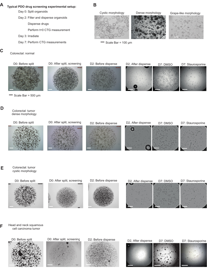
Figure 1: Organoids before and after dissociation, dispensing, and drug screening. (A) Typical schedule for PDO drug screening experiment. Depending on the experimental question and proliferation behavior of organoid models, this may require adaptation. (B) Representative brightfield (10x) images of organoids with cystic, dense, and grape-like morphology, prepared for screening purposes. Scale bars = 100 µm. (C-F) Representative brightfield (2.5x) images. (C) Cystic normal colorectal PDOs; (D) dense CRC PDOs; (E) cystic CRC PDOs; and (F) HNSCC PDOs. From left to right: day 0 (D0): before split; D0: after splitting (high density, 50% ECM); D2: before filtering and dispensing; D2: after filtering and dispensing into 384-well plate (250 organoids/well, 5% ECM); and after 5 days incubation in either 0.8% DMSO or 5 µM staurosporine, respectively, as negative and positive controls. Scale bars = 500 µm. Abbreviations: PDO = patient-derived organoid; CRC = colorectal carcinoma; HNSCC = head and neck squamous cell carcinoma; ECM = extracellular matrix; CTG = CellTiter-Glo 3D Cell Viability Assay; DMSO = dimethyl sulfoxide. Please click here to view a larger version of this figure.
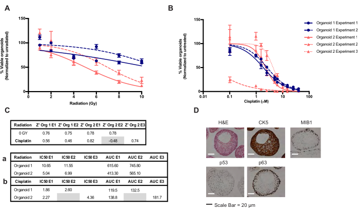
Figure 2: Representative results of a chemo- irradiation- therapy screen using HNSCC organoids. (A) Radiotherapy sensitivity in two HNSCC organoid cultures, where Y-axis displays % of viable organoids normalized to non-irradiated; X-axis displays increasing dosage of radiation (Gy). Error bars represent triplicate wells expressed as SEM; two biological repeat experiments were performed for experiment 1 (continuous line) and experiment 2 (dotted line). (B) Chemotherapy sensitivity to cisplatin of the same organoid cultures from (A), where Y-axis displays % viable organoids normalized to vehicle-only; X-axis displays increasing concentrations of cisplatin (µM). Error bars represent triplicate wells expressed as SEM; two biological repeat experiments were performed for experiment 1 (continuous line) and experiment 2 (dotted line). For experiment 2, organoid 2, one outlier (112.7%) of the triplicate wells was present for 0.1 µM cisplatin and was excluded from the analysis. (C) Table depicting the Z' for radiation and cisplatin screens. AUC and IC50 values generated from the irradiation (a) and cisplatin (b) treatments for each experiment and each organoid culture. (D) Representative images of organoid culture 1 illustrate tumor identity of the culture. From left to right: H&E stain, CK5, proliferative marker MIB1 (Ki67), wildtype p53 expression, and basal expression of p63. Scale bars = 20 µm. Abbreviation: E = experiment; SEM = standard error of the mean; HNSCC = head and neck squamous cell carcinoma; AUC = area under the curve; IC50 = concentration that results in 50% inhibition of organoid viability; H&E = hematoxylin & eosin; CK5 = Cytokeratin 5. Please click here to view a larger version of this figure.
| Morphology | Dissociation method | Org. size (µm) | # orgs/ well 384-plate | # orgs per mL for 40 µL per well | # orgs per in 25 mL (full 384-well plate) |
| Cystic | Mechanical shearing | 20-100 | 250 | 6250 | 156250 |
| Compact | TryplE + mechanical shearing | 20-40/70 | 500 | 12500 | 321500 |
| Grapelike | Gentle shearing | <40 | 1000 | 25000 | 625000 |
| Org.: short for organoid | |||||
Table 1: Recommended starting points for screen optimization.
Supplemental Figure S1: Example of experimental setup. Example of a 384-well drug screening plate layout, using the D300e software. In this example, 4 organoid cultures (rows A-D, E-H, I-L, M-P) are exposed to 10-step concentration gradients of cisplatin (red), carboplatin (green), and a fixed dose of staurosporine (yellow). Black triangles in bottom left corner of each well represent normalization wells to the highest-class concentration of drug, in this case Tween-20, as both cisplatin and carboplatin are aqueous solutions dissolved in Tween-20 for dispensing. In this example, 8 positive control wells (yellow) and 28 negative control wells (white) are included for each organoid line. As all wells are in use, edge-effects should be avoided by using permeable plate seals. Please click here to download this File.
Discussion
This article and video describe how to perform medium-throughput drug screening using PDOs. This protocol can, with optimization, be adopted to screen organoids derived from different tissue types from those described here. Determining the ideal passage timeframe prior to the screen is important as this will vary for individual organoid cultures and depend on the tissue type. The density and size of organoids seeded per well is an important factor to optimize as faster growing models will require more space within the well, and size differences may result in more variation. To ensure that only the test compounds affect organoid viability, it is important to make sure that untreated (negative control) organoids do not suffer from any deprivation during the course of the experiment. As untreated organoids will increase unhampered in volume compared to the treated populations, this assay assesses both treatment-induced cell death as well as inhibition of cell proliferation. Using this method, compounds and treatments, such as radiotherapy, can therefore be applied to organoid models to investigate in vitro responses in a three-dimensional format.
In this protocol, we make use of the CTG assay that uses ATP levels as a proxy for cell viability. Although this assay, in most instances, robustly reports the number of viable cells at the end of the experiment, it is important to realize that this assay could also be affected by severe changes in cell metabolism. Other viability assays are available that are dependent on, e.g., total DNA, protease activity, or leakage of lactate dehydrogenase, and may function as alternative or additive readouts in this assay.
Here, we discuss some additional recommendations and steps for optimization and upscaling of organoid drug screening experiments, as well as some considerations for drug screen analysis.
First, a challenge to performing (larger) drug screening is expanding the organoid culture enough to obtain a sufficient number of organoids to perform the experiment. It is therefore important to realize that many organoids tend to stick to untreated plastic, resulting in potential loss of critical cell mass. We recommend using low-retention plastics wherever possible and pre-wetting pipets/filters with aDF+++ or washing buffer prior to using them with concentrated organoid suspension.
Second, as increasing the handling time of organoids can be detrimental to their viability, we recommend working as efficiently as possible. In our experience, prolonged incubation of organoids in suspension while preparing them for dispensing negatively impacts their viability to a greater degree when kept on ice as compared to when kept at room temperature. Adding ROCK inhibitor during organoid preparation increases their viability. Moreover, in our experience, a healthy culture at the start of an experiment is absolutely required for a meaningful outcome.
Third, when processing multiple organoid models at once, there is a risk of swapping and contaminating the cultures. We therefore recommend only handling a single organoid model at a time. This could mean that multiple researchers need to work in parallel when processing larger amounts of organoid models or larger volumes of fewer organoid models. The identity of the organoid models should be verified through SNP analysis15 prior to and after screens and comparing the data to early passage and/or patient blood SNP data.
Some (targeted) therapies/compounds potentially interact/compete with growth factors/ECM present in the medium. Ideally, these potential interactions need to be identified, and the concentrations of these growth factors need to be minimized and tightly controlled to ensure that results are consistent. An example of such an interaction is epidermal growth factor (EGF)- and EGF receptor (EGFR)-targeting therapies, such as Cetuximab and Panitumumab, wherein high EGF concentrations in the medium potentially suppress EGFR expression16 or can compete with compound-EGFR binding. Ways to identify these interactions include performing a titration of the compound and a control compound against a titration of ECM or growth factors intended for the drug screen, or if the compound targets a membrane receptor (e.g., EGFR), using flow cytometry to assess receptor expression and confirm whether the ECM or compound affects antibody binding. Adjust the growth factor or ECM concentration such that there is no specific inhibition by the targeting compound compared to inhibition by the control compound.
In addition, we make the following recommendations regarding the experimental (plate) setup. First, use at least three technical replicates in each plate and ideally, two biological replicates for each screen. As seeding by most liquid handlers happens per row, we suggest putting replicates in columns to allow the detection of potential seeding issues. Second, we suggest including at least 6 (ideally >9) negative (vehicle) control wells and at least 3 (ideally >6) positive control wells on each plate. Lower numbers will likely negatively impact the Z' of the experiment. If multiple organoid models and/or solvents are used throughout the plate, include controls for each one. Ideally, the maximum concentration of solvent is 0.8% v/v for DMSO and 3% v/v for PBS/0.3% Tween. Greater than 1% and 5% solvent, respectively, will induce cell death and thus decrease the quality of screening results. Third, for accurate IC50 calculations, it is recommended to measure the organoid response to at least 9 drug concentrations, of which 2 fall in the upper plateau and 2 in the lower plateau, leaving 5 concentrations in the sigmoid phase17.
Users must note that the 384-well (or any multi-well) plate setup is sensitive to edge-effects (i.e., different results in the edges of the plate due to evaporation of medium). Practices to prevent this from affecting the results are the following: ensure a good humidification of the CO2 incubator (>85%), place the plates in the back of the incubator and prevent repetitive opening and closing, fill the outer wells with (cell-less) medium (water/PBS does not prevent the effect), or use permeable plate seals. Note that dispensing machines can only select per two rows in a 384-well format, meaning 4 rows are lost when dismissing the outer two tubes.
Recommendations for optimization for screening using novel organoid models are as follows. First, different organoid cultures have different growth kinetics and different basal ATP levels, resulting in different CTG readings. We therefore recommend optimizing the number of organoids per well for each organoid type (recommended range: 100-1,000 organoids/well), using Table 1 for reference. For optimization, assess Z' by measuring negative and positive controls with different organoid numbers.
Moreover, monitor the morphology of the organoids at the end of the screening period, and ensure the protocol results in healthy-looking organoids in the negative controls; any sign of deprivation during the course of the experiment should be avoided. It is important to note that different numbers of organoids/well can affect drug sensitivity. Further, different compound incubation times can affect the optimal amount of organoids/well. If organoids tend to fuse, using a higher percentage of ECM (v/v, up to 10%) could be beneficial. Additionally, some organoid models do not cope well with low ECM concentrations. If negative controls do not show regular proliferation or exhibit a changed morphology compared to normal expansion conditions, one should consider using 10% (v/v) ECM instead. It is worth mentioning that high ECM concentration can influence compound/antibody binding and effectiveness, lysis efficiency, and therefore activity in the CTG assay and subsequently, IC50 values. Once the screening assay performs well in smaller (pilot) screens, it is possible to increase the throughput of the assay.
Considerations to take into account when upscaling the screening assay are as follows. First, proliferation and differentiation of organoids are influenced not only by handling, but also by batch differences in ECM and growth factors. For screens performed over time to be comparable, it is important to ensure that sufficient amounts of growth factors, medium, and ECM from the same batch (comparable quality) are available to perform the complete screen. Additionally, we recommend including 1-2 organoid cultures that are used in every drug screen. These organoid cultures can act as controls to check and ensure reproducibility across drug screens. Shearing of organoids becomes less efficient in the presence of high concentrations of organoids and extracellular ECM. Increasing the number of organoids in a tube can therefore lead to a more than proportional increase in handling time. As prolonged handling of the organoids is often detrimental to their viability, the use of a dispase at critical steps can greatly improve the quality of the resulting organoid cultures. The D300e drug dispenser described in this protocol is a very flexible system and suited for smaller screens with only a few screening plates and a limited number of compounds. The software is however limited to drug addition to four screening plates per run, and the stock compounds need to be manually added to the cassette for each run.
When performing a screen with large numbers of plates or large compound libraries, it could be worthwhile using a liquid handler instead. Within the described protocol, there is space to add up to 5 µL of compound diluted in medium to each well without compromising the readout (taking solvent limits into account).
Downscaling of throughput is possible as well. This protocol can easily be adapted to a lower-throughput screen using a 96-well plate format. Both the cell dispenser and the robot drug printer described here are adaptable to dispense in a 96-well plate format. Manual pipetting is possible, but should be accompanied by careful quality assurance as there are higher chances of mistakes and of organoids being less evenly dispensed. Where the 384-well plate format uses 40 µL per well, doubling this in a 96-well plate format would serve as a good starting point; however, this should be further optimized prior to the screen.
The following points should be considered regarding drug screen data analysis. First, we recommend always taking a quick glance at the data by colorizing the raw data min/max, as seeding and edge-effects will become easily apparent this way. Second, to reiterate, Z' is an important metric to assess the dynamic range of each drug screen assay13 and should be calculated for each plate. Excluding drug screen results with a Z' lower than 0.3 and using data with a Z' > 0.5 is recommended. For the Z' to be informative, it is important that all organoids in the positive control wells have died. Third, it is recommended to always check curve-fitting after IC50 calculations. If curve-fitting appears difficult, the AUC parameter should be analyzed instead. Both IC50 and AUC reflect the effects of the tested drugs on both cell proliferation and cell death. Alternatively, organoid growth can be taken out of this equation by calculating the proliferation rate for normalization. The so-called GR metric requires an extra measurement at day 2 (described in section 7) and allows for easier comparison between fast- and slow-growing organoid line responses. However, for many drugs, proliferation effects are also to be expected, and these are dismissed by looking at this metric. Therefore, careful analysis of multiple metrices allows for the best output of this type of experiments.
Disclosures
MP and QXL are full-time employees of Crown Bioscience. RO and SB are full-time employees of Hubrecht Organoid Technology (HUB). HC is inventor on several patents related to organoid technology; his full disclosure is given at https://www.uu.nl/staff/JCClevers/. ED is inventor on a patent related to HN organoid technology. HC is founder of OrganoidZ, which employs organoids for drug development.
Acknowledgements
We thank Annemarie Buijs, Xiaoxi Xu, and Federica Parisi for discussions and valuable input, and Ingrid Boots and Marjolijn Gross for technical assistance.
Materials
| Name | Company | Catalog Number | Comments |
|---|---|---|---|
| Required equipment | |||
| 384-well bioluminescence platereader; e.g. Tecan Spark 10M plate reader | Tecan | ||
| Brightfield microscope with large field of view lens (2.5x) | |||
| Digital dispenser; e.g. Tecan D300e | Tecan | Drug dispensing | |
| 6 MV photon beam irradiator | Elekta model Synergy, Elekta Sweden | ||
| Liquid handler with large nozzle (“standard tube”) cassettes; | |||
| e.g. Multidrop Combi Reagent Dispenser | Thermo Scientific | ||
| Plastic container with plate holder insert for radiotherapy | Home-made | ||
| Spark control method editor software | |||
| Standard tissue culturing equipment (LAF cabinet, incubator, centrifuge, waterbath, etc) | |||
| Required materials | |||
| 1.5 mL plastic tubes | |||
| 15- and 50-mL plastic tubes | |||
| 5, 10- and 25-mL sterile plastic pipets | |||
| 6-well cell culture plates | |||
| Black 384-well ultra-low-attachment clear-bottom plate; e.g.. Corning 384 flat black | Corning | 4588 | |
| Breathe-Easy sealing membrane | Merck | pre-cut polyurethane medical-grade membrane with acrylic adhesive | |
| Glasstic slide | 10-chambered slide with hemocytometer grid | ||
| Multidrop Combi Reagent Dispenser standard tube dispensing cassette | Thermo Scientific | ||
| Plugged Pasteur’s pipet of which the tip has been tightened in a flame | |||
| Reversible 20/40/70/100 µm filters: PluriStrainer | Pluriselect | e.g. 43-50020-03 | |
| Sterile P1000, P200, P20 and P2 pipet tips and low-retention filter tips ( e.g. Sapphire tips) | Greiner | 750266 | |
| T8 Plus and D4 Plus casettes | HP/Tecan | ||
| Required reagents | |||
| 100 x Glutamax | L-glutamine substitute | ||
| 1 M HEPES | |||
| 30% (v/v) Tween-20 diluted in PBS | |||
| 70% EtOH | |||
| Advanced-DMEM/F12 | Thermo Scientific | 12634-010 | |
| CellTiter-Glo 3D cell viability assay | Promega | G9681 | |
| Compounds to test screen, including Staurosporin or other positive control | |||
| Dispase II | Sigma-Aldrich | D4693 | |
| DMSO | |||
| ECM for CRC: growth-factor reduced Matrigel, phenol-free | Corning | 356231 | |
| ECM for HNSCC PDOs: BME, Cultrex RGF Basement membrane extract, Type R1 | R&D Systems | 3433-005-R1 | |
| Expansion growth medium (specific for each organoid type) | |||
| Organoid growth factors (specific for each organoid type) | |||
| PBS | |||
| Pen/Strep (100 U/mL) | |||
| ROCK inhibitor: Y-27632 | Abmole | M1817 | |
| TrypLE | |||
| Required Software Packages: | |||
| GraphPad Prism | |||
| Microsoft Excel |
References
- Tiriac, H., et al. Organoid profiling identifies common responders to chemotherapy in pancreatic cancer. Cancer Discovery. 8 (9), 1112-1129 (2018).
- Sachs, N., et al. A Living biobank of breast cancer organoids captures disease heterogeneity. Cell. 172 (1-2), 373-386 (2018).
- Yan, H. H. N., et al. A comprehensive human gastric cancer organoid biobank captures tumor subtype heterogeneity and enables therapeutic screening. Cell Stem Cell. 23 (6), 882-897 (2018).
- Ganesh, K., et al. A rectal cancer organoid platform to study individual responses to chemoradiation. Nature Medicine. 25 (10), 1607-1614 (2019).
- Ooft, S. N., et al. Patient-derived organoids can predict response to chemotherapy in metastatic colorectal cancer patients. Science Translational Medicine. 11 (513), (2019).
- Vlachogiannis, G., et al. Patient-derived organoids model treatment response of metastatic gastrointestinal cancers. Science. 359 (6378), 920-926 (2018).
- Yao, Y., et al. Patient-derived organoids predict chemoradiation responses of locally advanced rectal cancer. Cell Stem Cell. 26 (1), 17-26 (2019).
- de Witte, C. J., et al. Patient-derived ovarian cancer organoids mimic clinical response and exhibit heterogeneous inter- and intrapatient drug responses. Cell Reports. 31 (11), 107762(2020).
- van de Wetering, M., et al. Prospective derivation of a living organoid biobank of colorectal cancer patients. Cell. 161 (4), 933-945 (2015).
- Driehuis, E., et al. Oral mucosal organoids as a potential platform for personalized cancer therapy. Cancer Discovery. 9 (7), 852-871 (2019).
- Sachs, N., et al. Long-term expanding human airway organoids for disease modeling. The EMBO Journal. 38 (4), 100300(2019).
- Driehuis, E., Kretzschmar, K., Clevers, H. Establishment of patient-derived cancer organoids for drug-screening applications. Nature Protocols. 15 (10), 3380-3409 (2020).
- Zhang, J. -H., Chung, T. D. Y., Oldenburg, K. R. A simple statistical parameter for use in evaluation and validation of high throughput screening assays. Journal of Biomolecular Screening. 4 (2), 67-73 (1999).
- Hafner, M., Niepel, M., Chung, M., Sorger, P. K. Growth rate inhibition metrics correct for confounders in measuring sensitivity to cancer drugs. Nature Methods. 13 (6), 521-527 (2016).
- Chen, X., Qian, W., Song, Z., Li, Q. -X., Guo, S. Authentication, characterization and contamination detection of cell lines, xenografts and organoids by barcode deep NGS sequencing. NAR Genomics and Bioinformatics. 2 (3), (2020).
- Driehuis, E., et al. Patient-derived head and neck cancer organoids recapitulate EGFR expression levels of respective tissues and are responsive to EGFR-targeted photodynamic therapy. Journal of Clinical Medicine. 8 (11), 1880(2019).
- Sebaugh, J. L. Guidelines for accurate EC50/IC50 estimation. Pharmaceutical Statistics. 10 (2), 128-134 (2011).
Reprints and Permissions
Request permission to reuse the text or figures of this JoVE article
Request Permission