Here, we describe a method of generating glioblastoma (GBM) organoids from primary patient specimens or patient-derived cell cultures and maintaining them to maturity. These GBM organoids contain phenotypically diverse cell populations and recreate tumor microenvironments ex vivo.
Method Article
Maintaining Human Glioblastoma Cellular Diversity Ex vivo using Three-Dimensional Organoid Culture
In This Article
Summary
Abstract
Glioblastoma (GBM) is the most commonly occurring primary malignant brain cancer with an extremely poor prognosis. Intra-tumoral cellular and molecular diversity, as well as complex interactions between tumor microenvironments, can make finding effective treatments a challenge. Traditional adherent or sphere culture methods can mask such complexities, whereas three-dimensional organoid culture can recapitulate regional microenvironmental gradients. Organoids are a method of three-dimensional GBM culture that better mimics patient tumor architecture, contains phenotypically diverse cell populations, and can be used for medium-throughput experiments. Although three-dimensional organoid culture is more laborious and time-consuming compared to traditional culture, it offers unique benefits and can serve to bridge the gap between current in vitro and in vivo systems. Organoids have established themselves as invaluable tools in the arsenal of cancer biologists to better understand tumor behavior and mechanisms of resistance, and their applications only continue to grow. Here, details are provided about methods for generating and maintaining GBM organoids. Instructions of how to perform organoid sample embedding and sectioning using both frozen and paraffin-embedding techniques, as well as recommendations for immunohistochemistry and immunofluorescence protocols on organoid sections, and measurement of total organoid cell viability, are all also described.
Introduction
Glioblastoma (GBM) is the most commonly occurring primary brain tumor with a grim prognosis of approximately 15 months from diagnosis1. Treatments that are effective in preclinical studies can often be poorly effective in patients2,3. Poor clinical response is attributed to many factors, including the microenvironmental heterogeneity of GBM and complex intra-tumoral interactions. These can be difficult to recreate in the laboratory setting with traditional adherent or sphere culture methods4. The presence of a subset of self-renewing cancer stem cells (CSCs) within GBM may also contribute to this complexity5,6. CSCs are crucial for tumor propagation and maintain tumor growth by promoting active angiogenesis, cancer invasion, and resistance to therapies including radiation7,8,9. CSCs are not uniformly distributed throughout tumors but rather are enriched within specific microenvironments, including the perivascular niche and perinecrotic regions, which each provide distinct molecular regulation of their cellular states10,11,12,13,14. CSCs are not passive recipients of microenvironmental cues but instead possess the ability to remodel their own microenvironments7,15,16. The microenvironment of a CSC can promote maintenance of stem cell state in response to pressures such as nutrient scarcity, pH, and hypoxia17,18,19,20, suggesting the importance of these conditions in a model system. Recapitulation of the diverse cellular microenvironment within tumors is therefore critical to understanding therapeutic resistance and identifying novel therapies.
Three-dimensional culture has increased in popularity in recent years21,22. Organoids have been used in other types of cancer, and the primary goal of maintaining cells as organoids is to allow for growth of heterogeneous cell populations (many of which may normally be outcompeted in more homogeneous sphere culture) and spatial diversity, seen as regional tumor microenvironments with genetic specificity4,23,24,25,26. There are many methods for three-dimensional culture of cancer cells, which each have advantages and disadvantages27,28,29. Organoid culture is not intended to be a replacement for traditional adherent or sphere culture. It is best used as a complementary technique to two-dimensional methods when there are specific questions where the interaction between cell microenvironment and tumor cell responses is critical.
This article describes reliable and repeatable methods to generate GBM organoids from either primary patient samples or patient-derived cultures. We address two different objectives for three-dimensional organoid culture: (1) establishing organoids from primary patient tissue, with maximal engraftment potential regardless of uniformity, or (2) growing uniform organoids for more quantitative experimental use. When establishing a primary specimen as organoids, it is not necessary to filter for single cells or count cells, because keeping maximum cell numbers and types to establish the initial culture is a priority. When growing organoids for comparative experiments, however, single-cell filtration and cell counting are needed to ensure replicate organoids are comparable for experimental consistency. This protocol details how to establish organoid cultures and create uniform organoids, as well as refined methods for embedding and preserving organoids and standard cell culture experiments, including immunohistochemistry, immunofluorescence, and assessment of total cell viability in GBM organoids.
Protocol
All steps of the protocol(s) detailed below were developed and conducted in accordance with Cleveland Clinic Institutional Review Board (IRB) Protocol #2559 and Institutional Biosafety Committee (IBC) approval #1711. Spheres and organoids are cultured in "Neurobasal Media Complete" (NBMc). See Table 1 for instructions.
1. Making organoid molds
- Take the wax paper off the parafilm sheet (roughly 4 cm x 4 cm in size) and place it between two sterile 96-well polymerase chain reaction (PCR) plates.
- Perform these steps in the culture hood and take special care to keep the "inside" of the parafilm (the side that is covered by the paper) clean. This clean side should make the concavity of the indentations.
- Apply even pressure to the top 96-well PCR plate to form small dimples in the parafilm. The goal is to have dimples be roughly 2 mm in depth without creating holes in the parafilm.
- Separate the two 96-well PCR plates gently. The dimpled parafilm will stick to the top plate. Place this on dry ice for about 30 s.
- After the parafilm has been on dry ice for 30 s, use sterile forceps to pull the parafilm off the top 96-well PCR plate.
- When removing the parafilm, use a quick motion rather than being very "careful". Frozen parafilm is easier to remove than warmed parafilm, which can cause the dimples to invert.
- Place the completed parafilm mold in a covered, sterile 10 cm cell culture dish. Molds can be made in advance and stored if sterility is maintained.
2. Macrodissection of the patient tissue specimen
- In a sterile culture dish (10 cm cell culture plates), use two sterile razor blades to finely mince the patient specimen while applying even pressure.
NOTE: Mincing the specimen is easiest with about 500 µL of NBMc on the culture dish. Attempt with razor blades to mince pieces as finely as possible; ideally, individual pieces are 1 mm3 or smaller. - Transfer the finely minced tumor pieces to a 15 mL centrifuge tube using a cut p1000 pipette tip.
NOTE: Whenever handling organoids, it is important to only use cut p1000 pipette tips. These can be cut using a sharp razor to desired opening size (between 5 and 8 mm roughly) and autoclaved. - Add 2 mL of room temperature (RT) cell detachment solution (Table of Materials) and place it in a 37 °C, 5% CO2 incubator for approximately 10 min.
NOTE: Observe and mix every few minutes. Some cell detachment solutions or specimens may need varying incubation times. If clumping appears, which can indicate DNA release due to cell lysis, proceed to the next step immediately. - Add 8 mL of NBMc media to neutralize the cell detachment solution, and spin for 3 min at 65 x g.
- Aspirate the supernatant and resuspend the tissue in 1-2 mL of NBMc.
3. Generating organoids from primary patient tissue
NOTE: The goal when making organoids from primary patient tissue is to establish three-dimensional culture. Do not filter for single cells or count cells, but keep the initial cell load as uniform as possible using visual inspection. It is normal to have heterogeneity in the growth and establishment of initial organoids. Each organoid will be 20 µL in volume (16 µL of laminin-rich extracellular matrix (lrECM) and 4 µL of tissue suspended in NBMc, from step 2.5). Instructions can be adjusted for the number of organoids intended; the goal is to typically form around 20-30 organoids from primary patient specimens.
- In an ice bucket or cold block, place the lrECM and a small centrifuge tube. Place the appropriate amount of lrECM (16 µL x X number of intended organoids) into the small centrifuge tube.
- Add appropriate volume of tissue suspension from step 2.5 (4 µL x number of intended organoids) to the centrifuge tube on ice.
- Carefully pipette 20 µL of the lrECM/cell suspension mixture onto parafilm molds; this will form a pearl-like droplet.
- Be sure to mix the lrECM/cell suspension mixture thoroughly; the cells tend to settle easily within the lrECM, resulting in heterogeneous organoids.
- Keep the lrECM/cell suspension mixture on ice. If lrECM warms, it can polymerize and compromise organoid formation. Be sure to cool the pipette tip every two to three organoids to prevent lrECM polymerization. Do not introduce air bubbles into organoids (avoid "double pushing" the pipette).
- Once the desired number of organoids are pipetted onto parafilm mold in a 10 cm cell culture plate, cover with plate lid and incubate at in a cell culture incubator at 37 °C, 5% CO2 for 1-2 h.
- After the organoids solidify, use NBMc media to flush them gently off the parafilm mold and into a new, sterile 10 cm culture plate with 20 mL of NBMc total. Using a p1000 tip works best to flush the organoids off the mold; they will slide off gently.
NOTE: When organoids are flushed from the molds into cell culture plates, they should be visible in the media as small pink spheres. If the organoids appear to have fallen apart or shattered in the media, this indicates an earlier issue with lrECM having polymerized, and organoids may still grow but will be very unlikely to be uniform in size. - Place the 10 cm culture dish in a cell culture incubator at 37 °C, 5% CO2 (without shaking) for 4 days.
NOTE: Placing organoids on an orbital shaker in the initial days can cause them to fall apart. Be sure they are not shaking until day 4. - After 4 days, exchange the media and place on an orbital shaker at 80 RPM in a cell culture incubator at 37 °C, 5% CO2.
- Exchanging media with immature organoids is challenging because they are difficult to visualize. Tilt the cell culture dish and wait for at least 20 s; the organoids will settle at the bottom and allow the media from above to be removed with a glass or plastic 10-20 mL pipette slowly.
- Pay careful attention to the collection of organoids at the bottom; if they appear stirred up with the force of media removal, it is best to pause and allow them to resettle. As organoids mature and are easier to visualize, this process becomes less nuanced.
NOTE: Sometimes, placing a piece of dark paper beneath the cell culture plate can help visualize immature organoids. It is advised not to use a Pasteur pipette with vacuum suction to remove media as it is very easy for the organoids to be sucked up and lost this way. - When organoids are first established, they do not consume media as rapidly as mature organoids. Beginning with 50% media exchanges reduces unnecessary media use and reduces the chance of accidentally damaging or aspirating new organoids during the media exchange process.
4. Generating organoids from established GBM sphere, adherent, or organoid culture
NOTE: The goal here is to make organoids that are uniform in size and cell quantity for use in comparative experiments, so use a single-cell filter and count cells to ensure this.
- Preparing single-cell suspension from GBM sphere culture.
NOTE: Sphere cultures of GBM cells are maintained in NBMc media.- Place spheres in a 15 mL centrifuge tube and spin at 120 x g for 5 min.
- Remove the supernatant and add 2 mL of RT cell detachment solution. Place in a 37 °C incubator for 3 min.
- Add 8 mL of NBMc media to neutralize the cell detachment solution. Strain through a single cell strainer (70 µm) and spin for 5 min at 120 x g.
- Remove the supernatant from the tube and resuspend the remaining cells in ~1 mL of NBMc. Count the cells (using cell impermeant stain) and skip to step 4.4.
- Preparing single-cell suspension from GBM adherent culture.
- Remove the used media from the plate, add 2 mL of RT cell detachment solution to the plate, and place in a 37 °C incubator for 3 min.
- Confirm under the microscope that the cells are detached from the plate.
- Add 8 mL of NBMc media to neutralize cell detachment solution. Strain through a single-cell strainer (70 µm).
NOTE: Single cell straining is optional when working with adherent culture. - Spin for 5 min at 120 x g. Remove the supernatant from the tube and resuspend the remaining cells in ~1 mL of NBMc. Count the cells (using cell impermeant stain) and skip to step 4.4.
- Preparing single-cell suspension from GBM organoid culture.
- Transfer the organoids using a cut p1000 pipette tip to a 10 cm culture plate and carefully remove as much residual media as possible.
- Using two sterile razors, carefully mince organoids finely. With the cut p1000 tip, move the minced organoids to a 15 mL centrifuge tube and add ~2-3 mL of NBMc media.
- Spin at 120 x g for 3 min and remove the supernatant (recommend using a pipette tip rather than vacuum suction for this part).
- Add 2 mL of cold cell detachment solution for 10 min.
NOTE: Cell detachment solution should be used directly from 4 °C, not warmed first. This appears to soften any remaining matrigel and aids in cell recovery. - Then move to a 37 °C incubator for 10-20 min, observing and mixing every few minutes. If clumping appears, which can indicate cell lysis, proceed to the next step immediately.
- Add 8 mL of NBMc media to dilute the cell detachment solution. Strain through a single-cell strainer (70 µm) and spin for 5 min at 120 x g.
- Remove the supernatant from the tube and resuspend the remaining cells in ~1 mL of NBMc. Count the cells (using cell impermeant stain) and proceed to step 4.4.
- Making organoids from a single-cell suspension
- In an ice bucket or cold block, place the lrECM and a small centrifuge tube. Place the appropriate amount of lrECM (16 µL x X number of intended organoids) into the small centrifuge tube.
- Create a mixture of cells in a total volume (4 µL x X number of intended organoids) that will contain 20,000 cells/organoid and add this to the small centrifuge tube with lrECM on ice.
- Carefully pipette 20 µL of the lrECM/cell suspension mixture onto parafilm molds; this will form a pearl-like droplet.
- Be sure to mix the lrECM/cell suspension mixture thoroughly, as the cells tend to settle easily within the lrECM, and this will result in heterogeneous organoids.
- Keep the lrECM/cell suspension mixture on ice. If lrECM warms, it can polymerize and compromise organoid formation.
- Be sure to cool the pipette tip every two to three organoids to prevent lrECM polymerization. Do not introduce air bubbles into organoids (avoid "double pushing" the pipette tip).
- Once the desired number of organoids are pipetted onto parafilm mold in a 10 cm culture plate, incubate at 37 °C for 1-2 h in a cell culture incubator.
- After organoids solidify, use NBMc media to flush them gently off the parafilm mold and into a new, sterile 10 cm culture plate with 20 mL of NBMc total. Using a p1000 tip works best to flush organoids off the mold; they will slide off gently.
NOTE: About 15-20 organoids per 10 cm culture dish is recommended. - Place the 10 cm culture dish in an incubator (without shaking) for 4 days.
- After 4 days, exchange the media and place on an orbital shaker at 80 RPM in the cell culture incubator. Exchange media every 2-3 days thereafter.
- Exchanging media with immature organoids is challenging because they are difficult to visualize. Tilt the cell culture dish and wait for at least 20 s; the organoids will settle at the bottom and allow the media from above to be removed with a large opening glass pipette slowly.
- Pay careful attention to the collection of organoids at the bottom; if they appear stirred up with the force of media removal, pause and allow them to resettle. As organoids mature and are easier to visualize, this process becomes less nuanced.
NOTE: Sometimes, placing a piece of dark paper beneath the cell culture plate can help visualize immature organoids. Do not use a Pasteur pipette with vacuum suction to remove media as it is very easy for the organoids to be sucked up and lost this way. - When organoids are first established, they do not consume media as rapidly as mature organoids. Begin with 50% media exchanges to reduce unnecessary media use and reduce the chance of accidentally damaging or aspirating new organoids during the media exchange process.
5. Cryoembedding
- Place each organoid in an individual 1.5 mL tube (using cut p1000 tip) containing 1 mL of 4% paraformaldehyde (PFA). Store organoids overnight at 4 °C. If embedding organoids in paraffin, proceed to section 6.
- After overnight fixation in 4% PFA, wash the organoids in 1x phosphate-buffered saline (PBS) three times.
- Transfer the organoid to a new 1.5 mL tube (using cut p1000 pipette tip) containing 1 mL of 30% sucrose in water and store at 4 °C overnight.
- Add a small amount (typically 1-2 mL) of optimum cutting temperature compound (OCT) to a cryomold, covering the bottom and filling about 1/3 to 1/2 of the depth of the mold.
- Transfer a single organoid to cryomold using cut p1000 pipette tip. This will result in some media transferring with the organoid, which is unavoidable. Use a smaller pipette tip to carefully remove surrounding media without disturbing the organoid.
NOTE: This can be difficult to visualize, but slow pipetting will demonstrate a clear difference in densities between media and OCT, so this is done "by feel" to some extent). - Place the cryomold on a tray of dry ice. OCT will begin freezing, becoming opaque in the process.
- Add additional OCT to completely cover the organoid, filling the rest of the volume of the cryomold. Blocks can be stored at -20 °C for short-term storage for several days or at -80 °C for long-term storage indefinitely.
6. Paraffin embedding
- Sort organoids by size (small organoids are below 3 mm, large organoids are over 3 mm). Transfer each organoid to a histology cassette using a cut p1000 pipette tip and process into paraffin wax according to either Table 2 or Table 3.
NOTE: The size/configuration of the histology cassette is up to the user. - Transfer organoids from the cassettes to embedding molds with 1-2 mL of melted paraffin wax and chill it until the wax is semisolid.
- Once the wax is partially solidified, add more wax to the top of the mold. Place the labeled cassette top over the mold and transfer this to a cold plate. Continue cooling until the wax is completely solid.
- When the wax has solidified, section the block using a microtome. Alternatively, store at RT or 4 °C for later sectioning.
7. Immunofluorescence (IF)
- De-paraffinize and rehydrate 5-12 μm paraffin-embedded tissue sections using a slide staining dish, according to the following steps.
NOTE: If using sections from OCT-embedded organoids, we recommend using 12 μm sections. Remove OCT by shaking slide in PBS for 30 min. Skip to step 7.2.- Incubate for 5 min in xylene. Repeat this two more times. Then incubate for 10 min in 100% ethanol. Repeat this once more.
- Incubate for 10 min in 95% ethanol. Repeat once more. Wash the sections in distilled water for 5 min, two times.
- For antigen unmasking, submerge slides in 1x citrate unmasking solution (Table of Materials) and microwave at sub-boiling temperature for 10 min. Make sure not to let the solution boil.
NOTE: This is best achieved if microwaved initially for ~2 min until boiling occurs, then lowering power and watching to ensure the solution does not boil over. The preferred unmasking temperature is just below 100 °C, ideally 98 °C. - Let the slides cool for 30 min at RT in the 1x citrate unmasking solution.
NOTE: This is the same 1x citrate solution; the solution need not be replaced at this step. - Wash the slides in distilled water for 5 min, two times.
- Wash the slides in 1x TBST (Tris-buffered saline with 0.1% Tween 20) buffer for 5 min.
- Remove the slides from TBST and carefully dry them around tissue sections using a laboratory cleaning wipe while being careful not to let the tissue section dry. Once the slide is sufficiently dry, circle tissue sections with a hydrophobic barrier pen.
- Block each section with 100-400 µL of 10% serum blocking solution for 1 h at RT. Choose the serum blocking solution based upon the secondary antibody. For example, if using a secondary antibody made in donkey, use a 10% normal donkey serum in 1x TBST.
- Remove the blocking solution and then add 100-400 µL of primary antibody diluted to desired concentration in the blocking solution.
- At 4 °C, incubate the sections with the primary antibody overnight.
- Remove the primary antibody solution and wash the slides in 1x TBST for 5 min, three times.
- Add 100-400 µL of secondary antibody (1:1000 dilution in blocking solution, or per the manufacturer's directions) to each section and incubate for 1.5 h at RT.
- Wash the slides in 1x TBST for 5 min, two times. Then wash the slides in 1x PBS for 5 min.
- Remove the slides from PBS and dry them around tissue sections using a laboratory cleaning wipe. Add a few drops of liquid curing mountant and mount a glass coverslip carefully. Once the slides are dry, store at -20 °C protected from light until ready for imaging.
8. Immunohistochemistry
- De-paraffinize and rehydrate the paraffin-embedded tissue sections using a slide staining dish, according to the following steps.
NOTE: If using sections from OCT-embedded organoids, remove OCT by shaking the slide in PBS for 30 min. Skip to step 8.2.- Incubate for 5 min in xylene. Repeat two more times.
- Incubate for 10 min in 100% ethanol. Repeat once more.
- Incubate for 10 min in 95% ethanol. Repeat once more.
- Wash sections in distilled water for 5 min, two times.
- For antigen unmasking, submerge slides in 1x citrate unmasking solution and microwave at sub-boiling temperature for 10 min. Make sure to not let the solution boil.
- Let slides cool for 30 min at RT in the 1x citrate unmasking solution.
- Wash the slides for 5 min in distilled water, three times.
- Incubate the slides for 10 min in 3% hydrogen peroxide.
- Wash the slides for 5 min in distilled water twice.
- Wash the slides for 5 min in 1x TBST.
- Remove the slides from 1x TBST and then use the corner of a laboratory cleaning wipe to carefully dry around tissue sections.
- Circle the tissue sections with a hydrophobic barrier pen once the slide is dry.
- Place 100-400 µL of the blocking solution on each section within the hydrophobic barrier pen circle at RT for 1 h. Use either 10% normal donkey serum (NDS) or 0.75% bovine serum albumin (BSA) diluted in 1x TBST.
- Next, remove the blocking solution and add 100-400 µL of the primary antibody to each section. Dilute this primary antibody to the desired concentration using the appropriate manufacturer's diluent.
- Incubate the sections at 4 °C overnight. Remove the primary antibody solution and wash the slides in 1x TBST for 5 min, three times.
- Add 100-400 µL of IHC detection reagent to each section and incubate for 1 h at RT. Choose the IHC detection reagent according to the primary antibody.
- Wash each section with 1x TBST for 5 min, two times, followed by 1x PBS for 5 min once.
- Prepare 3,3-Diaminobenzidine (DAB) solution according to the manufacturer's directions. Add 100-400 µL of DAB solution to each tissue section and monitor closely under a microscope. Between 1-10 min will provide acceptable staining intensity; be sure to note this time and keep consistent for all tissue sections.
- After desired staining intensity is reached, immerse the slides in distilled water.
- Perform hematoxylin counterstain and slide dehydration for mounting according to the instructions in Table 4.
- Remove the slide from the xylene substitute (Table of Materials) and wipe off extra liquid around the tissue section using a laboratory cleaning wipe. Use a small amount of permanent mounting medium to mount coverslip over tissue section and allow to dry.
9. Measurement of total cell viability
- Transfer each organoid to a 2 mL tube and, using a small pipette tip, carefully remove all excess media around the organoid (see Figure 3 for the schematic of this procedure).
NOTE: Cell viability assays must be performed with organoids that were made with identical numbers of cells. This experiment should not be performed on organoids made directly from patient specimens from surgery, from cutting alredy formed organoids, or other cases where single cells were not filtered and counted. - Prepare the luminescent cell viability assay reagent and 1x PBS in 1:1 ratio and add 500 µL to each tube.
- Use a p1000 pipette tip to aggressively pipette up and down to break down the organoid and let sit for 5 min. The organoid should be somewhat dissociated and softer by now; repeat mixing with the p1000 pipette tip again.
- The goal is to have 100 µL total volume per well of a 96-well plate. Add 25 µL of the mixture from the step 9.3 (will have enough for multiple technical replicates) and add 75 µL of remaining luminescent cell viability assay and PBS mixture.
- Place the plate on a shaker for 2 min and then incubate for 20 min at RT.
- Read the plate using a luminescence setting on a plate reader and collect the data (see Figure 4).
Results
Figure 1 shows early organoid growth seen via light microscopy at 10x magnification. Figure 1A shows migration and invasion of single cells through lrECM in the center view. The cells will continue to expand and 'colonize' the lrECM, and they will appear more dense and eventually opaque by visual inspection. Figure 1B shows several mature organoids (at 7 weeks) without magnification, relative to the size of a dime.
Figure 2 demonstrates immunohistochemical staining of GBM organoids for phospho-histone H3, a marker of active proliferation. Most highly proliferative cells are seen in the organoid perimeter compared to the organoid core. Positive staining will have a brown/copper appearance.
Figure 3 describes the process to homogenize and measure total cell number in GBM organoids using a 3D-specific luminescent cell viability assay. Due to the high number of cells present in GBM organoids, the larger organoid structure is initially homogenized by triturating in luminescent assay reagent. Then fractions of the total organoid lysate are loaded into individual wells and diluted with additional luminescent assay reagent prior to incubation and reading on an appropriate multi-well plate reader.
Figure 4 represents DMSO (common vehicle) control data for organoids. Plotted data demonstrate intra-organoid and inter-organoid consistency. Luminescent viability data will typically be normalized to controls for each specimen when generating experimental data.
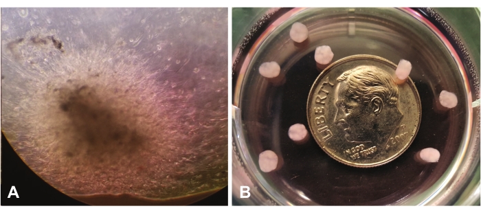
Figure 1: View through a light microscope (10x). (A) Early organoid growth demonstrating migration/invasion of GBM cells throughout the laminin-rich extracellular matrix. (B) Mature organoids relative to the size of a dime. Please click here to view a larger version of this figure.
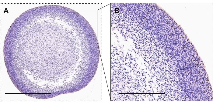
Figure 2: Immunohistochemistry of GBM organoids. (A, B) GBM organoids showing phospho-histone H3 stained cells for active proliferation. Scalebars are 600 µm and 300 µm for A and B, respectively. Please click here to view a larger version of this figure.
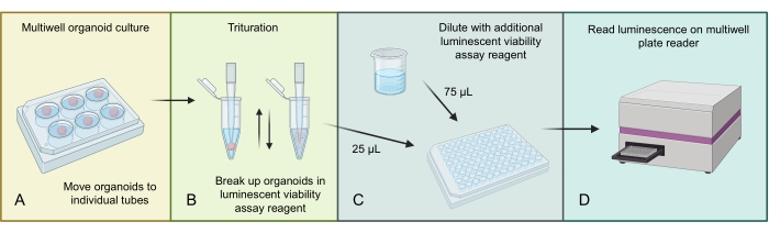
Figure 3: Luminescent cell viability protocol for organoids. (A) Move individual organoids to small centrifuge tubes and remove excess media. (B) Add 500 µL of 1:1 PBS and luminescent cell viability assay mixture to each tube and pipette aggressively to break down the organoid. (C) Add 25 µL of this organoid mixture and 75 µL of the same 1:1 PBS and luminescent cell viability assay mixture to each well of a 96-well plate. (D) Place on a shaker for 2 min, followed by incubation for 20 min at RT, and then read luminescence on a plate reader. Figure made using BioRender.com. Please click here to view a larger version of this figure.
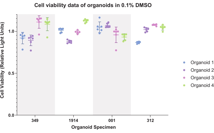
Figure 4: Cell viability. Cell viability data for organoids in 0.1% dimethyl sulfoxide (DMSO) for six technical replicates of four organoids for four patient specimens. Please click here to view a larger version of this figure.
| Component | Quantity |
| Neurobasal medium minus phenol red | 500 mL |
| B-27 supplement minus vitamin A (50x) | 10 mL |
| Antibiotic-antimycotic (100x) | 5 mL |
| Sodium pyruvate (100 mM) | 5 mL |
| Glutamine in 0.85% NaCl (200 mM) | 5 mL |
| Recombinant human FGF basic (250 µg/mL) | 20 µL |
| Recombinant human EFG protein (250 µg/mL) | 20 µL |
| Phenol red | 500 µL |
Table 1: Neurobasal medium complete (NBMc) formulation
| Reagent | Time |
| 50% Ethanol | 3 min |
| 75% Ethanol | 3 min |
| 95% Ethanol | 3 min |
| 95% Ethanol | 4 min |
| 100% Ethanol | 2 min |
| 100% Ethanol | 3 min |
| 100% Ethanol | 4 min |
| Xylene substitute | 2 min |
| Xylene substitute | 3 min |
| Xylene substitute | 4 min |
| Paraffin wax | 15 min |
| Paraffin wax | 15 min |
Table 2: Processing schedule for small organoids (below 3 mm in diameter)
| Reagent | Time |
| 50% Ethanol | 6 min |
| 75% Ethanol | 6 min |
| 95% Ethanol | 5 min |
| 95% Ethanol | 8 min |
| 100% Ethanol | 5 min |
| 100% Ethanol | 5 min |
| 100% Ethanol | 8 min |
| Xylene substitute | 5 min |
| Xylene substitute | 5 min |
| Xylene substitute | 8 min |
| Paraffin wax | 30 min |
| Paraffin wax | 30 min |
Table 3: Processing schedule for large organoids (above 3 mm in diameter)
| Reagent | Time |
| Hematoxylin | 2 min |
| Running diH2O | 2 min |
| Nuclear hematoxylin clarifying reagent | 1 min |
| Running diH2O | 1 min |
| Bluing Reagent | 1 min |
| Running diH2O | 2 min |
| 70% ethanol | 1 min |
| 100% ethanol | 1 min |
| 100% ethanol | 1 min |
| Xylene substitute | 2 min |
| Xylene substitute | 2 min |
Table 4: Hematoxylin counterstain
Discussion
GBM organoids are a complementary culture method to traditional spheres that include greater cellular and microenvironmental heterogeneity4,22,30. Although more time and resource-intensive, organoid culture can offer valuable insight into intra-tumoral behavior and mechanisms of drug resistance.
GBM is driven by a population of CSCs5,31, and these methods were developed to allow continued growth and self-renewal of this CSC population. Epidermal growth factor (EGF) and fibroblast growth factor (FGF) are known to enhance stem cell maintenance and growth and provide active receptor tyrosine kinase (RTK) signaling. The formation of heterogeneous cellular populations and distinct tumor microenvironments within GBM tumors relies on supporting CSC behaviors. The selection of an lrECM mimics the laminin-rich brain environment and supports the cells in organoid culture to self-organize and migrate by invasion. Although some groups have established organoid culture without the use of an lrECM or EGF/FGF enriched media24,28, which may offer a more time-efficient manner of this culture method and stronger selection of oncogenic signaling to drive growth, these methods were chosen to optimize the pro-stem cell environment to best establish the cellular heterogeneity of organoids. Both cerebral organoids and GBM organoids have been made with lrECM in the literature previously21,32,33. Although we have established data regarding the tumor populations found within organoids and the spatial variation, less is known about non-tumor populations within the organoids and how long they survive from the original patient specimens. Certain IHC stains (such as CD45) may provide this data, and could be an interesting point of research in the future with organoids.
Knowing the intended use for organoid culture is important for selecting appropriate methods. Establishing organoids from primary specimens versus growing uniform organoids for specific experiments have slightly different procedures. Having an appreciation for how organoids mature and visually fill in the lrECM scaffold is important for being able to allocate proper time and resources towards organoid culture. Sparse regions of cells in organoids will slowly expand and grow to fill the lrECM, which can take anywhere from 2-8 weeks, depending on the specimen's behavior. This rate of growth is somewhat intrinsic to each specimen; it is preserved across different batches of organoids and fairly consistent with the relative rate of sphere growth. Organoids can be maintained for over 1 year and retain tumor formation capabilities on xenograft into mice; however, it is recommended to grow them with a distinct purpose as to not waste lab resources (both materials and time)4. Organoid growth has been tested in multiple well sizes and formats, and shows that a 10 cm plate is the ideal setting for maintaining optimal cell viability, followed by a 6-well plate with three organoids per well34. Organoids consume more media compared to their two-dimensional culture counterparts, and using a smaller well format does not lead to proper maintenance. For example, one well of a 96-well format plate does not have enough space or media volume relative to the size of an organoid to sustain organoid growth.
As organoids establish, being observant is important for increasing success. Initially, organoids will consume media slowly, but as they become denser and more mature, they will consume media more quickly. Adding phenol red to the media can help serve as an indicator of media consumption. When the media is more yellow, it may prompt us to adjust the feeding pattern, whether it is to exchange a higher volume of media, increase the total media amount in the plate, divide organoids amongst multiple cell culture plates to keep up with their growth, or even adjust the timeline for experiments.
In many ways, organoids are an inefficient way of conducting cancer research. They involve long time scales, and are expensive and resource-heavy compared to GBM sphere culture. However, compared to patient-derived xenografts, an alternative method for recreating cellular and microenvironmental diversity, they are more straightforward, less expensive, and controllable. Selection of when to best use organoids is important for cancer researchers. They are not intended to replace traditional sphere or adherent culture and not to replace xenograft models. Organoids can, when applied to the right scientific question, combine the benefits of both these systems and may allow us to observe tumor cell biology that would otherwise remain hidden. The scientific community is only beginning to understand what learning opportunities organoids offer, but it is clear they will be an invaluable tool in the future for understanding the complex biology of GBM.
Disclosures
The authors have no conflicts to disclose.
Acknowledgements
We would like to thank Dr. Justin Lathia for his invaluable advice and ongoing support. We also thank Katrina Fife, Lisa Wallace, and Maya Camhi for their excellent technical support.
Materials
| Name | Company | Catalog Number | Comments |
|---|---|---|---|
| 3,3-Diaminobenzidine (DAB) tablets | MP Biomedicals | 08980681 | 3,3-Diaminobenzidine (DAB) tablets |
| 96-well PCR plates | ThermoFisher Scientific | 96-well PCR plates | |
| Accutase | ThermoFisher Scientific | SCR005 | Cell detachment solution |
| Antibiotic-antimycotic | ThermoFisher Scientific | 15240062 | Antibiotic-antimycotic |
| B-27 supplement minus vitamin A | ThermoFisher Scientific | 12587001 | B-27 supplement minus vitamin A (50x) |
| GelCode Blue Stain Reagent | ThermoFisher Scientific | 24590 | Bluing reagent |
| Cell strainer (70 µm) | CellTreat Scientific | 229483 | Cell strainer (70 µm) |
| CellTiter-Glo 3D | Promega | G9681 | Luminescent cell viability assay |
| Clarifier 2 | ThermoFisher Scientific | 7301 | Nuclear hematoxylin clarifying reagent |
| Clear-Rite 3 | ThermoFisher Scientific | 6901 | xylene substitute |
| Epredia Gill 2 Hematoxylin | ThermoFisher Scientific | 72504 | Hematoxylin |
| Glutamine in 0.85% NaCl | ThermoFisher Scientific | 35050061 | Glutamine in 0.85% NaCl (200 mM) |
| Matrigel | ThermoFisher Scientific | 354234 | Laminin-enriched extracellular matrix |
| Mini PAP pen | ThermoFisher Scientific | 008877 | Hydrophobic barrier pen |
| Mounting medium | ThermoFisher Scientific | 22-050-102 | Mounting medium |
| Neurobasal media minus phenol red | ThermoFisher Scientific | 12349015 | Neurobasal media minus phenol red (500 mL) |
| Normal donkey serum (NDS) | Jackson ImmunoResearch | 017-000-121 | Normal donkey serum (NDS) |
| Paraformaldehyde 4% in PBS | ThermoFisher Scientific | AAJ19943K2 | Paraformaldehyde 4% in PBS |
| Phenol red | Sigma | P0290 | Phenol red (0.5%) |
| ProLong Gold Antifade Mountant with DAPI | ThermoFisher Scientific | P10144 | Liquid curing mountant |
| Recombinant human EGF protein | R&D systems | 236-EG-01M | Recombinant human EGF protein (250 µg/mL) |
| Recombinant human FGF basic | R&D systems | 4144-TC-01 | Recombinant human FGF basic (250 µg/mL) |
| SignalStain Antibody Diluent | Cell Signaling | 8112 | Antibody diluent |
| SignalStain Boost IHC Detection Reagent | Cell Signaling | 8114 | Immunohistochemistry detection reagent |
| SignalStain Citrate Unmasking Solution | Cell Signaling | 14746 | Citrate unmasking solution |
| Single edge razor blade | Uline | H-595B | Single edge razor blade |
| Sodium pyruvate | ThermoFisher Scientific | 11360070 | Sodium pyruvate (100 mM) |
| Tissue-Tek Cryomolds | VWR | 25608-916 | Disposable cryomolds |
| Tissue-Tek O.C.T. Compound | VWR | 25608-930 | Optimal cutting temperature compound |
| Trypan Blue Stain | ThermoFisher Scientific | T10282 | Cell impermeant stain (0.4%) |
| Xylene | ThermoFisher Scientific | X3P-1GAL | Xylene |
References
- Stupp, R., et al. Radiotherapy plus concomitant and adjuvant temozolomide for glioblastoma. New England Journal of Medicine. 352 (10), 987-996 (2005).
- Heffernan, J. M., Sirianni, R. W. Modeling microenvironmental regulation of glioblastoma stem cells: a biomaterials perspective. Frontiers in Materials. 5, 7(2018).
- Zanders, E. D., Svensson, F., Bailey, D. S. Therapy for glioblastoma: is it working. Drug Discovery Today. 24 (5), 1193-1201 (2019).
- Hubert, C. G., et al. A Three-dimensional organoid culture system derived from human glioblastomas recapitulates the hypoxic gradients and cancer stem cell heterogeneity of tumors found in vivo. Cancer Research. 76 (8), 2465-2477 (2016).
- Lathia, J. D., Heddleston, J. M., Venere, M., Rich, J. N. Deadly teamwork: neural cancer stem cells and the tumor microenvironment. Cell Stem Cell. 8 (5), 482-485 (2011).
- Meyer, M., et al. Single cell-derived clonal analysis of human glioblastoma links functional and genomic heterogeneity. Proceedings of the National Academy of Sciences of the United States of America. 112 (3), 851-856 (2015).
- Bao, S., et al. Stem cell-like glioma cells promote tumor angiogenesis through vascular endothelial growth factor. Cancer Research. 66 (16), 7843-7848 (2006).
- Yu, S. C., Bian, X. W. Enrichment of cancer stem cells based on heterogeneity of invasiveness. Stem Cell Reviews and Reports. 5 (1), 66-71 (2009).
- Bao, S., et al. Glioma stem cells promote radioresistance by preferential activation of the DNA damage response. Nature. 444 (7120), 756-760 (2006).
- Vescovi, A. L., Galli, R., Reynolds, B. A. Brain tumour stem cells. Nature Reviews. Cancer. 6 (6), 425-436 (2006).
- Zhu, T. S., et al. Endothelial cells create a stem cell niche in glioblastoma by providing NOTCH ligands that nurture self-renewal of cancer stem-like cells. Cancer Research. 71 (18), 6061-6072 (2011).
- Calabrese, C., et al. A perivascular niche for brain tumor stem cells. Cancer Cell. 11 (1), 69-82 (2007).
- Charles, N., et al. Perivascular nitric oxide activates notch signaling and promotes stem-like character in PDGF-induced glioma cells. Cell Stem Cell. 6 (2), 141-152 (2010).
- Seidel, S., et al. A hypoxic niche regulates glioblastoma stem cells through hypoxia inducible factor 2 alpha. Brain. 133, 983-995 (2010).
- Yan, K., et al. Glioma cancer stem cells secrete Gremlin1 to promote their maintenance within the tumor hierarchy. Genes & Development. 28 (10), 1085-1100 (2014).
- Cheng, L., et al. Glioblastoma stem cells generate vascular pericytes to support vessel function and tumor growth. Cell. 153 (1), 139-152 (2013).
- Flavahan, W. A., et al. Brain tumor initiating cells adapt to restricted nutrition through preferential glucose uptake. Nature Neuroscience. 16 (10), 1373-1382 (2013).
- Hjelmeland, A. B., et al. Acidic stress promotes a glioma stem cell phenotype. Cell Death and Differentiation. 18 (5), 829-840 (2011).
- Bar, E. E., Lin, A., Mahairaki, V., Matsui, W., Eberhart, C. G. Hypoxia increases the expression of stem-cell markers and promotes clonogenicity in glioblastoma neurospheres. The American Journal of Pathology. 177 (3), 1491-1502 (2010).
- Li, Z., et al. Hypoxia-inducible factors regulate tumorigenic capacity of glioma stem cells. Cancer Cell. 15 (6), 501-513 (2009).
- Azzarelli, R., Ori, M., Philpott, A., Simons, B. D. Three-dimensional model of glioblastoma by co-culturing tumor stem cells with human brain organoids. Biology Open. 10 (2), 056416(2021).
- Linkous, A., et al. Modeling patient-derived glioblastoma with cerebral organoids. Cell Reports. 26 (12), 3203-3211 (2019).
- da Silva, B., Mathew, R. K., Polson, E. S., Williams, J., Wurdak, H. Spontaneous glioblastoma spheroid infiltration of early-stage cerebral organoids models brain tumor invasion. SLAS Discovery: Advancing Life Sciences R & D. 23 (8), 862-868 (2018).
- Jacob, F., et al. A Patient-derived glioblastoma organoid model and biobank recapitulates inter- and intra-tumoral heterogeneity. Cell. 180 (1), 188-204 (2020).
- Boretto, M., et al. Patient-derived organoids from endometrial disease capture clinical heterogeneity and are amenable to drug screening. Nature Cell Biology. 21 (8), 1041-1051 (2019).
- Huang, L., et al. Ductal pancreatic cancer modeling and drug screening using human pluripotent stem cell-and patient-derived tumor organoids. Nature Medicine. 21 (11), 1364-1371 (2015).
- Ledur, P. F., Onzi, G. R., Zong, H., Lenz, G. Culture conditions defining glioblastoma cells behavior: what is the impact for novel discoveries. Oncotarget. 8 (40), 69185-69197 (2017).
- Lenin, S., et al. A drug screening pipeline using 2D and 3D patient-derived in vitro models for preclinical analysis of therapy response in glioblastoma. International Journal of Molecular Sciences. 22 (9), 4322(2021).
- Rybin, M. J., Ivan, M. E., Ayad, N. G., Zeier, Z. Organoid models of glioblastoma and their role in drug discovery. Frontiers in Cellular Neuroscience. 15 (4), 605255(2021).
- Shakya, S., et al. Altered lipid metabolism marks glioblastoma stem and non-stem cells in separate tumor niches. Acta Neuropathologica Communications. 9 (1), 101(2021).
- Lee, J., et al. Tumor stem cells derived from glioblastomas cultured in bFGF and EGF more closely mirror the phenotype and genotype of primary tumors than do serum-cultured cell lines. Cancer Cell. 9 (5), 391-403 (2006).
- Bian, S., et al. Genetically engineered cerebral organoids model brain tumor formation. Nature Methods. 15 (8), 631-639 (2018).
- Lancaster, M. A., Knoblich, J. A. Generation of cerebral organoids from human pluripotent stem cells. Nature Protocols. 9 (10), 2329-2340 (2014).
- Sundar, S. J., et al. Three-dimensional organoid culture unveils resistance to clinical therapies in adult and pediatric glioblastoma. Translational Oncology. 15 (1), 101251(2021).
Reprints and Permissions
