This protocol details the establishment of enteroids, a three-dimensional intestinal model, from fetal intestinal tissue. Immunofluorescent imaging of epithelial biomarkers was used for model characterization. Apical exposure of lipopolysaccharides, a bacterial endotoxin, using microinjection technique induced epithelial permeability in a dose-dependent manner measured by the leakage of fluorescent dextran.
Method Article
Testing Epithelial Permeability in Fetal Tissue-Derived Enteroids
Opens in a new tab
In This Article
Summary
Loading...
$$\rightleftharpoonup{xx}$$
$$\longleftharp{xx}$$,
$$\longrightharp{xx}$$,
Abstract
Loading...
$$\rightleftharpoonup{xx}$$
$$\longleftharp{xx}$$,
$$\longrightharp{xx}$$,
Human fetal tissue-derived enteroids are emerging as a promising in vitro model to study intestinal injuries in preterm infants. Enteroids exhibit polarity, consisting of a lumen with an apical border, tight junctions, and a basolateral outer layer exposed to growth media. The consequences of intestinal injuries include mucosal inflammation and increased permeability. Testing intestinal permeability in vulnerable preterm human subjects is often not feasible. Thus, an in vitro fetal tissue-derived intestinal model is needed to study intestinal injuries in preterm infants. Enteroids can be used to test changes in epithelial permeability regulated by tight junction proteins. In enteroids, intestinal stem cells differentiate into all epithelial cell types and form a three-dimensional structure on a basement membrane matrix secreted by mouse sarcoma cells. In this article, we describe the methods used for establishing enteroids from fetal intestinal tissue, characterizing the enteroid tight junction proteins with immunofluorescent imaging, and testing epithelial permeability. As gram-negative dominant bacterial dysbiosis is a known risk factor for intestinal injury, we used lipopolysaccharide (LPS), an endotoxin produced by gram-negative bacteria, to induce permeability in the enteroids. Fluorescein-labeled dextran was microinjected into the enteroid lumen, and serial dextran concentrations leaked into the culture media were measured to quantify the changes in paracellular permeability. The experiment showed that apical exposure to LPS induces epithelial permeability in a concentration-dependent manner. These findings support the hypothesis that gram-negative dominant dysbiosis contributes to the mechanism of intestinal injury in preterm infants.
Introduction
Loading...
$$\rightleftharpoonup{xx}$$
$$\longleftharp{xx}$$,
$$\longrightharp{xx}$$,
Preterm infants are exposed to frequent and prolonged inflammation that puts them at an increased risk for intestinal injury resulting in long-term disability or death1. Research in this area is challenged by the limited ability to conduct experiments on vulnerable preterm infants. Moreover, a lack of suitable models has hampered the comprehensive study of the premature intestinal environment2. Existing in vitro and in vivo models have failed to comprehensively represent the premature human intestinal environment. Specifically, single epithelial fetal cell lines may not form tight junctions, and animal models exhibit different inflammatory and immunological responses than human preterm infants. With the discovery of Wnt signaling as a primary pathway in the proliferation and differentiation of intestinal crypt stem cells and the novel Lgr5+ tissue stem cells, intestinal tissue-derived organoids such as enteroids and colonoids were established as in vitro models3,4,5. Using this technology, it is possible to create and utilize three-dimensional (3D) enteroid models that are developed from whole tissue or biopsy of an intestine to study epithelial responses to the intestinal environment6,7.
In contrast to typical intestinal cell lines grown in culture, enteroids exhibit polarity with a lumen connected by tight junction proteins8. This allows for exposure to the basolateral border in the growth media, as well as luminal microinjection to assess the apical border. Further, enteroids display similar genetic, physiologic, and immunologic characteristics as the human epithelium9,10. Fetal tissue-derived enteroids allow for the examination of the role of prematurity on epithelial function. The unique characteristics of enteroids can resemble the preterm intestinal environment more closely9. Tissue-derived enteroids can be used to test for tight junction integrity as a monolayer form or as 3D structures embedded in a solidified basement membrane protein mixture. A microinjection technique is required for the latter form if an apical exposure is desired. The measurement of epithelial responses in enteroid models includes gene expression by RNA sequencing, biomarkers by enzyme-linked immunoassay (ELISA), or advanced imaging techniques. The technique presented here provides another feasible option for measuring gross permeability with fluorometry.
Intestinal injury in preterm infants has a multifactorial pathogenesis that includes the imbalance of the gut microbial community. Enteroids can provide excellent models to study certain aspects of preterm intestinal diseases such as necrotizing enterocolitis that involve epithelial functions11. Enteroids display similar characteristics as the human fetal intestine10. Exposing enteroids to lipopolysaccharide (LPS), an endotoxin produced by gram-negative bacteria, in the culture media as a basolateral exposure induces gene expression that can lead to increased inflammation and intestinal permeability7. This study aims to evaluate the changes in gross epithelial permeability after apical exposure to bacterial products such as LPS. The results may provide insight into the microbe-epithelial interactions involved in the pathogenesis of intestinal injury. The method designed to test gross permeability requires microinjection setup and skill.
Protocol
Loading...
$$\rightleftharpoonup{xx}$$
$$\longleftharp{xx}$$,
$$\longrightharp{xx}$$,
The human tissue collection was approved by the University of Washington Institutional Review Board (study ID: STUDY380 and CR ID: CR3603) and performed by the Birth Defects Research Laboratory. The Birth Defects Research Laboratory was supported by NIH award number 5R24HD000836 from the Eunice Kennedy Shriver National Institute of Child Health and Human Development. The specimens were collected from consenting participants and sent as de-identified and without any health information. The small intestine specimens were stored in ice-cold Dulbecco's phosphate-buffered saline (DPBS) and mailed overnight to the receiving laboratory.
1. Reagent preparation
NOTE: See the Table of Materials for a list of reagents and catalog numbers; the recommended volumes are for a 6-well plate unless mentioned otherwise.
- Prepare 0.1% bovine serum albumin (BSA) by dissolving 50 mg of BSA powder in 50 mL of phosphate-buffered saline (PBS) with 100 µg/mL of a broad-spectrum antibiotic primocin to coat all the plasticware and tips that come in contact with tissues or enteroids.
- To coat with BSA, place non-filtered pipet tips in a 50 mL conical tube with enough BSA to cover the tips, add 1-2 mL of BSA to cover the Petri dish surface, or fill 15 mL conical tubes with BSA for at least 20-30 min at room temperature prior to usage.
- Prepare DPBS media by mixing 50 mL of DPBS with 100 µg/mL primocin and 50 µg/mL gentamicin. Prepare this media with and without 2.5 µg/mL amphotericin.
- Prepare enteroid growth media by mixing 2 mL of organoid growth medium, 100 µg/mL primocin, 10 µM Y27632, and 2.5 µM CHIR99021. Make fresh media daily and warm it in a cell culture incubator at 37 °C.
NOTE: The dilution factor for Y27632 is 1:250. For CHIR99021, dilute the CHIR stock solution to 1:100 in warm media and then to 1:40 in enteroid growth media to achieve a 1:4000 dilution. - Prepare basement membrane matrix mix by adding to the stock basement membrane matrix 10 µM Y27632 and 2.5 µM CHIR99021. Make a total of 285 µL of the final basement membrane matrix mix at an 8 mg/mL protein concentration for one well of a 6-well plate.
NOTE: Basement membrane matrix needs to be ice-cold to stay in liquid form and will solidify at warmer temperatures. The protein concentration of the stock basement membrane matrix determines the volume required to make an 8 mg/mL final protein concentration using the equation:
Volume 1 × Concentration 1 = Volume 2 × Concentration 2 - Prepare 4% paraformaldehyde (PFA) by diluting the 32% stock PFA in PBS by 1:8, and store at room temperature. Make 0.5 mL for each well of enteroids to be fixed.
- Prepare blocking buffer by adding 5% goat serum and 0.5% Triton X-100 in PBS. Prepare 1 mL for the first antibody per well and 0.5 mL for the additional antibody per well.
NOTE: Use serum from the same species as the host of the secondary antibodies - Prepare primary and secondary antibodies, diluted in blocking buffer at the concentration recommended by the manufacturer for each antibody.
- Prepare 5 μg/mL Dmidino-2-phenylindole (DAPI) by diluting to 1:200 from 1 mg/mL stock solution in PBS. Make 0.5 mL per well of stained enteroids.
- Prepare 70% glycerol in PBS and make 0.5 mL for mounting the enteroids onto microscope slides.
- Prepare 5 mg/mL dextran-FITC (Fluorescein-Isothiocyanate) by diluting the 25 mg/mL stock solution in PBS by a 1:5 ratio. Make 20 μL for microinjection.
- Prepare lipopolysaccharides (LPS) and dextran-FITC by reconstituting the LPS powder in PBS to a 5 mg/mL stock solution. Dilute stock solutions of LPS and dextran in PBS to 0.1 mg/mL and 0.5 mg/mL LPS and 5 mg/mL dextran-FITC. Make 20 μL for microinjection.
- Prepare ethylene glycol-bis(β-aminoethyl ether)-N,N,N',N'-tetraacetic acid (EGTA) by making a stock solution of 0.2 M by dissolving EGTA powder in distilled water and adjusting the pH to approximately 8 using hydrochloric acid (HCl). Prepare a testing concentration of 2 mM in culture media by performing a 1:100 dilution.
2. Specimen collection
- Once the samples are received, perform epithelial cell isolation and plating within 24 h of the specimen collection.
3. Intestinal epithelial cell plating in basement membrane matrix from whole specimens
NOTE: This procedure follows modified protocols from the Translational Tissue Modeling Laboratory at the University of Michigan12,13,14. Using growth media with a high Wnt factor yields consistent results for enteroid establishment. Once the enteroids are established, use the same media with a high Wnt factor to drive the enteroids to spherical shapes for microinjection.
- Transfer the intestine segments to a 60 mm x 15 mm Petri dish with ice-cold DPBS with amphotericin and soak for 20 min on ice. While the tissue soaks, identify the regions (duodenum, jejunum, or ileum) of the small intestine and remove the connective tissue and blood vessels with forceps and scissors. Wash out the lumen content as needed using a small 23-25 G needle and a 3 mL syringe without disrupting the epithelium.
- Cut the intestine segments into 3-5 mm pieces using a scalpel and place 1-2 pieces (for plating in one well of a 6-well plate) in a 35 mm x 15 mm Petri dish that contains 0.5-1 mL of organoid growth medium on ice.
- Using dissecting micro scissors and forceps, cut the intestinal tube longitudinally. Use the forceps tip to scrape the epithelial cells from the fascia under a 10x stereo microscope. Remove the fascia and swirl the dish to break up the clumps of cells.
- Use a 20 μL pipette cut tip to transfer the cells and media at the increment of 5-10 μL to the basement membrane matrix mix to make a 285 μL solution at an 8 mg/mL matrix protein concentration. Pipet up and down slowly 20x to mix cells in basement membrane matrix on ice using a 200 μL cut tip.
- Place five 50 μL strips of the cell basement membrane matrix mix in one well of a pre-warmed 6-well plate kept on a warm foam brick. Incubate the plate in the cell culture incubator at 37 °C and 5% CO2 for 10 min to allow the basement membrane matrix to polymerize and harden.
- Add 2 mL of the enteroid growth media into the well of the solidified basement membrane matrix strips and place back inside the cell culture incubator. Change the media daily to boost enteroid growth. Check for intestinal stem cell differentiation under a 10x inverted microscope daily.
- After 10-14 days, pass the enteroids into one well of a 12-well or 6-well plate, depending on the enteroid density. Start changing the media every other day and pass in a 1:2 ratio every 5-7 days as the enteroids start to thrive.
- Remove the cells that do not differentiate into enteroids with the basement membrane matrix layer during passages. Create a frozen stock of enteroids with a low passage by freezing them in 80% growth media, 10% fetal bovine serum (FBS), and 10% dimethyl sulfoxide (DMSO), if needed.
4. Immunofluorescent staining of enteroids
NOTE: The process of fixing and staining takes 3 days. In this protocol, we fixed and stained for nuclei (DAPI), epithelial markers (villin, CDX2), lysozyme, mucin, and tight junction proteins (claudin 2, claudin 3, occludin, zonula occluden-1) using a modified protocol15. Fluorescent images were taken by a confocal microscope.
- Fixing
NOTE: This process is usually done at the same time as the enteroid passage or freezing procedure.- Place the enteroid plate on ice. Remove the growth media and gently wash 2x with cold DPBS.
- Identify the enteroids to be fixed and stained. Transfer them to a 12-well plate by carefully pipetting them with a 200 μL cut tip or using a 1 µL inoculating loop. Place enteroids in separate wells if staining with different antibodies is to be done.
- Remove as much liquid with a 200 μL tip as possible without disrupting the enteroids. Add 0.5 mL of 4% PFA and incubate for 30 min at room temperature. Swirl the plate occasionally to facilitate detachment of the enteroids from the basement membrane matrix.
- Examine grossly to determine the detachment of the enteroids from the basement membrane matrix. Carefully aspirate and discard the liquid PFA without disrupting the enteroids. Wash with PBS 3x to remove PFA and any residual basement membrane matrix
NOTE: Aspirating the liquid under a stereo microscope can be helpful for avoiding the enteroids. Fixed enteroids can be stored in PBS at 4 °C for up to 1 month.
- Blocking
- Carefully remove residual PBS with a 200 μL pipette, without disrupting the enteroids. Add 0.5 mL of blocking buffer and incubate for 2 h at room temperature.
- Remove and discard the blocking buffer with a 200 μL pipette, being careful to avoid disrupting the enteroids.
- Antibody staining
- Add 500 µL of primary antibody in blocking buffer to each well with enteroids. For primary antibodies and for lysozyme, the dilution factor is 1:100, for CDX2 the dilution factor is 1:100, and for villin the dilution factor is 1:50. Incubate at 4 °C overnight.
- On the following day, remove the antibody solution and wash 3x with PBS. Allow 10-15 min of incubation time for each wash.
- Repeat steps 4.3.1.-4.3.2. for the secondary antibody with a dilution factor of 1:400.
- Add 500 µL of DAPI at 5 µg/mL in PBS and incubate at room temperature for 15 min. After incubation, wash 3x with PBS, and allow 10-15 min of incubation time for each wash
- Mounting
- Remove as much PBS as possible. Wash the stained enteroids with 70% glycerol 1x.
- Lift the enteroids out of the plate using a 1 µL inoculating loop or a cut 200 μL tip and mount with 70% glycerol onto a glass microscope slide.
- To preserve the 3D structure of the enteroids, ensure a 0.5-1 mm space between the bottom glass slide and the coverslip. Use cut-outs of thin silicon rubber sheet or glass coverslip to create a space between the glass slide and the coverslip. The enteroids can now be imaged with a confocal microscope.
5. Preparation for microinjection of enteroids
NOTE: The microinjection protocol is a modified protocol from Hill et al.16 to fit the resources and setting. Some of the preparation steps need to be completed a few days in advance.
- Setting up the microinjection setup
- Set up the microinjector apparatus either inside a biosafety cabinet or on a clean counter, depending on the objective of the experiments, as follows: a stereo microscope for viewing, a micromanipulator with X-, Y-, and Z-axis control nobs mounted on a heavy stand next to the microscope, a micropipette holder mounted on the micromanipulator arm, a microinjector tubing connecting the micropipette holder and a syringe with a three-way stopcock, then to a pneumatic pump, and a wall air source connected to the pump (Figure 1).
- Disinfect the microinjection apparatus with 70% ethanol prior to experimental use and afterward. Test out the positioning of the microscope and micromanipulator to fit the desired physical specifications, including moving the axis knobs to ensure the micropipette can reach the targeted specimen.
- Preparation of the micropipette
NOTE: Perform these steps in advance.- Use a micropipette puller to pull the glass capillary tubes into the micropipettes.
- Under the stereo microscope, place the micropipette on a horizontal micropipette holder (Figure 2), focus on the tips, and cut the tips using a pair of micro scissors while looking through the microscope eyepieces. Prepare about 10-15 micropipettes at a similar tip size for each experiment.
- Test the tip size by pulling up 0.5-1 μL of liquid and count how many pumps are needed to empty the volume. Test several tip sizes to find the appropriate size for the experiment. An appropriate tip size ejects approximately 10-30 nL volume per pump.
- Store the cut glass micropipettes in a clean box or tube without damaging the tips.
NOTE: Pre-cut micropipettes are available commercially.
- Enteroid preparation
- Place a plate of mostly large and spherical enteroids onto ice. Replace the old growth medium with fresh medium.
- Gently dissociate the basement membrane matrix dots from the plate with a cell lifter. Swirl the gel dots from side to side on ice to break up the basement membrane matrix without disturbing the enteroids.
- Select around 10-15 spherical enteroids with a cut 20 µL pipette tip under a stereo microscope and add to the basement membrane matrix to make 285 µL of 8 mg/mL protein concentration mixture.
- Pipet up and down gently with a cut 200 µL tip to mix the enteroids without breaking them. Plate five 50 µL gel dots in the center of a round 35 mm x 10 mm cell culture Petri dish or one with similar dimensions.
- Incubate the enteroid gel dots at 37 °C for 10 min to solidify. Then, add 1 mL of fresh growth medium to the dish. Incubate the enteroids for at least 2-3 days for stabilization and until the enteroids reach an appropriate size of 0.5-1 µL.
NOTE: It is important to have a dish with a low wall for easier access to the enteroids and a small diameter for less volume of growth medium.
- Microinjection of dextran-FITC and tested material
- Turn the three-way stopcock off to the pneumatic pump. Fill the micropipette with the injected material, in this case dextran-FITC with or without LPS.
- Prepare a Petri dish covered with a strip of transparent film. Place two to four 1 µL dots of injected material on the film.
- Use the micromanipulator to drive the tip of the micropipette to just inside the liquid but above the film under the visualization of a stereo microscope. Gently pull on the syringe to withdraw the injected material into the micropipette.
- Fill the micropipette with 2-4 µL of injected material and push on the syringe to remove any air at the tip of the micropipette. Inspect the injected material column in the micropipette to make sure there is no air pocket. Record the volume of injected material that withdrew inside the micropipette.
NOTE: The liquid is visible in a greenish color due to dextran-FITC. - Turn on the air source connected to the pneumatic pump, which provides an air pressure of approximately 60 psi. Turn on the pump and set the pump duration to 10-15 ms. Turn the stopcock on the syringe to open the line from the pump to the micropipette.
NOTE: The longer pump duration allows for more material to be released per pump. It is recommended to use the same pump duration and micropipettes with similar tip sizes for the entire experiment for estimation of the injected volume. - Remove the enteroids from the cell culture incubator and place the dishes on top of a warm foam brick in a covered container to limit light exposure after microinjection.
- Place the Petri dish with enteroids under the stereo microscope and move the micromanipulator knobs to place the tip of the micropipette at about a 35°-45° angle to the horizontal surface (Figure 3). This requires some practice in advance.
- Visualize and map out the enteroids in each Petri dish under the microscope to make a plan for injecting around 3-5 spherical enteroids per dish.
- Once the tip of the micropipette is close to the liquid surface, look into the microscope eyepieces to advance the tip toward the targeted enteroid.
- Puncture the enteroid with the micropipette tip by advancing the z-axis knob using a precise slow movement. The enteroid wall will depress and pop back once the tip goes through the enteroid surface, at which point the advancement should stop.
- Tap on the pump pedal with one foot and fill the enteroid with the greenish dextran-FITC solution until it is slightly expanded. Record the number of pumps to calculate the pumped volume and dextran concentration.
- Withdraw the tip of the micropipette after finishing microinjection and move to the next enteroid. With practice, the process can go smoothly.
- If the tip breaks, replace it with another micropipette with a similar tip size. Pull enough volume of injected material for all the enteroids in the same exposure group and change the micropipette between injected materials to avoid cross-contamination.
- Place the injected enteroid dish under cover to avoid light exposure.
- Dextran-FITC collection and measurement
- After the microinjection procedure, immediately remove the culture media. Wash with fresh growth media 2x to remove any residual dextran-FITC. Add fresh media and record the time as time 0 post microinjection.
NOTE: You may need a second person to carry out this step without disrupting the microinjection process. - Collect 200 µL of culture media in an opaque 0.5 mL microfuge tube and then replace the removed culture media with the same volume of fresh growth media at 1 h, 2 h, 4 h, 6 h, 8 h, 10 h, and 12 h post microinjections.
NOTE: The time interval may vary depending on the experiment. With a basolateral exposure, the replacement media should contain the exposure at the same concentration. - Store the collected culture media at 4 °C and measure the dextran concentration within 24 h. Store leftover media at −80 °C for other analyses. At the end of the culture media collection, harvest the enteroids for RNA extraction or imaging, if needed.
- Measure dextran-FITC concentrations with a fluorescence microplate reader. Construct a standard curve for each plate using serial dilutions and by fitting a linear regression line as shown in Figure 4.
- Correct the absorbance for the removed volume of culture media containing dextran that was replaced with fresh growth media without dextran as follows: removal of 200 µL out of 2 mL of culture media is 10% by volume.
Corrected absorbance at 2 h = (absorbance at 1 h x 10%) + measured absorbance at 2 h
Absorbance of the first corrected medium does not need correction. Then, calculate the dextran concentration from the corrected absorbance value using the standard curve equation. - For normalization, divide the dextran concentration by the total number of pumps for each Petri dish then multiply by the largest total number of pumps per dish.
NOTE: All exposures are performed in triplicates (three Petri dishes per exposure with 3-5 microinjected enteroids per dish). The mean values of the triplicates are compared between exposures using a Student's t-test.
- After the microinjection procedure, immediately remove the culture media. Wash with fresh growth media 2x to remove any residual dextran-FITC. Add fresh media and record the time as time 0 post microinjection.
Results
Loading...
$$\rightleftharpoonup{xx}$$
$$\longleftharp{xx}$$,
$$\longrightharp{xx}$$,
We established an enteroid cell line from donated ileum fetal tissue following modified protocols provided by our collaborators at the University of Michigan Translational Tissue Modeling Laboratory12. The enteroid cell line tested negative for Mycoplasma infection. The enteroids stained positive for villin, CDX2, lysozyme, mucin, claudin, occludin, and zonula-occluden 1 (Figure 5), confirming their small intestine epithelial origin. The enteroids were microinjected with 4 kDa dextran-FITC at 5 mg/mL in PBS (Figure 6). The test exposures were LPS at different concentrations, 0.1 mg/mL and 0.5 mg/mL, mixed with dextran-FITC for apical exposure. As a positive control, we used 2 mM EGTA and added it to the culture media at 4 h post microinjection of dextran. EGTA is a calcium chelator that increases the permeability of tight junctions. The negative controls were microinjected with dextran-FITC in PBS alone. For basolateral exposure, the replacement media for serial culture media collection had the same concentration of the exposure (i.e., 2 mM EGTA). The results show a clear increase in dextran concentration in culture media after the addition of EGTA in comparison to the negative control, PBS. Apical exposure of LPS induced higher permeability of dextran starting approximately 8 h post exposure in a concentration-dependent manner (Figure 7).

Figure 1: Microinjector setup. The setup includes a stereo microscope, micromanipulator mounted on a magnetic stand on a heavy steel base plate, a micropipette holder connected to a syringe with a three-way stopcock, and a pneumatic pump. The wall air supply provides the air pressure, which is regulated by the pump to generate a consistent pump volume. Please click here to view a larger version of this figure.
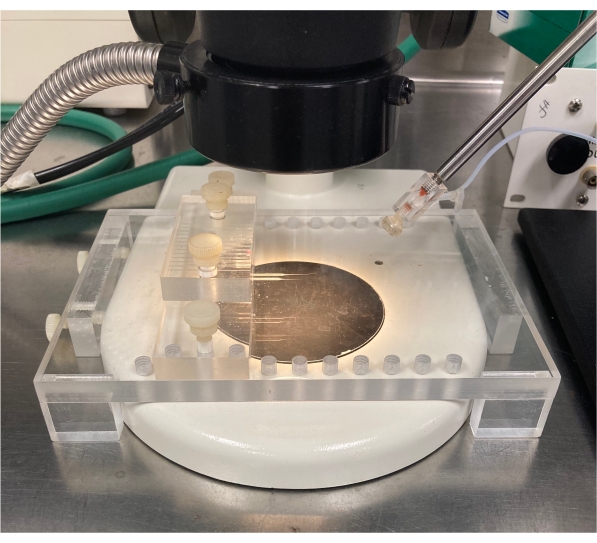
Figure 2: Horizontal micropipette holder. This plastic platform is designed to hold the micropipette after pulling of the glass capillary tube for cutting the tip. Please click here to view a larger version of this figure.
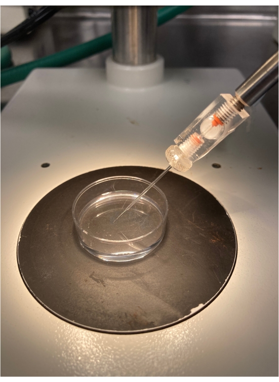
Figure 3: Micropipette positioning. An angle of 35°-45° is ideal for injecting enteroids located in the middle of the Petri dish. Please click here to view a larger version of this figure.
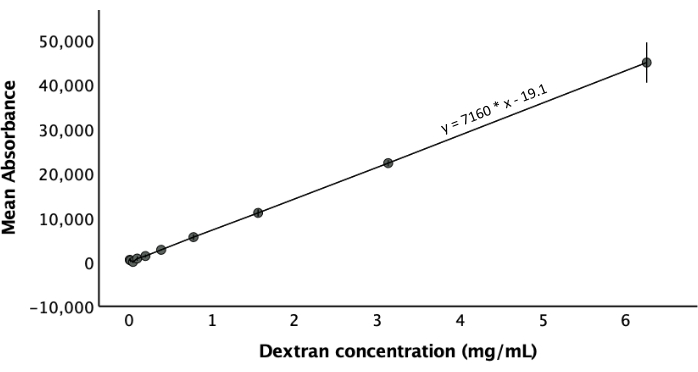
Figure 4: Dextran standard curve. The curve was constructed with fluorescent absorbance of 10 serial dilutions of dextran in duplicates. A linear regression line was fitted to generate a regression equation. The error bars are SEM. Please click here to view a larger version of this figure.
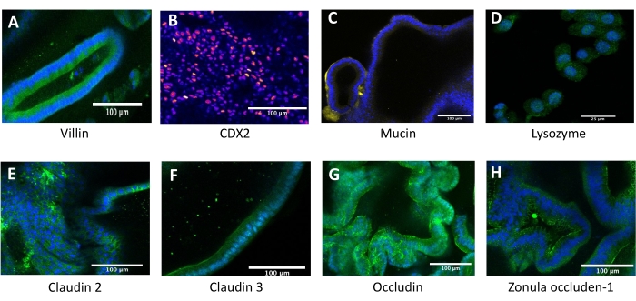
Figure 5: Fluorescent biomarkers of enteroids. DAPI for nucleus stain is blue. The following biomarkers are shown: (A) villin, (B) CDX2, (C) lysozyme, (D) mucin, (E) claudin, (F) occluding, and (G) zonula-occluden 1. Please click here to view a larger version of this figure.
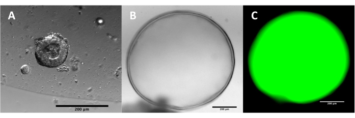
Figure 6: Enteroids at different stages. (A) An enteroid at 7-10 days old is small and with a thick wall. (B) An enteroid that is ready for microinjection with a large size, a lumen, and a think wall, and (C) fluorescent dextran-FITC inside an enteroid 2 days after microinjection. Please click here to view a larger version of this figure.
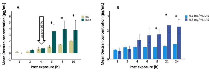
Figure 7: Permeability of dextran-FITC post microinjection. (A) Measured dextran levels in the media were higher after the addition of EGTA compared to PBS. (B) Microinjected 0.5 mg/mL LPS induced greater leakage of dextran from the enteroid lumen into the media than microinjected 0.1 mg/mL LPS did beginning at 8 h post-injection. *p-value < 0.05, error bars are SEM, n = 3, a Student's t-test was used to calculate significance. Please click here to view a larger version of this figure.
Discussion
Loading...
$$\rightleftharpoonup{xx}$$
$$\longleftharp{xx}$$,
$$\longrightharp{xx}$$,
This protocol details the establishment of enteroids from fetal intestinal tissue, as well as model characterization with immunofluorescent staining and epithelial permeability testing. The permeability of the enteroids was tested using a microinjection technique and serial time course measurements of leaked dextran-FITC concentration in the culture media. The novelty of this protocol is the apical exposure that more closely resembles human intestinal physiology compared to basolateral exposure in the culture media7. In previous studies, Hill et al. utilized serial imaging and calculation of the fluorescence intensity over time16. Ares et al. exposed the basolateral membrane of an epithelial model to LPS and then compared the cellular gene expression pattern7. In comparison, we use apical exposure of the tested reagents and then examine potential alterations in gross permeability by measuring serial concentrations of leaked dextran in the culture media. Our method also allows for serial comparative analysis of cytokines produced by epithelial cells in culture media and gene expression by collecting cellular mRNA. LPS has been commonly used to study intestinal injury in animal and in vitro models because of its ability to induce permeability and inflammation7,17. When LPS was tested in this model, epithelial permeability was differentiated by exposure concentration. This protocol can be expanded to study other disease pathologies using different microinjected materials and outcome measurements.
Critical steps in this protocol include establishing enteroids from fetal intestinal tissues, enteroid characterization, and the microinjection technique. The integrity of this study depends on accurate cell sampling. Using anatomical landmarks and blood vessels is helpful to ensure the selection of small intestinal cells. Due to the robust growth and differentiation of fetal intestinal stem cells, an extensive procedure to isolate the stem cells from other epithelial cells is not necessary. After the enteroids have been established, it is important to confirm the characteristics of the model by staining for proteins and cell markers. In this protocol, the enteroids were stained for enterocytes using villin and CDX2, Paneth cells using lysozyme, and goblet cells using mucin, all the cells which are found within the small intestine epithelium18,19,20. In contrast to traditional single cell lines that display one cell type, enteroids establish all cell types from the intestinal progenitor stem cells8. The staining portion of this protocol can be modified for the specific cellular markers of interest. Crucial to the reliability of this protocol is the microinjection technique. Consistency of the micropipette tips can be verified by measuring the volume per pump using dextran-FITC solution and visualizing the diameter of the tips under the microscope. Due to the risk of contamination, the same micropipette cannot be used for more than one exposure. Additionally, the shape and growth of the enteroids can be influenced by the contents of their growth media. We found that the spherical rather than cauliflower-shaped enteroids provided better models for microinjection. The spherical shape may be induced by a greater Wnt factor in the media21.
This protocol largely depends on the performer's skill in microinjection to reduce variations, especially in time-sensitive measurements. Variations can be minimized by having the same experienced performer with consistent techniques, using the same cell origin to avoid genetic variation, testing at the same passage to remove maturity bias, and growing the cells in the same type of media for similar cell differentiation. The components of the growth media may induce varying differentiation of stem cells in vitro rather than in an in vivo environment. For example, in vivo, lysozyme is not expressed until weeks 22-24 of gestational development when Paneth cells form and become functional22. However, we were able to detect lysozyme in our enteroids established from 10-week fetal intestines. This method can limit the number of tested exposures at one time due to the high technical skill required for microinjection. The dextran leakage from the microinjection puncture hole can affect the assessment of permeability. To eliminate this effect, triple washes immediately after the microinjection are recommended to remove residual dextran from the procedure. The dextran concentration in the media should be measured hourly for 4-6 h post microinjection. Experiments with a significant rise in dextran concentration within 2-4 h post injection should be excluded from the final analysis.
This method has several advantages. It requires a lower cost and fewer resources in comparison to enteroid-derived monolayers on transwell. Additionally, it can be expanded to other exposures such as live bacteria or viruses to study the initial interaction between gut microbes and the epithelium. The enclosed lumen of the enteroid can maintain a stable growth of microinjected live bacteria without contamination of the growth media13. Unlike the monolayer being exposed to growth media and incubation oxygen, the enclosed lumen is a tight, isolated space. An enclosed lumen allows for no communication to the growth media and a luminal oxygen content that is lower over time with live bacterial growth13.
The use of fetal intestinal tissue more accurately depicts the intestinal epithelium of preterm infants as compared to adult intestinal stem cells or animal models7. Further, the polarity of enteroids allows for both apical and basolateral exposures and measurements23. The enteroids form an enclosed lumen with lower oxygenation concentration, which more closely mimics the oxygenation concentration of the intestines24. In contrast to more technologically advanced protocols16, the use of gross media measurements allows for greater accessibility of this technique. The experiment demonstrated that epithelial leakage can be induced by apical exposure to LPS and is concentration-dependent. Since this method examines the changes in leaked concentration of dextran, it is useful for detecting gross functional changes in tight junctions. Messenger RNA collection and sequencing of the exposed enteroids and western blot analysis can complement analysis of the gross functional changes. This model studies intestinal epithelium integrity in a system that highly resembles the preterm environment and, thus, can be used to gain a better understanding of preterm intestinal injuries and other disease pathologies.
Disclosures
Loading...
$$\rightleftharpoonup{xx}$$
$$\longleftharp{xx}$$,
$$\longrightharp{xx}$$,
All authors declare that they have no conflicts of interest to disclose.
Acknowledgements
Loading...
$$\rightleftharpoonup{xx}$$
$$\longleftharp{xx}$$,
$$\longrightharp{xx}$$,
We thank Dr. Ian Glass and the personnel at the Birth Defects Research Laboratory at the University of Washington for sharing the fetal tissues. We also thank Dr. Michael Dame and Dr. Jason Spence at the Translational Tissue Modeling Laboratory at the University of Michigan for their endless support and guidance throughout the process.
Materials
| Name | Company | Catalog Number | Comments |
|---|---|---|---|
| Amphotericin 250 uL/mL | Gibco | 15-290-026 | |
| Anti-CDX-2 [CDX2-88] 0.5mL concentrated Mouse, IgG, monoclonal | Biogenex | MU392A-5UC | |
| Anti-Claudin 2 antibody (ab53032) | abcam | ab53032 | |
| Anti-Claudin 3 antibody (ab15102) | abcam | ab15102 | |
| Anti-LYZ antibody produced in rabbit | Millipore Sigma | HPA066182-100UL | |
| Anti-Mucin 2/MUC2 Antibody (F-2): sc-515032 | Santa Cruz | sc-515032 | |
| Anti-Villin, Clone VIL1/4107R 0.5mL concentrated Rabbit, IgG, monoclonal | Biogenex | NUA42-5UC | |
| Bovine Serum Albumin (BSA) | Millipore Sigma | A8806 | |
| Cell culture plates, CytoOne 12-well non-treated plates | USA Scientific Inc | 50-754-1395 | |
| Cell culture plates, CytoOne 6-well non-treated plates | USA Scientific Inc | 50-754-1560 | |
| Centrifuge with 15 mL tube buckets | Eppendorf | 05-413-110 | |
| CHIR 99021 | Tocris Bioscience | 4423 | |
| Confocal microscope | Olympus FV1200 | N/A | Or a similar microscope |
| Conical centrifuge tubes, 15 ml | Falcon | 05-527-90 | |
| Cover glass for microscope slides | Fisher Scientific | 12-544-DP | |
| Disposable scalpels | Mopec | 22-444-272 | |
| Dmidino-2-phenylindole (DAPI) Solution (1 mg/mL) | Fisher Scientific | 62248 | |
| Dulbecco's Phosphate-buffered Saline (DPBS, 1X) | Fisher Scientific | AAJ67802K2 | |
| Ethylene glycol-bis(β-aminoethyl ether)-N,N,N′,N′-tetraacetic acid tetrasodium salt (EGTA) | Millipore Sigma | E8145-10G | |
| Fluorescein isothiocyanate dextran (Dextran-FITC) 4 kDa | Millipore Sigma | 46944 | |
| Fuorescence microplate reader | Agilent BioTek | Synergy HTX | |
| Gentamicin 50 mg/mL | Gibco | 15-750-060 | |
| Glass capillary tubes, single-barrel borosilicate, 1x0.5mm, 6" (cut in half before pulling) | A-M systems | 626500 | |
| Goat anti-Mouse IgG (H+L) Highly Cross-Adsorbed Secondary Antibody, Alexa Fluor Plus 594 | Fisher Scientific | A32742 | |
| Goat anti-Rabbit IgG (H+L) Highly Cross-Adsorbed Secondary Antibody, Alexa Fluor Plus 488 | Fisher Scientific | A32731 | |
| Goat Serum | Fisher Scientific | 16210064 | |
| ImageJ software | NIH | N/A | https://imagej.nih.gov/ij/download.html |
| IntestiCult Organoid Growth Medium (Human) | Stem Cell Technologies | 06010 | |
| Lipopolysaccharides from Escherichia coli O111:B4 | Millipore Sigma | L4391-1MG | |
| Magnetic stand | World Precision Instruments | M10 | |
| Matrigel Basement Membrane Matrix, LDEV-free, 10 mL | Corning | 354234 | protein concentration > 9 mg/mL preferably |
| Micro forceps | Fisher Scientific | 13-820-078 | |
| Micro scissors | Fisher Scientific | 08-953-1B | |
| Micromanipulator | World Precision Instruments | M3301 | |
| Micropipette puller | World Precision Instruments | SU-P1000 | Or a similar equipment |
| Microscope slides | Fisher Scientific | 22-034486 | |
| Occludin Polyclonal Antibody | Fisher Scientific | 71-1500 | |
| Paraformaldehyde 32% aqueous solution | ELECTRON MICROSCOPY SCIENCES | RT 15714 | |
| Petri Dishes, 35x10 mm | Fisher Scientific | 150318 | |
| Petri Dishes, 60x15 mm | Fisher Scientific | 12-565-94 | |
| Phosphate Buffered Saline (PBS) | Fisher Scientific | 10010031 | |
| PicoPump foot switch | World Precision Instruments | 3260 | |
| Pipette tips, non-filtered, 1000 uL | Fisher Scientific | 21-402-47 | |
| Pipette tips, non-filtered, 20 uL | Fisher Scientific | 21-402-41 | |
| Pipette tips, non-filtered, 200 uL | Fisher Scientific | 21-236-54 | |
| Pneumatic PicoPump system | World Precision Instruments | SYS-PV820 | or a similar picopump system |
| Primocin 50 mg/mL, 10x1 ml vial | InvivoGen | ant-pm-1 | |
| Steel base plate | World Precision Instruments | 5052 | |
| Stereo microscope | Zeiss stemi 350 | Or a similar microscope | |
| ThermoSafe PolarPack Foam Bricks | Sonoco | 03-531-53 | |
| Triton X-100 | Millipore Sigma | T8787 | |
| Wall air supply | N/A | N/A | |
| Y-27632 dihydrochloride | Tocris Bioscience | 1254 | |
| ZO-1 Polyclonal Antibody | Fisher Scientific | 61-7300 |
References
Loading...
$$\rightleftharpoonup{xx}$$
$$\longleftharp{xx}$$,
$$\longrightharp{xx}$$,
- Humberg, A., et al. Preterm birth and sustained inflammation: Consequences for the neonate. Seminars in Immunopathology. 42 (4), 451-468 (2020).
- Kim, J., Koo, B. K., Knoblich, J. A. Human organoids: Model systems for human biology and medicine. Nature Reviews Molecular Cell Biology. 21 (10), 571-584 (2020).
- Pinto, D., Gregorieff, A., Begthel, H., Clevers, H. Canonical Wnt signals are essential for homeostasis of the intestinal epithelium. Genes & Development. 17 (14), 1709-1713 (2003).
- Barker, N., et al. Identification of stem cells in small intestine and colon by marker gene Lgr5. Nature. 449 (7165), 1003-1007 (2007).
- Sato, T., et al. Single Lgr5 stem cells build crypt-villus structures in vitro without a mesenchymal niche. Nature. 459 (7244), 262-265 (2009).
- Mahe, M. M., Sundaram, N., Watson, C. L., Shroyer, N. F., Helmrath, M. A. Establishment of human epithelial enteroids and colonoids from whole tissue and biopsy. Journal of Visualized Experiments. (97), e52483(2015).
- Ares, G. J., Buonpane, C., Yuan, C., Wood, D., Hunter, C. J. A novel human epithelial enteroid model of necrotizing enterocolitis. Journal of Visualized Experiments. (146), e59194(2019).
- Stewart, C. J., Estes, M. K., Ramani, S. Establishing human intestinal enteroid/organoid lines from preterm infant and adult tissue. Methods in Molecular Biology. 2121, 185-198 (2020).
- Finkbeiner, S. R., et al. Transcriptome-wide analysis reveals hallmarks of human intestine development and maturation in vitro and in vivo. Stem Cell Reports. 4 (6), 1140-1155 (2015).
- Senger, S., et al. Human fetal-derived enterospheres provide insights on intestinal development and a novel model to study necrotizing enterocolitis (NEC). Cellular and Molecular Gastroenterology and Hepatology. 5 (4), 549-568 (2018).
- Alganabi, M., Lee, C., Bindi, E., Li, B., Pierro, A. Recent advances in understanding necrotizing enterocolitis. F1000 Research. 8, (2019).
- Dame, M. K., et al. Human colonic crypts in culture: Segregation of immunochemical markers in normal versus adenoma-derived. Laboratory Investigation. 94 (2), 222-234 (2014).
- Hill, D. R., et al. Bacterial colonization stimulates a complex physiological response in the immature human intestinal epithelium. eLife. 6, 29132(2017).
- Tsai, Y. H., et al. A method for cryogenic preservation of human biopsy specimens and subsequent organoid culture. Cellular and Molecular Gastroenterology and Hepatology. 6 (2), 218-222 (2018).
- Su, K., Asbrock, N., Chu, V., Hoffmann, S., Hewitt, P. In Vitro Differentiation of Human iPS Cells Into Colon Organoids in Serum-Free Cell Culture Conditions. Technology Networks. , https://www.technologynetworks.com/cell-science/white-papers/in-vitro-differentiation-of-human-ips-cells-into-colon-organoids-in-serum-free-cell-culture-328450 (2019).
- Hill, D. R., Huang, S., Tsai, Y. H., Spence, J. R., Young, V. B. Real-time measurement of epithelial barrier permeability in human intestinal organoids. Journal of Visualized Experiments. (130), e56960(2017).
- Schoultz, I., Keita, A. V. The Intestinal Barrier and Current Techniques for the Assessment of Gut Permeability. Cells. 9 (8), 1909(2020).
- Kumar, N., et al. The lineage-specific transcription factor CDX2 navigates dynamic chromatin to control distinct stages of intestine development. Development. 146 (5), (2019).
- Walker, R. W., Clemente, J. C., Peter, I., Loos, R. J. F. The prenatal gut microbiome: Are we colonized with bacteria in utero. Pediatric Obesity. 12, Suppl 1 3-17 (2017).
- Spence, J. R., et al. Directed differentiation of human pluripotent stem cells into intestinal tissue in vitro. Nature. 470 (7332), 105-109 (2011).
- Shin, W., et al. Spatiotemporal gradient and instability of Wnt induce heterogeneous growth and differentiation of human intestinal organoids. iScience. 23 (8), 101372(2020).
- Lueschow, S. R., McElroy, S. J. The Paneth cell: The curator and defender of the immature small intestine. Frontiers in Immunology. 11, 587(2020).
- In, J. G., Foulke-Abel, J., Clarke, E., Kovbasnjuk, O. Human colonoid monolayers to study interactions between pathogens, commensals, and host intestinal epithelium. Journal of Visualized Experiments. (146), e59357(2019).
- Dheer, R., Young, V. B. Stem-cell-derived models: Tools for studying role of microbiota in intestinal homeostasis and disease. Current Opinion in Gastroenterology. 37 (1), 15-22 (2021).
Reprints and Permissions
Request permission to reuse the text or figures of this JoVE article
Request Permission