Here, we present a protocol to treat the great saphenous vein (GSV) and the small saphenous vein (SSV) affected by severe pathological reflux, using an original sclerosing and embolizing cyanoacrylate-based glue composed of N-butyl cyanoacrylate and methacryloxy sulfolane (NBCA+MS).
Method Article
Occlusion of the Great and Small Saphenous Vein Using Copolymeric Glue Based on N-Butyl Cyanoacrylate and Methacryloxy Sulfolane
* These authors contributed equally
In This Article
Erratum Notice
Important: There has been an erratum issued for this article. View Erratum Notice
Summary
Abstract
We present the preliminary results of a longitudinal observational study aimed at evaluating the effectiveness and safety at different short- and long-term follow-ups of vascular occlusion of the great saphenous vein (GSV) and small saphenous vein (SSV) affected by severe pathological reflux, using an innovative modified cyanoacrylate surgical glue composed of N-butyl cyanoacrylate and methacryloxy sulfolane (NBCA+MS). Ninety patients, prospectively recruited for 1 year, underwent the study with EcoColor-Doppler (ECD) to evaluate the maximum diameters of the GSV and SSV in the orthostatic position and the reflux time (RT). An RT greater than 0.5 s was considered pathologic. Clinical, etiology, anatomy, and pathophysiology (CEAP) assessment was used for the complete evaluation of each patient in the study. All the patients were treated by NBCA+MS glue to obtain vein occlusion and observed before treatment (baseline; T0), within 6 h after treatment (T1), 1 month after treatment (T2), 3 months after treatment (T3), 6 months after treatment (T4), and 1 year after treatment (T5). Chi-square (χ) analysis was performed to evaluate the effectiveness and safety of the treatment. All the patients participated in the entire duration of the study. Complete occlusion was maintained in 100% of patients at T1, 98.9% at T2 and T3, and 97.8% at T4 and T5 (p < 0.001). None of the patients suffered from post-surgical thrombosis. No blue hyperpigmentation, or paresthesia was observed during the entire observation period. Immediately after treatment, 7.7% of patients needed painkillers; 1 week after treatment, 100% of patients returned to normal life. Vascular occlusion of the great or small saphenous vein using NBCA+MS glue is a safe procedure with persistent benefits after a 1 year follow-up. This procedure can be performed with local anesthesia, allowing a quick return to normal life. Thanks to its low invasiveness, the treatment is not painful.
Introduction
Varicose veins are a symptom of venous disease that may lead to severe complications1. The great saphenous vein (GSV) and small saphenous vein (SSV) are superficial veins, separated from the deep veins by a series of valves. These valves ensure that blood flows from the superficial system to the deep system, thus preventing backflow2. The incapacity of these valves results in varicose veins, which affects 16% of men and 29% of women3. Varicose veins are engorged, tortuous veins, that become enlarged, swollen, dilated, and overfilled with blood, often appearing blue or dark purple and tender to the touch2,3. In the veins of the lower limbs, healthy valves physiologically allow a small retrograde blood flow just before the valve closes. However, in a pathological condition, valve closure is absent or incompetent, thus increasing the time the blood needs to flow back4.
Furthermore, because varicose veins may rupture or develop into varicose ulcers on the skin in severe conditions, treatment always has to be considered5. After careful clinical evaluation and instrumental study through EcoColor-Doppler (ECD) examination, patients with saphenofemoral (SF) and/or saphenous-popliteal (SP) junctional incontinence, with pathological refluence along the GSV and/or SSV axis, along with the presence or absence of collateral extra-saphenous varices, are addressed by doctors for the treatment of saphenous occlusion.
The therapeutic management of chronic venous disease can be conservative or invasive6. Examples of conservative treatments include lifestyle changes, compression therapy, or pharmacotherapy (phlebotropic drugs)5,6,7. These treatments are not sufficient in the presence of severe varicose veins; in these cases, surgery is the only solution5,6,7. Traditionally, ligature and stripping of the saphenous veins have largely been used; however, today, less invasive treatments are preferred5. Varicose veins can be treated under eco-graphic guidance using foam sclerotherapy (ultrasound-guided foam sclerotherapy [UGFS]), endovenous thermal ablation using an endovenous laser (EVL) or radiofrequency (RF)8, and recently glue9. The latter allows for the obliteration of the damaged vein. Vein occlusion using N-butyl cyanoacrylate (NBCA)-based glues has the same, if not superior, therapeutic effectiveness compared to other endovascular methods like EVL and RF, for example, through lower anesthetic invasiveness (100% of treatments completed in a single local anesthesia)10. The treatment allows for a quick patient discharge, within 3-4 h after the procedure, and rapid recovery of daily and work activities, without post-procedural pain10. Furthermore, the use of glue has no diameter-related limitations, which is a problem in the case of large diameters (10-18 mm) for some of the above-mentioned techniques10.
This naturalistic study aims to assess the effectiveness of this method to obtain vascular occlusion of the GSV and SSV, affected by severe pathological reflux using an innovative modified cyanoacrylate NBCA+MS (N-butyl cyanoacrylate and methacryloxy sulfolane) surgical glue, at different short-term (6 h and 1 month post-treatment) and long-term (3 months, 6 months, and 1 year post-treatment) follow-ups, as well as its safety in terms of the occurrence of glue-related adverse events.
Protocol
The research was conducted in three different Italian Centers: Humanitas Research Hospital - Gavazzeni (BG), Montallegro Clinic (GE), and Biomedical Institute (GE) from February 2020 to February 2021, and approved by the hospital's Ethical Committee or Internal Review Board (IRB). The research was conducted in respect of the Helsinki rules for human rights and GCP. All patients were informed about the risk of the procedure and signed a written consent before being included in the study. Refer to Supplementary File 1 for the overview of the procedure.
1. Preparation
- Prepare the room and the operating table with all the materials listed in the Table of Materials. As it is commonly used in human surgical interventions, all the materials are packaged and sterilized at the origin by each manufacturer.
- Place 5% dextrose solution into a bowl and immerse a 3 mL and a 10 mL syringe, a needle (18 G), and either a 1 mL or 2 mL vial (see step 3.2.5) containing NBCA+MS surgical glue.
2. Anesthesiology technique and surgical preparation of the patient (Figure 1)
- Completely depilate the leg to be treated, from the groin to the foot.
- Disinfect the leg to be treated with an alcoholic solution of 2% chlorhexidine di-gluconate.
- Perform an ECD in the orthostatic and clinostatic positions. Set the instrument through the manufacturer's software, which provides various presets.
NOTE: The venous vascular preset is used for this procedure, and the ultrasound probe is set at 7.5 MHz.- First, position the patient upright on a step in front of the operator and map the entire venous vascular tree through the ultrasound probe.
- Subsequently, place the patient on the operating table in the supine position and perform the vascular scanning again. This preoperative procedure is necessary for a complete clinical evaluation of the vessels.
- Map out the pathway of venous incompetence on the skin through ECD using a dermographic pen (Figure 1).
- Place the patient in the supine position for GSV treatment or the prone position for SSV treatment.
- Disinfect the leg again with an alcoholic solution of 2% chlorhexidine di-gluconate.
- Prepare the sterile field with a surgical drape.
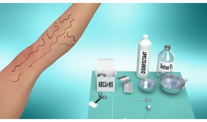
Figure 1: Preparation steps. On the right side, the figure shows the operating table preparation with the tools needed for the procedure, such as disinfectant, 5% dextrose solution, syringes, and an NBCA+MS surgical glue vial. The figure shows a schematic mapping of venous incompetence on the left side. Specifically, this is carried out under ECD guidance using a dermographic pen directly on the patient's skin. Please click here to view a larger version of this figure.
3. Surgical technique
- Time of vascular study (Figure 2)
- Perform the ultrasound-guided study of the lengths and diameters of the veins to be treated as described in step 2.3. Place the patient in the clinostatic position and perform the vascular study through the ultrasound probe.
- Evaluate the reflux time (RT) with the ultrasound, which defines the degree of incompetence of the terminal valve. The physiological values of RT are 0.1 s < RT < 0.5 s, while RT > 0.5 s indicates a pathological condition4. Carry out the classification of severity by evaluation of the Galeandro quartile11.
- Using the ECD, identify the junction between the epigastric vein and preterminal valve for the treatment of the GSV (Figure 2), or the saphenous-popliteal junction for the treatment of the SSV.
- Operative time (Figure 3)
- Induce local anesthesia with 1-1.5 mL of 10 mg/mL mepivacaine in the area of needle insertion.
- Perform ultrasound-guided catheterization of the saphenous vein (GSV/SSV) according to the Seldinger technique12 (using an angiographic needle, a guidewire, and an introducer) 14-16 cm downstream of the relevant junction (Figure 3A).
- Percutaneously insert a 6Fr vascular introducer into the vessel with the help of a guidewire (Figure 3B).
- Under ultrasound guidance, place the tip of the introducer 2-3 cm from the bifurcation of the epigastric vein and preterminal valve (for GSV treatment) (Figure 3C) or 2-4 cm below the saphenous-popliteal junction (for SSV treatment).
- Draw 1 mL of NBCA+MS surgical glue in one syringe for veins with a diameter between 8-10 mm, or 2 mL of NBCA+MS surgical glue for veins over 10 mm.
- Draw 7-10 mL of 5% dextrose solution into one syringe.
- Connect the two syringes to the stopcock of the washing catheter of the introducer (Figure 3D).
- Injection time (Figure 4)
- Compress the leg with the ultrasound probe at the level of the bifurcation immediately downstream of the bifurcation itself to stop the blood flow in the saphenous vein (Figure 4A).
NOTE: Compression with the ultrasound probe must be strong and maintained throughout the procedure. - Flush the dead volume of the introducer with dextrose solution (about 2 mL).
- Close the dextrose solution inlet through the stopcock and inject 1 mL of NBCA+MS surgical glue (Figure 4B).
- Through the stopcock, close the NBCA+MS surgical glue inlet and inject the dextrose solution to push the NBCA+MS surgical glue into the vein (Figure 4C).
- At the same time, retract the introducer until it is removed (Figure 4C) to perform a retrograde release of the liquid, adhesive, occlusive, and sclerosing agent (NBCA+MS). Perform a 10 cm occlusion for the GSV or a 7-10 cm occlusion for the SSV.
- Compress the leg with the ultrasound probe at the level of the bifurcation immediately downstream of the bifurcation itself to stop the blood flow in the saphenous vein (Figure 4A).
- Closure time (Figure 5)
- Immediately perform manual compression with the palm, starting from the insertion site and along the course of the vein for 3-5 min. At the same time, always keep the compression with the ultrasound probe at the level of the junction (Figure 5).
- Conclusive time
- At the end of the compression, raise the hand and verify that there is no bleeding from the insertion site.
- Perform an ECD (step 2.3) to check that there is no more blood flow in the saphenous vein (GSV/SSV).
- Under ultrasound guidance, occlude the varicose collateral veins by direct puncture with polidocanol foam, according to the normal and consolidated clinical practice for this treatment13,14.
- Perform an eccentric compression of the saphenous axis with latex strips or rolls of gauze, wrapping the leg from the groin to the knee.
- With the patient still in the clinostatic position, apply a Class II compression stocking (28-30 mmHg) with an open toe, fitting it like a normal pantyhose.
- At the end of the procedure, mobilize the patient immediately, making him take a few steps over a distance of 10-15 m.
- Before discharge, after 2 h, perform an ECD check.
- Discharge the patient with pain relief/anti-inflammatory therapy as needed (i.e., 200 mg of Ibuprofen, one to three times a day).
- Recommend the patient to wear the compression stocking day and night for 10 days, then only during the day for another 30 days.
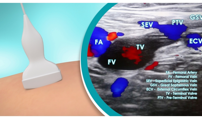
Figure 2: Vascular study. ECD identification of the junction between the epigastric vein and the preterminal valve for the treatment of the GSV. The magnification highlights the ECD image showing the femoral artery (FA), femoral vein (FV), superficial epigastric vein (SEV), great saphenous vein (GSV), external circumflex vein (ECV), terminal valve (TV), and preterminal valve (PTV). Please click here to view a larger version of this figure.

Figure 3: Operative steps. (A) Ultrasound-guided catheterization of the saphenous vein (GSV/SSV) according to the Seldinger technique: a needle is percutaneously inserted 14-16 cm below the junction. Subsequently, a guidewire is inserted through the needle, which is then removed (not shown in the figure). (B) A 6Fr vascular introducer is inserted with the help of the guidewire. At this point, the guidewire is removed and the introducer is left in position (C) Under ultrasound guidance, the introducer tip is positioned 2-3 cm from the bifurcation of the epigastric vein and the preterminal valve (for GSV treatment). The femoral vein (FV), superficial epigastric vein (SEV), and great saphenous vein (GSV) are highlighted in the picture (D) One syringe loaded with 1 mL of NBCA+MS surgical glue and one syringe loaded with 7 mL of 5% dextrose solution are connected to the stopcock of the introducer washing catheter. Please click here to view a larger version of this figure.

Figure 4: Injection steps. (A) Compression of the leg using the ultrasound probe at the level of the bifurcation immediately downstream of it to stop the blood flow in the saphenous vein (GSV/SSV). (B) Closure of the 5% dextrose solution inlet and injection of 1 mL of NBCA+MS surgical glue (black arrow) (C) Closure of the NBCA+MS surgical glue inlet and injection of the 5% dextrose solution to inject the NBCA+MS surgical glue into the vein (black arrow). At the same time, the introducer is retracted to perform a retrograde injection of the NBCA+MS (red arrow). Please click here to view a larger version of this figure.
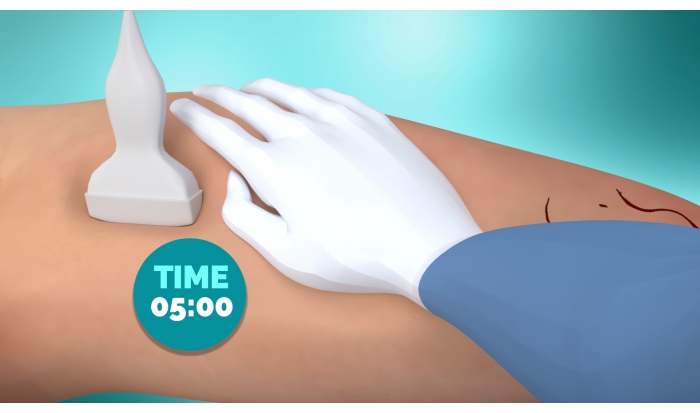
Figure 5: Closure time. After removal of the introducer, 5 min of manual compression with the palm of the hand is performed at the level of the insertion site and along the course of the vein. Compression with the ultrasound probe is maintained at the level of the bifurcation during the 5 min manual compression. Please click here to view a larger version of this figure.
Results
Ninety patients (62 women and 28 men), with a mean age of 59.8 + 12.8 years, were selected for this observational research. For each patient, the following information was collected: age, sex, major and minor systemic disease, drug usage, and voluptuary habits (e.g., smoking) (Table 1); diagnostic data of the disease (Table 2); data about the condition of the vein at the different observational-points, presence of ulcerations, or negative outcomes (Table 3); time to return to normal activities and work (Table 4); patients' sensation of "heavy and painful legs" by the Numeric Pain Rating Scale (Table 5).
Six observation points were chosen: T0 = baseline, before treatment; T1 = immediately after treatment (within 6 h); T2 = 1 month after treatment; T3 = 3 months after treatment; T4 = 6 months after treatment; T5 = 1 year after treatment. The main evaluated outcome was the occlusion of the vein.
Table 1 summarizes the comorbidities observed in our sample; some patients were suffering from more than one disease. 14.4% of patients (13 subjects) were under treatment with anticoagulant therapy, six subjects took acetylsalicylic acid (ASA), three subjects took novel oral anticoagulants (NOACs), two subjects took traditional oral anticoagulants, and two subjects took low-molecular-weight heparin.
The subjects underwent ECD in orthostatic and clinostatic postures at the first consultation (T0) and in all consecutive follow-ups (T1-T5). All patients with at least second-degree varicose veins of the lower limbs were enrolled in the study. Traditional CEAP classification was used because it allowed for a complete evaluation of the clinical aspect, etiology of the disease, and anatomical and pathophysiological classification15. Maximum diameters of the GSV and SSV in the orthostatic position and the RT were evaluated by ECD. In the lower limb veins, non-pathological valves allow for a small retrograde flow of blood just before the valve closes. In contrast, in a pathological condition, valve closure is incompetent or totally absent, increasing the time it takes for the blood to flow back4. The degree of the incompetence of a valve is described by the RT. Specifically, RT values between 0.1 s and 0.5 s are physiological, while RT values > 0.5 s represent a pathological condition4. The classification of the severity was done by Galeandro's quartile assessment11.
Exclusion criteria included the presence of both deep and superficial active thrombosis and the simultaneous presence of neoplastic disease. All patients underwent the same procedure, performed by the same surgical team. The procedure was performed under local anesthesia.
NBCA+MS surgical glue was used to generate the occlusion. NBCA+MS is an original surgical glue, Conformité Européenne (CE) marked, developed and authorized to be used for endovascular applications. NBCA is combined with another monomer, methacryloxy-sulpholane (MS), to generate a more plastic polymer whose milder exothermic reaction (45 °C) results in better biocompatibility with less inflammation and histotoxicity16.
The NBCA+MS surgical glue was applied for the occlusion of GSV and/or SSV and incontinent perforating voluminous veins, while polidocanol foam was used only for extra-saphenous varices by percutaneous injection.
During the early control with ECD (T1), in more than 70% of cases, a caudal extension of the occlusion beyond 10 cm was found. This can be attributed to the related polymerization times of the NBCA+MS surgical glue, which begins to react with the blood after 2 s from its release into the vessel and can therefore expand for a short distance.
Results are expressed in terms of prevalence (%) calculated on the number of patients. Chi-square was used to evaluate nominal data by comparing the different follow-ups (T1, T2, T3, T4, and T5). All 90 patients completed the five follow-ups and were included in the final analysis. Following CEAP classification, 57 patients were in class 2, 23 in class 3, and 10 in class 6 (Table 2). A total of 79 patients (88.8%) were treated to occlude GSV and 11 for SSV (11.2%). Mean GSV caliber/diameter in the orthostatic posture was 12.4 mm (SD: 3.6; 95% CI: 7-20 mm) and mean SSV caliber/diameter was 11.4 mm (SD: 3; 95% CI: 5-15 mm).
All patients had an RT > 10 s; 60 were in the second quartile, 24 were in the third quartile, and six were in the fourth quartile. Of the patients, 66.7% (60 patients) needed two ampoules of glue to completely fill the vessels, corresponding to 2 ml of product. In the rest, (33.3%, 30 patients), a single vial (1 mL of glue) was sufficient to obtain a total vein occlusion (Table 3). A caudal extension of the thrombosis was observed in 62.2% (57 patients) and absent in 37.8% (33 subjects).
All patients obtained the complete occlusion of the treated vein at T1, one patient (1.1%) had a re-canalization of the vessel at T2, one patient at T3 (1.1%), two patients (2.2%) at T4, and two patients (2.2%) at T5 (Table 3) (: p < 0.0001). None of the patients presented a re-canalization of the vessel >50% of its diameter, and none showed blood reflux in the orthostatic posture (Table 3). None of the patients were affected by post-surgical thrombosis. No other adverse events (i.e., blue hyperpigmentation or paresthesia) occurred during the observation period (Table 3).
We found that the patients were satisfied with the treatment. The NPRS (Numeric Pain Rating Scale) showed a score of 3 in seven patients, while the rest of the scores were between 0 and 2. The seven patients with score 3, indicative of pain, were treated with 200 mg of Ibuprofen, one to three times a day as needed, for 4 days. The resolution of leg pains was maintained for all the follow-up times (Table 5), and 70% of patients (63 subjects) had a notable reduction of leg edema. All patients returned to their normal routine in 2 days, and all restarted their work activity (light or heavy) within 1 week (Table 4).
| Sample size | 90 | |
| Sex | 62 women | |
| Age (mean) | 59.8 ± 12.8 | |
| Comorbidity (n) | Heart Attack | 40 |
| Diabetes | 16 | |
| Cardiomiopathy | 25 | |
| COPD | 16 | |
| Chronic Kidney Disease | 4 | |
| Fibrillar Atrial Disease | 4 | |
| Surgery of Mitral Valve | 1 | |
| Drugs used (n) | ASA | 6 |
| NOACs | 3 | |
| Traditional oral anticoagulants | 2 | |
| Low-molecular weight heparin | 2 | |
| Alcool and/or psychotropic substances user (n) | 0 |
Table 1: Demographic and anamnestic data. Patient recruitment for the observational study. All patients with at least second-degree varicose veins of the lower limb were enrolled in the study. For each patient, the following information was collected: age, sex, major and minor systemic disease, drug usage, and voluptuary habits. Abbreviations: n = number of subjects; COPD = chronic obstructive pulmonary disease; ASA = acetylsalicylic acid; NOACs = novel oral anticoagulants.
| Mean GSV caliber (mm) at T0 | 12.4 | |
| SD | 3.6 | |
| 95% CI | 7–20 | |
| Mean SSV caliber (mm) at T0 | 11.4 | |
| SD | 3 | |
| 95% CI | 5–15 | |
| RT > 10 s (%) at T0 | 100 | |
| First quartile | 0 | |
| Second quartile | 60 | |
| Third quartile | 24 | |
| Fourth quartile | 6 | |
| CEAP classification (n) at T0 | Class 2 | 57 |
| Class 3 | 23 | |
| Class 6 | 10 | |
| Treatment for GSV (%) | 88.8 |
Table 2: Diagnostic data. For each patient, ECD was performed before the treatment to acquire the following baseline parameters: mean GSV caliber (mm) at T0, mean SSV caliber (mm) at T0, RT >10 s (%) at T0, CEAP classification (n) at T0, and treatment for GSV (%). Abbreviations: GSV = great saphenous vein; SSV = small saphenous vein; T0 = baseline, before treatment; SD = standard deviation; CI = confidence interval; RT = reflux time; CEAP = clinical, etiological anatomical and pathophysiological; n = number of subjects.
| T1 | T2 | T3 | T4 | T5 | |||||
| 1 mL of glue used for the occlusion (n) | 30 | ||||||||
| 2 mL of glue for the occlusion (n) | 60 | ||||||||
| Occlusion of the vein (n) | 90 | 89 | 89 | 88 | 88 | ||||
| Re-canalization of the vessel >50% of its diameter | 0 | 0 | 0 | 0 | |||||
| Blood reflux in orthostatic posture | 0 | 0 | 0 | 0 | |||||
| Presence of ulceration (n) | 0 | 0 | 0 | 0 | 0 | ||||
| Post-surgical thrombosis (n) | 0 | 0 | 0 | 0 | 0 | ||||
| Paresthesia (n) | 0 | 0 | 0 | 0 | 0 | ||||
| Hyperpigmentation (n) | 0 | 0 | 0 | 0 | 0 | ||||
Table 3: Follow-up data. All the patients completed the five follow-ups and were included in the final analysis. The table shows data about the condition of the vein at the different post-treatment observational points and the presence of complications. A total of 33.3% of patients needed 1 ml of (NBCA+MS) surgical glue to completely fill the vessel; 66.7% of patients (n = 60) needed 2 ml of (NBCA+MS) surgical glue to completely fill the vessel. All patients obtained the complete occlusion of the treated vein at T1. One patient (1.1%) had a re-canalization of the vessel at T2, one patient (1.1%) at T3, two patients (2.2%) at T4, and two patients (2.2%) at T5 (: p < 0.0001). None of the patients presented a re-canalization of the vessel >50% of its diameter, and none of them showed blood reflux in the orthostatic posture. None of the patients were affected by post-surgical thrombosis. No other adverse events occurred during the entire observation period. Abbreviations: T1 = immediately after treatment (within 6 h); T2 = 1 month after treatment; T3 = 3 months after treatment; T4 = 6 months after treatment; T5 = 1 year after treatment; n = number of subjects.
| n = 90 | |
| Return to normal life (days) | 2 |
| Return to work (days) | 7 |
Table 4: Recovery time. The amount of time, expressed in days, the patients needed to return to normal life and restart work activities. Abbreviations: n = number of subjects; d = days.
| 0 | 1 | 2 | 3 | 4 | 5 | 6 | 7 | 8 | 9 | 10 | |
| T1 | 83 | 0 | 0 | 7 | 0 | 0 | 0 | 0 | 0 | 0 | 0 |
| T2 | 90 | 0 | 0 | 0 | 0 | 0 | 0 | 0 | 0 | 0 | 0 |
| T3 | 90 | 0 | 0 | 0 | 0 | 0 | 0 | 0 | 0 | 0 | 0 |
| T4 | 90 | 0 | 0 | 0 | 0 | 0 | 0 | 0 | 0 | 0 | 0 |
| T5 | 90 | 0 | 0 | 0 | 0 | 0 | 0 | 0 | 0 | 0 | 0 |
Table 5: Pain assessment. Evaluation of the pain intensity at each follow-up time point, according to the Numeric Pain Rating Scale (NPRS), an 11-point numeric scale in which 0 indicates no pain and 10 is the worst possible pain. Numbers in the table represent the number of subjects. Abbreviations: T1 = immediately after treatment (within 6 h); T2 = 1 month after treatment; T3 = 3 months after treatment; T4 = 6 months after treatment; T5 = 1 year after treatment.
Supplementary File 1: Overview of the procedure. Please click here to download this File.
Discussion
The outcomes of this preliminary research show that the described endovenous management of varicose veins by this new method using NBCA+MS surgical glue is a safe technique that allows for a persistent occlusion of the vein(s) at 1 year follow-up, with the resolution of painful legs and peripheral edema. The use of this procedure with the modified glue-indicated and authorized to embolize vessels-allowed for a quick recovery, and patients returned to their usual activities within 1 week after surgery.
The most critical and delicate steps of the procedure are those related to identifying the exact point where the catheter must be inserted to obliterate the saphenous vein (GSV/SSV). A precise ultrasound mapping is necessary to know where the glue must be released, avoiding being too proximal or distal to the saphenofemoral and/or saphenous-popliteal junction, which would make the procedure partially ineffective. A prior hemodynamic study of the saphenous axis must be performed to identify the degree of reflux and any incontinent branches and collateral refluxes.
The GSV was affected by varicose disease more than the SSV; however, the number of glue ampoules was never decided based on the vessel caliber/diameter, but rather always on the function of vein dilatation. In most cases, 2 mL of glue was needed to occlude the vein, because most of the patients had very dilated veins (>10 mm in caliber/diameter); 1 mL of glue was sufficient to completely occlude the vein with a diameter <10 mm in one-third of our patients. After the occlusion via NBCA+MS surgical glue, the various collateral veins were occluded through direct injection of polidocanol foam. One of the most recurrent side effects of polidocanol injections is postoperative pain. Specifically, there is no known minimum volume of polidocanol; it can cause pain even when injected in very low doses17 since it induces inflammatory wall damage. Thus, the postoperative pain experienced following polidocanol injections is independent of its dosage17,18. Moreover, the sensation of pain is patient-dependent. In the present study, none of the patients presented severe negative outcomes (i.e., paresthesia or ecchymosis), and only 7.7% of them needed painkillers to treat postoperative pain.
Cyanoacrylate-based glues have strong embolic or ablative capacities. From a histopathological point of view, the NBCA+MS surgical glue used here, thanks to its strong adhesive ability19,20, induces an instant occlusion of the vessel; when the glue comes into contact with the blood, it polymerizes rapidly21, thanks to the presence of hydroxyl ions. It forms a cast made of blood and adhesive, a plastic thrombus that adheres firmly to the vessel walls, occluding it permanently16,22.
In over 70% of cases, in the early control with ECD, we found that the caudal extension of the occlusion obtained from the NBCA+MS glue always went well beyond 10 cm of the trans-catheter deposition due to its density, which is similar to water. Since the venous flow is blocked, the cranial extension of the polymerization and the related phenomena of "glue-induced thrombosis" did not occur. In fact, compression with the ultrasound probe immediately downstream of the saphenous bifurcation occluded the lumen of the vein and, consequently, the flow in the cranial direction. Therefore, the high adhesiveness and biochemical properties of the NBCA+MS surgical glue cause not only injuries and mechanical collapses with the adhesion of the vessel walls, but also a pro-coagulant action in the treated vessel.
The choice to use the co-monomeric NBCA+MS surgical glue was both due to its described different endovascular behavior with respect to pure cyanoacrylate (NBCA)-based glues13 and its several documented and authorized indications in interventional radiology. It has been applied as a liquid embolic agent to permanently occlude vascular abnormalities such as cerebral and spinal arteriovenous malformations23,24, to perform embolization of the portal vein25,26, pre-operative renal-tumor27, endoleaks of the abdominal aorta28,29, and for bleeding control30, and in endoscopy as a sclerosing agent to treat gastroesophageal varices31,32.
However, thanks to the combination of safe and widely tested vein catheterization, NBCA+MS surgical glue, and vein compression after glue injection, we obtained better results here in terms of both vascular occlusion10 and safety10,33,34,35,36 compared to the aforementioned studies. Moreover, none of the patients in this study presented slight or severe post-surgical complications in the short and long term (as shown in Table 3).
Complete occlusion of the leg veins occurs in the supine position with a pressure of 20-25 mm Hg37,38 with the ultrasound probe, which is the necessary and sufficient force to compress the vessels and keep them effectively occluded. This pressure exerted in the procedure has never aroused discomfort in any of the patients treated.To refine and standardize the compressive phase of the method, a pneumatic or mechanical system, which can be calibrated on the bases of the individual diameters and the individual vascular tracts to be occluded, could be designed.
This technique does not involve particular procedural variations, thanks to the in-depth and detailed study of the patient's vascular system in the diagnostic phase. The only technique-related issue that may occur is the inability of catheterizing the vessel. In this case, the procedure cannot be done, and it is necessary to change the type of sclerosing procedure. One limitation of this technique is excessive patient body weight (over 100 kg), which does not allow for effective compression of the limb with the ultrasound probe to block venous flow in the saphenous veins.
Compared to other existing methods34,35,36, we believe this procedure is simpler and faster, is more standardizable thanks to the hemodynamic study, and possesses comparable results. In our personal experience, we have noticed that patients experience less pain, both intra- and post-procedurally, than when other sclerosing techniques are used (e.g., laser, radiofrequency, and other cyanoacrylates).
Although the preliminary results of this protocol are very encouraging, additional studies on larger sample sizes are necessary to confirm the validity of the novel method illustrated here. Given the reassuring results of this study, we hypothesize that in the future this technique could be used to implement embolization and venous occlusion in other pathological venous districts, through the use of more flexible and thinner catheters.
Disclosures
The authors have nothing to disclose.
Acknowledgements
Thanks to all collaborators of the Department of Cardiovascular Surgery, Angiology Functional Unit of Humanitas Gavazzeni Hospital, Bergamo, Montallegro Clinic, Genova, BioMedical Institute, Genova.
Materials
| Name | Company | Catalog Number | Comments |
|---|---|---|---|
| 5% Dextrose injection | BBraun Milano S.p.A., Milan, Italy | S5104-5384 | Dextrose solution for intravenous administration |
| Angiographic needle | Merit Medical, Utah, USA | AN18T72SH | Suitable needle for the Selding procedure |
| Atossisclerol 1% | Chemische Fabrik Kreussler & Co. GmbH, Wiesbaden, Germany | 22199071 | Polidocanol foam for venous sclerosis |
| Avanti | Cordis, Miami, Florida, USA | 504-606X | Vascular Introducer |
| BD microlance 3 | BD Company, Drogheda, Ireland | 304622 | Needle 18 G |
| BD syringe | BD Company, Drogheda, Ireland | 300912 | 10 mL syringe |
| BD syringe | BD Company, Drogheda, Ireland | 309658 | 3 mL syringe |
| Carbosen (10 mg/mL) | Galenica Senese, Siena, Italy | 33640640 | Mepivacaine solution 10 mg/mL |
| Clorexinal (2%) | Nuova Farmec, Verona, Italy | PF373 | Solution of 2% chlorhexidine and 70% isopropanol alcohol |
| Dermographic pen | Novatech SA, La Ciotat, France | 18996 | Sterile pen for writing on the patient's skin during surgery |
| Glubran 2 | GEM Srl, Viareggio, Italy | G-NB-2 | Synthetic cyanoacrylate liquid modified by addition of a monomer synthesized by the manufacture (NBCA+MS) |
| MyLabGamma | Esaote, Genova, Italy | 101741000 | Portable Ultrasound Machine |
| Sterile bowls | Delta Med Spa, Viadana (MN), Italy | 82.H8428.00 | Sterile bowls used for keep the tools for the procedure submerged in 5% dextrose solution |
| Sterile gauzes | Delta Med Spa, Viadana (MN), Italy | 82.H8428.00 | Sterile gauzes commonly used during surgery |
| Sterile surgical drape | MedLine, Firenze, Italy | CPU29017 CE | Sterile drapes commonly used during surgery |
References
- Hamdan, A. Management of varicose veins and venous insufficiency. Journal of the American Association. 308 (24), 2612-2621 (2012).
- Chwała, M., Szczeklik, W., Szczeklik, M., Aleksiejew-Kleszczyński, T., Jagielska-Chwała, M. Varicose veins of lower extremities, hemodynamics and treatment methods. Advances in Clinical and Experimental Medicine. 24 (1), 5-14 (2015).
- Tisi, P. V. Varicose veins. BMJ Clinical Evidence. 2011, 0212(2011).
- Labropoulus, N., et al. Definition of venous reflux in lower-extremity veins. Journal of Vascular Surgery. 38 (4), 793-798 (2003).
- de Mik, S. M., Stubenrouch, F. E., Legemate, D. A., Balm, R., Ubbink, D. T. Treatment of varicose veins, international consensus on which major complications to discuss with the patient: A Delphi study. Phlebology. 34 (3), 201-207 (2019).
- Raetz, J., Wilson, M., Collins, K. Varicose veins: Diagnosis and treatment. American Family Physician. 99 (11), 682-688 (2019).
- Piazza, G. Varicose veins. Circulation. 130 (7), 582-587 (2014).
- Pichot, O., De Maeseneer, M. Treatment of varicose veins: does each technique have a formal indication. Perspectives in Vascular Surgery Endovascular Therapy. 23 (4), 250-254 (2011).
- Belramman, A., Bootun, R., Lane, T. R. A., Davies, A. H. Endovenous management of varicose veins. Angiology. 70 (5), 388-396 (2019).
- alık, E. S., Arslan, Ü, Erkut, B. Ablation therapy with cyanoacrylate glue and laser for refluxing great saphenous veins-a prospective randomised study. Vasa. 48 (5), 405-412 (2019).
- Galeandro, A. I., et al. A three-dimensional electronic report of a venous echo color Doppler of the lower limbs: MEVeC®. Vascular Health Risk Management. 10, 549-555 (2014).
- Seldinger, S. I. Catheter replacement of the needle in percutaneous arteriography; a new technique. Acta Radiologica. 39 (5), 368-376 (1953).
- Alòs, J., et al. Efficacy and safety of sclerotherapy using polidocanol foam: a controlled clinical trial. European Journal of Vascular and Endovascular Surgery. 31 (1), 101-107 (2006).
- Li, N., Li, J., Huang, M., Zhang, X. Efficacy and safety of polidocanol in the treatment of varicose veins of lower extremities. A protocol for systematic review and meta-analysis. Medicine. 100 (8), (2021).
- Lurie, F., et al. The 2020 update of the CEAP classification system and reporting standards. Journal of Vascular Surgery: Venous and Lymphatic Disorders. 8 (3), 342-352 (2020).
- Levrier, O., et al. Efficacy and low vascular toxicity of embolization with radical versus anionic polymerization of n-butyl-2-cyanoacrylate (NBCA). An experimental study in the swine. Journal of Neuroradiology. 30 (2), 95-102 (2003).
- Todd, K. L., Wright, D. R. VANISH-2 Investigator Group. The VANISH-2 study: a randomized, blinded, multicenter study to evaluate the efficacy and safety of polidocanol endovenous microfoam 0.5% and 1.0% compared with placebo for the treatment of saphenofemoral junction incompetence. Phlebology. 29 (9), 608-618 (2014).
- Todd, K. L., et al. Durability of treatment effect with policdocanol endovenous microfoam on varicose vein symptoms and appearance (VANISH-2). Journal of Vascular Surgery Venous and Lymphatic Disorders. 3 (3), 258-264 (2018).
- Losi, P., et al. Cyanoacrylate surgical glue as an alternative to suture threads for mesh fixation in hernia repair. Journal of Surgical Research. 163 (2), e53-e58 (2010).
- Kull, S., et al. Glubran2 surgical glue: in vitro evaluation of adhesive and mechanical properties. Journal of Surgical Research. 157 (1), e15-e21 (2009).
- Cipolletta, L., et al. Acrylate glue injection for acutely bleeding oesophageal varices: A prospective cohort study. Digestive and Liver Disease. 41 (10), 729-734 (2009).
- Rivet, C., et al. Endoscopic treatment of gastroesophageal varices in young infants with cyanoacrylate glue: a pilot study. Gastrointestinal Endoscopy. 69 (6), 1034-1038 (2009).
- Wang, Y., Zhang, H., Ling, F. Coexistence of a single cerebral arteriovenous malformation and spinal arteriovenous malformation. Neurology India. 57 (6), 785-788 (2009).
- Liu, J., et al. Curative glubran 2 embolization of cerebral arteriovenous malformations patient selection and initial results. Interventional Neuroradiology. 20 (6), 722-728 (2014).
- Luz, J. H. M., Gomes, F. V., Coimbra, E., Costa, N. V., Bilhim, T. Preoperative portal vein embolization in hepatic surgery: a review about the embolic materials and their effects on liver regeneration and outcome. Radiology Research and Practice. , (2020).
- Lanza, E., et al. Survival analysis of 230 patients with unresectable hepatocellular carcinoma treated with bland transarterial embolization. PLOS ONE. 15 (1), e0227711(2020).
- Prigent, F. V., et al. Selective arterial embolization of renal angiomyolipomas with a n-butyl cyanoacrylate-lipiodol mixture: Efficacy, safety, short- and mid-term outcomes. Journal of Clinical Medicine. 10 (18), 4062(2021).
- Midulla, M., et al. Transcatheter transcaval embolization of a Type II endoleak after EVAR using a transseptal needle-sheath system. Vascular and Endovascular Surgery. 46 (5), (2012).
- Resch, T., Dias, N. Treatment of endoleaks: techniques and outcome. The Journal of Cardiovascular Surgery. 53 (1), 91-99 (2012).
- Abdulmalak, G., et al. Safety and efficacy of transcatheter embolization with Glubran 2 cyanoacrylate glue for acute arterial bleeding: a single-center experience with 104 patients. Abdominal Radiology. 43 (3), 723-733 (2017).
- Franco, M. C., et al. Efficacy and safety of endoscopic prophylactic treatment with undiluted cyanoacrylate for gastric varices. World Journal of Gastrointestinal Endoscopy. 6 (6), 254-259 (2014).
- Cipolletta, L., et al. Acrylate glue injection for acutely bleeding oesophageal varices: A prospective cohort study. Digestive and Liver Disease. 41 (10), 729-734 (2009).
- Hirsch, T. Varicose vein therapy and nerve lesions. Vasa. 46 (2), 96-100 (2017).
- Koramaz, I., et al. Ablation of the great saphenous vein with nontumescent n-butyl cyanoacrylate versus endovenous laser therapy. Journal of Vascular Surgery: Venous and Lymphatic Disorders. 5 (2), 210-215 (2017).
- Kollury, L., et al. Network meta-analysis to compare VenaSeal with other superficial venous therapies for chronic venous insufficiency. Journal of Vascular Surgery: Venous and Lymphatic Disorders. 8 (3), 472-481 (2020).
- Bissacco, D., Stegher, S., Calliari, F. M., Viani, M. P. Saphenous vein ablation with a new cyanoacrylate glue device: a systematic review on 1000 cases. Minimally Invasive Therapy and Allied Technologies. 28 (1), 6-14 (2019).
- Partsch, B., et al. Which pressure do we need to compress the great saphenous vein on the thigh. Dermatologic Surgery. 34 (12), 1726-1728 (2008).
- Mosti, G. Postinterventional compression in phlebology: Evidence and empirical observations. Presence of Varices After Operative Treatment: A Review (Part 2). 2 (1), 12-17 (2015).
Reprints and Permissions
Erratum
Formal Correction: Erratum: Occlusion of the Great and Small Saphenous Vein Using Copolymeric Glue Based on N-Butyl Cyanoacrylate and Methacryloxy Sulfolane
Posted by JoVE Editors on 2/07/2023. Citeable Link.
An erratum was issued for: Occlusion of the Great and Small Saphenous Vein Using Copolymeric Glue Based on N-Butyl Cyanoacrylate and Methacryloxy Sulfolane. The Author section was updated from:
Pecis Claudio*123
Bellandi-Alberti Pier Luca*23
Fumagalli Miriam*1
1Department of Cardiovascular Surgery, Angiology Functional Unit, Humanitas Gavazzeni Hospital
2Department of Cardiovascular Surgery, Angiology Functional Unit, Montallegro Clinic
3Department of Cardiovascular Surgery, Angiology Functional Unit, BioMedical Institute
* These authors contributed equally
to:
Claudio Pecis*123
Pier Luca Bellandi-Alberti*23
Miriam Fumagalli*1
1Department of Cardiovascular Surgery, Angiology Functional Unit, Humanitas Gavazzeni Hospital
2Department of Cardiovascular Surgery, Angiology Functional Unit, Montallegro Clinic
3Department of Cardiovascular Surgery, Angiology Functional Unit, BioMedical Institute
* These authors contributed equally
