The present protocol describes a rodent model of newborn hypoxic-ischemic injury for identifying early changes in cerebral tissue by gross morphology and magnetic resonance imaging. This has benefits over existing models, which can be used to study late injury but do not allow the evaluation of reproducible early changes.
Method Article
Early Pathological and Magnetic Resonance Detection of Cerebral Injury Using a Rat Model of Neonatal Hypoxic Ischemic Encephalopathy
Opens in a new tab
In This Article
Summary
Loading...
$$\rightleftharpoonup{xx}$$
$$\longleftharp{xx}$$,
$$\longrightharp{xx}$$,
Abstract
Loading...
$$\rightleftharpoonup{xx}$$
$$\longleftharp{xx}$$,
$$\longrightharp{xx}$$,
Perinatal hypoxic-ischemic encephalopathy (HIE) is an acute disease that may afflict newborns, resulting in variable long- and short-term neurodevelopmental outcomes. Early diagnosis is critical to identifying infants who may benefit from intervention; however, early diagnosis relies heavily on clinical criteria. No molecular or radiological tests have shown promise in detecting early cerebral injury. Studies have shown that magnetic resonance imaging (MRI) can show changes in both blood flow/ischemia and metabolic disruption. However, they have all been used to evaluate the secondary phase of the disease (>12 h) after the onset of the injury. Early diagnosis is critical to rapidly starting therapeutic hypothermia in eligible infants, which is currently recommended to be initiated within 6 h of birth. The rat model of hypoxic-ischemic injury was developed in 1981 and has been validated and used extensively to study changes in brain perfusion, cerebral injury markers, and morphology. However, it has primarily been used as a "late model", evaluating injury several days after the initial ischemic insult. The model has been known to have poor sensitivity in evaluating reliable and reproducible early cerebral changes. The objective of this study was to develop a reliable model to study early gross morphological and radiological markers of HIE using pathological staining and cerebral magnetic resonance imaging/magnetic resonance spectroscopy.
Introduction
Loading...
$$\rightleftharpoonup{xx}$$
$$\longleftharp{xx}$$,
$$\longrightharp{xx}$$,
Hypoxic ischemic encephalopathy (HIE) is a devastating condition resulting from various factors in newborn infants1. Perinatal asphyxia and/or the disruption of cerebral blood flow may result in focal or global ischemic changes in the brain2. The occurrence rate is approximately 1.6 in 1,000 live births but may be as high as 12.1 in 1,000 live births in developing countries3. This condition results in high mortality (20%-50%), while 25% of those who survive are likely to suffer from a long-term neural disability such as mental retardation, epilepsy, or cerebral palsy4. The only therapeutic intervention proven effective in mild to moderate injury is therapeutic hypothermia, which must be initiated within 6 h of birth5,6,7,8,9. While this may help prevent the metabolic changes that lead to secondary injury, there may also be potential for side effects such as hypotension, thrombocytopenia, prolonged coagulation time, intracranial hemorrhage, dysrhythmias, fat necrosis, and serum electrolyte imbalance4,5. Early diagnosis of HIE in babies is often difficult as the criteria are subjective and rely heavily on physical exam findings, which evolve over time. Magnetic resonance imaging may show changes reflective of injury several days to weeks after injury. However, morphologic changes in T1/T2 MRI can be normal in up to two-thirds of moderate encephalopathy, the category of infants most likely to benefit from therapeutic hypothermia10. As per recent reports, magnetic resonance spectroscopy (MRS) may show early changes correlating with neonatal HIE11. However, no standardization or validation has been performed to date.
Many investigators rely on animal models to evaluate potential diagnostic or therapeutic interventions for cerebrovascular injury. The most frequently used method to create an infarct is ligating rodents' middle cerebral artery12,13. While often used to study adult ischemic stroke, this is technically challenging in neonatal rodents due to the small size and the fragility of the pups at the age equivalent to human newborn disease. Furthermore, it does not represent the global cerebral ischemic changes likely to be seen in HIE. The Rice-Vanucci Model14 of unilateral carotid artery ligation in rats has been used since the 1980s as a cost-effective rodent model to study hypoxic-ischemic brain injury. However, there is large variability in early cerebrovascular changes and high mortality in earlier experiments. Most studies report the cerebral injury in long-term changes (i.e., after 24 h of injury), which are more consistent. This study aimed to develop an approach to evaluate early (within 6 h) molecular and radiological changes in a rat model of HIE. The protocol was designed to ensure ischemia at an early (term newborn equivalent) age and to increase the survival of the pups, especially during exposure to hypoxia. MRI/MRS were used to evaluate radiological evidence of altered flow, cerebral tissue changes, and metabolic changes within 6 h of injury. Gross morphological evaluation of the infarct areas was also performed. Further validation of the reproducibility was conducted by repeating the experiments in multiple litters.
Protocol
Loading...
$$\rightleftharpoonup{xx}$$
$$\longleftharp{xx}$$,
$$\longrightharp{xx}$$,
All the experimental procedures were approved by the Oklahoma Medical Research Foundation (OMRF) Institutional Animal Care and Use Committee (protocol IACUC #17-17). Pregnant female Sprague-Dawley rat pups at E14 were used for the present study. The animals were obtained from a commercial source (see Table of Materials).
1. Animal preparation
- Acclimatize the animals in the animal facility prior to delivery of the litters.
- Maintain all rats on a12 h light/darkcycleand feed standard ratchow.
- After delivery of the litters, keep the pups with their respective dams. Use both sexes for the experiments.
- At postnatal age 10 (P10), randomize the pups to either sham or HIE groups.
NOTE: The experimental timeline is depicted in Figure 1. All the experiments were performed on the same day at P10.
2. Carotid artery ligation (CAL) for the experimental HIE group
- Place the rat pups on a warming pad.
- Initiate anesthesia with 4% isoflurane in oxygen (0.6 LPM) using a nasal cone until the pinch reflex disappears. Drop the flow of gas to 0.5%-2% for the maintenance of anesthesia. Ensure the pups are unconscious without suppressing the respiratory drive.
- Mark the pups on the tail for identification and gently restrain them with tape on all four limbs.
- Shave the neck area and sterilize with 70% iodine-povidone solution swabs (see Table of Materials).
- Using an #11 blade, make a 1 cm midline neck incision through the skin. Carefully dissect the left parotid gland and fascia until the left carotid artery is exposed. Gently mobilize the vessel using hemostats to free it from the fascia.
- Using a small hemostat, carefully pass two 5-0 sutures (see Table of materials) around the vessel, 0.5 cm apart, and tie them tightly.
- Using small scissors, cut the artery in between the two sutures to ensure the discontinuity of blood flow. Close the skin and fascia with 5-0 silk sutures (see Table of Materials).
- Inject buprenorphine (10 units in 800 μL of sterile normal saline) intraperitoneally and another 800 μL of normal saline subcutaneously in the back of the neck to prevent dehydration.
NOTE: The procedure must be completed within 10-12 min. - Return the pups to the cages with their dams and allow the pups to awaken and recover for 1-2 h.
3. Sham surgical procedure for the control group
- Place the rat pups on a warming pad.
- Initiate anesthesia with 4% isoflurane in oxygen (0.6 LPM) using a nasal cone until the pinch reflex disappears. Drop the flow of gas to 0.5%-2% for the maintenance of anesthesia. Ensure the pups are unconscious without suppressing the respiratory drive.
- Mark the pups on the tail for identification and gently restrain them with tape on all four limbs.
- Shave the neck area and sterilize with 70% iodine-povidone solution swabs.
- Using an #11 blade, make a 1 cm midline neck incision through the skin and then close it with 5-0 silk sutures.
- Follow the same hydration, analgesia, and postoperative care as for the HI group (steps 2.8-2.9).
4. Hypoxia exposure for both the CAL and sham groups
- Prepare the clear plexiglass hypoxia chamber (see Table of Materials) by attaching tubing to the chamber lid to provide continuous airflow of 6 LPM of the hypoxia gas mixture (8% oxygen, 92% nitrogen).
- Place a blue absorbent pad in the chamber and immediately place the rat pups from both groups in the chamber. Allow the pups to remain in the hypoxia chamber for 45 min.
- Immerse the chamber in a water bath with continuously flowing warm water to keep the temperature set at 37 °C inside the chamber.
- Hydrate the pups with an oral saline solution via gavage of 600 μL before being placed in the hypoxia chamber and 600 μL at the end of the 45 min.
- Remove all the pups to the cages with their dams (experimental and sham) and allow them to recover for 2 h in a room next to the small animal imaging facility.
5. Magnetic resonance imaging and spectroscopy
- Perform MRI and MRS to identify and evaluate the radiological and metabolic markers 4 h after the end of carotid artery ligation. Perform the procedure under anesthesia with continuous cardiovascular monitoring at the small animal imaging facility.
- Anesthetize each animal (with 1.5% isoflurane and 0.7 L/min oxygen) and place it in the MR probe (see Table of Materials) in a supine position on a blue absorbent pad covering a heating pad. Monitor the respiration rate of the animals continuously using an abdominal pneumatic pillow (see Table of Materials).
- Use a head surface coil as a signal receiver and transmit radiofrequency pulses to the sample through a quadrature volume coil (72 mm inner diameter, see Table of Materials).
- Perform MRI to evaluate both the changes in cerebral blood flow (CBF) and the changes in water diffusion constants (ADC) following previously published methods15,16,17. Perform MRI morphology (T1 and T2), diffusion, and perfusion to determine the brain's most affected and least perfused areas.
NOTE: The mean values for perfusion and diffusion (ADC) in each group are compared between the ligated side and the control side (intact carotid artery side). - Perform MRS following previously published methods15,17 and analyze using an in-house coded program using Mathematica software (see Table of Materials).
- Scale the MR spectra in parts per million (ppm) by calibrating against the water peak (4.78 ppm). Identify the major brain metabolic peaks as N-acetylaspartate (NAA) at 2.02 ppm, choline (Cho) at 3.22 ppm, creatine (Cr) at 3.02 ppm, and myo-inositol at 3.53 ppm.
NOTE: The peak area measurements of the metabolites are used to calculate the following ratios: NAA to Cho (NAA/Cho), Cr to Cho (Cr/Cho), and Myo-Ins to Cho (Myo-Ins/Cho)15.
6. Serum and cerebral tissue analysis
- Perform blood sampling at 5.5 h following the carotid artery ligation or sham procedure according to previously published methods18.
- Anesthetize the pups again with 4% isoflurane.
- Using a sharp #11 blade, make an abdominal incision, followed by a diaphragmatic incision to expose the heart.
- Perform blood sampling via cardiac puncture as previously described18. Briefly, insert a 32 G needle on a 1 mL syringe into the right heart chamber and gently aspirate 1 mL of blood.
- Allow the whole blood to coagulate, followed by centrifugation at 1,000 x g for 15 min at 4 °C. The serum gets separated into clean microcentrifuge tubes.
- Decapitate the whole pup head for the gross assessment of cerebral pathology and then immerse it in ice for 2 min.
- Make an incision on the dorsal scalp from the base of the skull to the tip of the nose and peel the skull bones from around the brain. Remove the intact whole brain into a clean Petri dish.
- Mark the right side of the brain with a non-toxic marker. Position the brain with the cephalad surface upward so that both hemispheres are visible. Using an ice-cold razor blade, slice the brain into four equal sections parallel to the coronal plane.
- Immerse the brain sections in 2,3,5-triphenyltetrazolium chloride (TTC, see Table of Materials) solution in a Petri dish covered with foil to prevent photosensitization and incubate for 15 min at 37 °C.
NOTE: Infarcted areas are delineated as white areas devoid of red TTC stain. - If infarcts are subtle or difficult to detect, inject 0.5-1 mL of TTC (1%) in phosphate-buffered saline directly into the right heart after abdominal incision and thoracotomy, and allow to perfuse for 2 min prior to the decapitation of the pup.
- Store the brain tissue and serum at −80 °C if further analysis is required.
Results
Loading...
$$\rightleftharpoonup{xx}$$
$$\longleftharp{xx}$$,
$$\longrightharp{xx}$$,
The present protocol to produce and evaluate early cerebral changes after HIE was easy to implement and allowed gross pathological and radiological visualization of cerebral injury within 6 h of insult in rat pups at P10. The experimental plan is depicted in Figure 1. Both sexes were analyzed together, and 24 animals from five litters were examined in each group. Animal mortality was very low, with 99% survival of animals until the terminal experiments were performed.
Areas of ischemia are clearly visualized 6 h after CAL and hypoxia on brain sections stained with TCC. These lesions are seen as a clear white area of injury devoid of stain, depicted in Figure 2. No cardiac perfusion of TCC was necessary for any samples to visualize the injury. Many animals had multiple areas of infarcts denoted. The majority were on the cortical surface of the brain.
A decrease in MR perfusion on the ligated side of the brain compared to the perfusion on the contralateral or non-surgical side of the brain was seen. An 80% decrease in perfusion on the ligated side in the HIE group was detected 3 h after CAL and hypoxia. As expected, there was no difference between both sides of the brain in the sham group. Representative MR results are shown in Figure 3. A similar pattern was seen in the diffusion scan between the two sides of the brain. A 10% decrease in ADC was seen on the ligated side of the brain in the HIE group that was detected 3 h after CAL surgery. Again, there was no difference between both sides in the sham group.
The results of the MRS analysis are shown in Figure 4. MRS metabolites are normalized to creatine, the most stable of all the metabolites. There were no statistically significant differences between the groups, but there was a clear trend for N-acetylcysteine (NAA) to decrease by 30%, while myo-inositol increased more than two-fold in the HIE compared to the sham group. There was no change in either choline or taurine.

Figure 1: Experimental timeline for rat pups at P10. Both the experimental group and the sham control group are shown. Please click here to view a larger version of this figure.
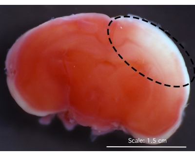
Figure 2: Representative image of a coronal brain section after staining with TCC. The area of the infarct shows the absence of staining (white), which is marked in the dotted line. Abbreviation: TCC = 2,3,5-triphenyltetrazolium chloride. Please click here to view a larger version of this figure.
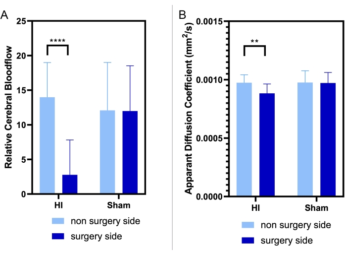
Figure 3: Representative MR results. (A) The MR perfusion and (B) apparent diffusion coefficient (ADC) of the carotid artery ligation (CAL) side of the brain in both groups (which has either the CAL in HI or just the neck incision in sham) compared to the perfusion of the contralateral or non-surgical side of the brain. The side of the brain with CAL has a statistically significant decrease in relative cerebral blood flow (RCBF) and ADC compared to the non-ligated side (n = 6 per group). The comparison of the CAL and sham groups was analyzed using unpaired two-tailed t-tests with **** p < 0.0001, ** p < 0.01. The error bars show the standard deviation (SD). Please click here to view a larger version of this figure.
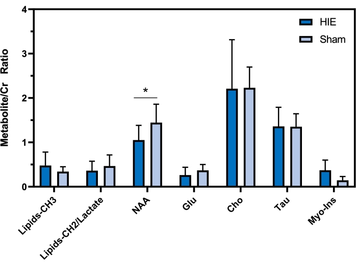
Figure 4: MRS analysis results. For MRS metabolites, there were no statistically significant differences between the groups, but there was a trend toward a decrease in NAA between the ligated and non-ligated sides. Myo-inositol increased more than two-fold in the HIE compared to the sham group. There was no change in either choline or taurine. Abbreviations: MRS = magnetic resonance spectroscopy; NAA = N-acetylcysteine; Cr = creatine; Cho = choline; Glu = glucose; Tau = taurine, Myo-Ins = myo-inositol. n = 6 per group. The comparison of the CAL and sham groups was analyzed using unpaired two-tailed t-tests with *p < 0.05. The error bars show the standard deviation (SD). Please click here to view a larger version of this figure.
Discussion
Loading...
$$\rightleftharpoonup{xx}$$
$$\longleftharp{xx}$$,
$$\longrightharp{xx}$$,
A research protocol in newborn rat pups was successfully designed to visualize and analyze early markers of cerebral injury in HIE. To date, there is a lack of objective assessment tools to detect early cerebral injury in the newborn population. After HI injury, there is a phase (1-6 h) in which the impairment of cerebral oxidative metabolism has the potential to partially recover before the failure of mitochondrial function19, which is irreversible. This latent phase is the therapeutic window for neuroprotective interventions such as therapeutic hypothermia6. Most published clinical trials advocate for the initiation of therapy within the first 6 h5,7, and most institutions do not offer this therapy past that time. However, cooling must be used selectively as there is a potential for serious adverse effects20. These include dysrhythmias, hypotension, platelet dysfunction and thrombocytopenia, persistent pulmonary hypertension, coagulation disturbances, and subcutaneous fat necrosis21. Therefore, the decision to begin treatment must be made selectively only for those infants who would have the most benefit.
The use of rodents in the study of HIE is both cost-efficient and easy to perform. A variety of postnatal days (PND) have been suggested to study cerebral ischemic injury in rats, ranging from P7 to P13. The original Rice-Vanucci model14 consisted of unilateral common carotid artery ligation in rat pups at P7. At 4-8 h later, the pups were exposed to 8% oxygen at 37 °C for 3.5 h. The brain tissue was then examined for morphological changes. A literature search revealed that most investigators use this model to evaluate markers of injury after 24 h or longer. The acquisition of consistent and reproducible results at shorter intervals has proven to be difficult. In addition, high mortality has been observed by investigators due to dehydration, as well as difficulty in maintaining body temperature during exposure to hypoxia. This protocol was designed to ensure improved survival of the rat pups, as well as a clear delineation of brain injury. This is critical to evaluate potential diagnostic tools and other early therapies for preventing encephalopathy. For these experiments, P10 in Sprague-Dawley rat pups was chosen as the timepoint, as this most closely represents the term newborn period while ensuring the survival of the animals. The maintenance of hydration is paramount. An oral electrolyte solution was gavage fed both before and after hypoxia, and a normal saline solution was injected intraperitoneally and subcutaneously. Another factor was the strict maintenance of body temperature at 37 °C. These key steps ensured >90% survival of the rat pups in both groups at the time of terminal evaluation.
Other authors have reported a wide variation in the degree of injury observed after unilateral carotid artery ligation. While this model successfully reproduces neuronal injury, it was also observed that the degree of injury is variable as the contralateral (unligated side of the brain) provides blood flow in variable amounts (via the circle of Willis) to restore blood flow to the ischemic area. However, the variation in injury severity correlates well with what is seen in human babies and serves as a relevant, clinically equivocal animal model to study disease.
The confirmation of infarcted areas is necessary by gross visualization as often areas of injury correlate with prognosis and may help identify areas of neuronal tissue for further analysis. For the visualization of infarcted neuronal tissue, 2,3,5-triphenyltetrazolium chloride (TTC) is a commonly used compound as it differentiates viable tissue from infarction macroscopically22,23. TCC is reduced to form a red formazan product mainly in the mitochondria. The intensity of the red stain correlates with the number and functional activity of mitochondria24; therefore, TTC-unstained brain tissue is infarcted and TTC-stained tissue viable. Gross morphological evaluation of brain tissue after TCC staining showed clear areas of infarct and injury. The pathological data shown did not require cardiac perfusion with TCC; however, it may be performed, especially when a more subtle injury is visualized.
Magnetic resonance imaging has been used extensively in human babies to determine the areas and severity of injury affected by infarcts and injury after HIE. However, most studies evaluated injury at a "late" stage (i.e., after 48 h of life)10,25,26. The recent emergence of MRS and its ability to target specific bio-metabolites affected during HIE has made it a valuable prognostic tool27 in evaluating these infants. Again, no studies have evaluated its use in the early stages of the disease. Using a combination of MRI/MRS techniques, these data show that early changes and injury markers are apparent in the rodent model, which may identify infants with cerebrovascular involvement and target them for early therapies. Results from this study show that N-acetylaspartate (NAA) levels are decreased in infarcted tissue. This is similar to previous studies on rats and humans at different stages after injury. For example, Looji et al. found a 30% decrease in NAA levels 5 h after injury in adult human subjects16. Lally et al. investigated MRS metabolite changes 4-14 days after injury in human newborns as prognostic factors for determining the neurodevelopmental outcome. They found that the population with neurodevelopmental delay had lower NAA than the less-affected subjects27. Cheong et al. found similar results on days 1-3 after injury28. In the present study, it is seen that a decrease in NAA levels can occur within 1 h after the HI injury and can be used as a very early marker of diagnosis.
Choline was found to be decreased early after injury in the HIE group. Our findings are similar to other studies that showed a mild decrease in choline in MR spectroscopy, which was also directly related to the severity of the neurodevelopmental outcome in the newborn with HI injury27,29. This contrasts with the findings of Guo et al., who observed an increased choline/Cr ratio in human subjects from 0-15 days of age5. However, levels of choline/Cr seem to vary according to age, sex, and time after the injury. Serial-timed MRS may be needed to study the variation in choline levels reported by different studies.
The only metabolite found to be increased in the current data from early MRS spectroscopy was myo-inositol. Myo-inositol is one of the least-studied metabolites in HIE-related MRS. Van de Looji et al.28 found that myo-inositol levels decreased 4 days after injury, but these levels increased significantly after that. These differences might be partially explained by the findings of Shibasaki et al., who linked the change in myo-inositol levels to the severity of HI injury29. On comparing myo-inositol expression in newborns sustaining HI injury with different outcomes, they found that, despite the early increase in myo-inositol expression, it drops within 2 weeks of injury, with this drop much more significant in the group severely affected by the HI injury. Therefore, this may be a useful marker of the severity of the injury.
In conclusion, maintaining hydration and body temperature were the key elements in successfully establishing a rat model to evaluate early cerebral injury in HIE. This will be an invaluable tool to help define markers and for the evaluation of early injury in term newborn infants. The early HIE rat model allows cerebral injury assessment by all modalities tested, including MRI, MRS, and gross pathological examination. Hopefully, these studies will be of value in the early identification of infants that will benefit the most from lifesaving therapies.
Disclosures
Loading...
$$\rightleftharpoonup{xx}$$
$$\longleftharp{xx}$$,
$$\longrightharp{xx}$$,
There are no conflicts of interest for any of the authors.
Acknowledgements
Loading...
$$\rightleftharpoonup{xx}$$
$$\longleftharp{xx}$$,
$$\longrightharp{xx}$$,
We thank the veterinary staff of the Oklahoma Medical Research Foundation for their expertise and assistance in modifying the animal care protocols.
Materials
| Name | Company | Catalog Number | Comments |
|---|---|---|---|
| 0.9% Normal saline | Fisher Scientific | Z1376 | |
| 2,3,5-triphenyltetrazolium chloride (TTC) | Millipore Sigma | T8877 | |
| Abdominal pneumatic pillow | SA Instruments, Inc., Stony Brook, NY | ||
| Absorbent Underpads with Waterproof Moisture Barrier, 58.4 x 91.4 cm, 680 mL | Fisher Scientific | 501060566 | |
| BD 30 G Needle and syringe | Fisher Scientific | Catalog No.14-826-10 | |
| Biospec 7.0 Tesla/30 cm horizontal-bore magnet small animal imaging system | Bruker Biospin, Ettlingen, Germany | ||
| Buprenorphine | Provided by veterenary medicine | ||
| Compact Thermometer with Probe | Fisher Scientific | S01549 | |
| Gas mixture 92% nitrogen 8% oxygen | Airgas | ||
| Head surface coil | Bruker BioSpin MRI Gmbh, Ettlingen, Germany | ||
| Isoflurane gas | Provided by veterenary medicine | ||
| Isotemp Immersion Circulator 2100 | Fisher Scientific | Discontinued | Immersed in water bath chamber with continous flowing water via tubing |
| Lead Ring Flask Weights | VWR | 29700-060 | Water bath weights to ensure rodent chamber stays submerged in water bath |
| Mathematica Software | Wolfram Research, Champaign, IL, USA | version 6.0 | |
| Pedialyte Electrolyte Solution, Hydration Drink, 1 Liter, Unflavored | Pedialyte | Obtained from CVS | |
| Phosphate-buffered saline (DPBS, 1X), Dulbecco's formula | Millipore Sigma | J67670.AP | |
| Plastic clear bucket | We used an old rodent housing cage- this is a good alternative: Cambro 182615CW135 Camwear Food Storage Box, 18" X 26" X 15", Model #:182615CW135 | ||
| Plexiglass Rodent Restraint Chamber | Pedialyte/CVS | Vetinary medicine provided a small chamber used to restrain rodents. Approximately 6x4x4 inches | |
| Pregnant Sprague Dawley rats at E14 | Charles River | Strain Code 400 | |
| Purdue Products Betadine Swabsticks | Fisher Scientific | 19-061617 | |
| Quadrature volume coil (72-mm inner diameter) | Bruker BioSpin MRI Gmbh, Ettlingen, Germany | ||
| Stoelting Silk Suture | Fisher Scientific | Catalog No.10-000-656 | |
| Vicryl 5-0 suture | Fisher Scientific | NC1985424 |
References
Loading...
$$\rightleftharpoonup{xx}$$
$$\longleftharp{xx}$$,
$$\longrightharp{xx}$$,
- Hypoxic-ischemic encephalopathy: A review for the clinician. JAMA Pediatrics. 169 (4), 397-403 (2015).">Douglas-Escobar, M., Weiss, M. D. Hypoxic-ischemic encephalopathy: A review for the clinician. JAMA Pediatrics. 169 (4), 397-403 (2015).
- Neonatal hypoxic-ischemic encephalopathy: A radiological review. Journal of Pediatric Neurosciences. 12 (1), 1-6 (2017).">Bano, S., Chaudhary, V., Garga, U. C. Neonatal hypoxic-ischemic encephalopathy: A radiological review. Journal of Pediatric Neurosciences. 12 (1), 1-6 (2017).
- Intrapartum-related neonatal encephalopathy incidence and impairment at regional and global levels for 2010 with trends from. Pediatric Research. 74, 50-72 (2013).">Lee, A. C., et al. Intrapartum-related neonatal encephalopathy incidence and impairment at regional and global levels for 2010 with trends from. Pediatric Research. 74, 50-72 (2013).
- Executive summary: Neonatal encephalopathy and neurologic outcome, second edition. Report of the American College of Obstetricians and Gynecologists Task Force on Neonatal Encephalopathy. Obstetrics & Gynecology. 123 (4), 896-901 (2014).">American College of Obstetricians and Gynecologists Task Force on Neonatal Encephalopathy. Executive summary: Neonatal encephalopathy and neurologic outcome, second edition. Report of the American College of Obstetricians and Gynecologists Task Force on Neonatal Encephalopathy. Obstetrics & Gynecology. 123 (4), 896-901 (2014).
- Whole-body hypothermia for term and near-term newborns with hypoxic-ischemic encephalopathy: A randomized controlled trial. Archives of Pediatrics and Adolescent. 165 (8), 692-700 (2011).">Jacobs, S. E., et al. Whole-body hypothermia for term and near-term newborns with hypoxic-ischemic encephalopathy: A randomized controlled trial. Archives of Pediatrics and Adolescent. 165 (8), 692-700 (2011).
- Mechanisms of hypothermic neuroprotection. Clinics in Perinatology. 41 (1), 161-175 (2014).">Drury, P. P., Gunn, E. R., Bennet, L., Gunn, A. J. Mechanisms of hypothermic neuroprotection. Clinics in Perinatology. 41 (1), 161-175 (2014).
- Hypothermia and other treatment options for neonatal encephalopathy: An executive summary of the Eunice Kennedy Shriver NICHD workshop. The Journal of Pediatrics. 159 (5), 851-858 (2011).">Higgins, R. D., et al. Hypothermia and other treatment options for neonatal encephalopathy: An executive summary of the Eunice Kennedy Shriver NICHD workshop. The Journal of Pediatrics. 159 (5), 851-858 (2011).
- Therapeutic hypothermia and hypoxia-ischemia in the term-equivalent neonatal rat: characterization of a translational preclinical model. Pediatric Research. 78 (3), 264-271 (2015).">Patel, S. D., et al. Therapeutic hypothermia and hypoxia-ischemia in the term-equivalent neonatal rat: characterization of a translational preclinical model. Pediatric Research. 78 (3), 264-271 (2015).
- Hypothermia augments neuroprotective activity of mesenchymal stem cells for neonatal hypoxic-ischemic encephalopathy. PLoS One. 10 (3), 0120893(2015).">Park, W. S., et al. Hypothermia augments neuroprotective activity of mesenchymal stem cells for neonatal hypoxic-ischemic encephalopathy. PLoS One. 10 (3), 0120893(2015).
- Early identification of brain injury in infants with hypoxic ischemic encephalopathy at high risk for severe impairments: Accuracy of MRI performed in the first days of life. BMC Pediatrics. 14, 177(2014).">Agut, T., et al. Early identification of brain injury in infants with hypoxic ischemic encephalopathy at high risk for severe impairments: Accuracy of MRI performed in the first days of life. BMC Pediatrics. 14, 177(2014).
- Early identification of hypoxic-ischemic encephalopathy by combination of magnetic resonance (MR) imaging and proton MR spectroscopy. Experimental and Therapeutic Medicine. 12 (5), 2835-2842 (2016).">Guo, L., et al. Early identification of hypoxic-ischemic encephalopathy by combination of magnetic resonance (MR) imaging and proton MR spectroscopy. Experimental and Therapeutic Medicine. 12 (5), 2835-2842 (2016).
- A new model of neonatal stroke: reversible middle cerebral artery occlusion in the rat pup. Pediatric neurology. 12 (3), 191-196 (1995).">Ashwal, S., Cole, D. J., Osborne, S., Osborne, T. N., Pearce, W. J. A new model of neonatal stroke: reversible middle cerebral artery occlusion in the rat pup. Pediatric neurology. 12 (3), 191-196 (1995).
- Transient middle cerebral artery occlusion model of neonatal stroke in P10 rats. Journal of Visualized Experiments. (122), e54830(2017).">Larpthaveesarp, A., Gonzalez, F. F. Transient middle cerebral artery occlusion model of neonatal stroke in P10 rats. Journal of Visualized Experiments. (122), e54830(2017).
- The influence of immaturity on hypoxic-ischemic brain damage in the rat. Annals of Neurology. 9 (2), 131-141 (1981).">Rice, J. E., Vannucci, R. C., Brierley, J. B. The influence of immaturity on hypoxic-ischemic brain damage in the rat. Annals of Neurology. 9 (2), 131-141 (1981).
- Sepsis-associated encephalopathy: A magnetic resonance imaging and spectroscopy study. Journal of Cerebral Blood Flow & Metabolism. 30 (2), 440-448 (2010).">Bozza, F. A., et al. Sepsis-associated encephalopathy: A magnetic resonance imaging and spectroscopy study. Journal of Cerebral Blood Flow & Metabolism. 30 (2), 440-448 (2010).
- Multiparametric assessment of the anti-glioma properties of OKN007 by magnetic resonance imaging. Journal of Magnetic Resonance Imaging. 31 (4), 796-806 (2010).">Garteiser, P., et al. Multiparametric assessment of the anti-glioma properties of OKN007 by magnetic resonance imaging. Journal of Magnetic Resonance Imaging. 31 (4), 796-806 (2010).
- Anti-inflammatory agent, OKN-007, reverses long-term neuroinflammatory responses in a rat encephalopathy model as assessed by multi-parametric MRI: implications for aging-associated neuroinflammation. Geroscience. 41 (4), 483-494 (2019).">Towner, R. A., et al. Anti-inflammatory agent, OKN-007, reverses long-term neuroinflammatory responses in a rat encephalopathy model as assessed by multi-parametric MRI: implications for aging-associated neuroinflammation. Geroscience. 41 (4), 483-494 (2019).
- A better method for terminal bleeding of mice. Lab Animal. 20 (1), 70-72 (1986).">Adeghe, A. J., Cohen, J. A better method for terminal bleeding of mice. Lab Animal. 20 (1), 70-72 (1986).
- Hypoxic-ischemic encephalopathy and mitochondrial dysfunction: Facts, unknowns, and challenges. Antioxiddants & Redox Signaling. 33 (4), 247-262 (2020).">Rodriguez, M., Valez, V., Cimarra, C., Blasina, F., Radi, R. Hypoxic-ischemic encephalopathy and mitochondrial dysfunction: Facts, unknowns, and challenges. Antioxiddants & Redox Signaling. 33 (4), 247-262 (2020).
- Experimental models of hypothermic circulatory arrest. Seminars in Pediatric Neurology. 6 (1), 48-54 (1999).">Vannucci, R. C., Towfighi, J. Experimental models of hypothermic circulatory arrest. Seminars in Pediatric Neurology. 6 (1), 48-54 (1999).
- Systemic complications and hypothermia. Seminars in Fetal and Neonatal Medicine. 15 (5), 270-275 (2010).">Sarkar, S., Barks, J. D. Systemic complications and hypothermia. Seminars in Fetal and Neonatal Medicine. 15 (5), 270-275 (2010).
- Qualitative and quantitative analysis of the progressive cerebral damage after middle cerebral artery occlusion in mice. Brain Research. 606 (2), 251-258 (1993).">Chiamulera, C., Terron, A., Reggiani, A., Cristofori, P. Qualitative and quantitative analysis of the progressive cerebral damage after middle cerebral artery occlusion in mice. Brain Research. 606 (2), 251-258 (1993).
- Triphenyltetrazolium chloride (TTC) as a marker for ischaemic changes in rat brain following permanent middle cerebral artery occlusion. Neuropathology and Applied Neurobiology. 17 (1), 61-67 (1991).">Hatfield, R. H., Mendelow, A. D., Perry, R. H., Alvarez, L. M., Modha, P. Triphenyltetrazolium chloride (TTC) as a marker for ischaemic changes in rat brain following permanent middle cerebral artery occlusion. Neuropathology and Applied Neurobiology. 17 (1), 61-67 (1991).
- Automated measurement of infarct size with scanned images of triphenyltetrazolium chloride-stained rat brains. Stroke. 27 (9), 1657-1662 (1996).">Goldlust, E. J., Paczynski, R. P., He, Y. Y., Hsu, C. Y., Goldberg, M. P. Automated measurement of infarct size with scanned images of triphenyltetrazolium chloride-stained rat brains. Stroke. 27 (9), 1657-1662 (1996).
- Magnetic resonance spectroscopy biomarkers in term perinatal asphyxial encephalopathy: From neuropathological correlates to future clinical applications. Current Pediatric Reviews. 10 (1), 37-47 (2014).">Robertson, N. J., Thayyil, S., Cady, E. B., Raivich, G. Magnetic resonance spectroscopy biomarkers in term perinatal asphyxial encephalopathy: From neuropathological correlates to future clinical applications. Current Pediatric Reviews. 10 (1), 37-47 (2014).
- Evolution of pattern of injury and quantitative MRI on days 1 and 3 in term newborns with hypoxic-ischemic encephalopathy. Pediatric Research. 74 (1), 82-87 (2013).">Gano, D., et al. Evolution of pattern of injury and quantitative MRI on days 1 and 3 in term newborns with hypoxic-ischemic encephalopathy. Pediatric Research. 74 (1), 82-87 (2013).
- Prognostic value of 1H-MRS in neonatal encephalopathy. Pediatric Neurology. 34 (5), 360-366 (2006).">da Silva, L. F., Hoefel Filho, J. R., Anes, M., Nunes, M. L. Prognostic value of 1H-MRS in neonatal encephalopathy. Pediatric Neurology. 34 (5), 360-366 (2006).
- Longitudinal MR assessment of hypoxic ischemic injury in the immature rat brain. Magnetic Resonance in Medicine. 65 (2), 305-312 (2011).">van de Looij, Y., Chatagner, A., Huppi, P. S., Gruetter, R., Sizonenko, S. V. Longitudinal MR assessment of hypoxic ischemic injury in the immature rat brain. Magnetic Resonance in Medicine. 65 (2), 305-312 (2011).
- Changes in brain metabolite concentrations after neonatal hypoxic-ischemic encephalopathy. Radiology. 288 (3), 840-848 (2018).">Shibasaki, J., et al. Changes in brain metabolite concentrations after neonatal hypoxic-ischemic encephalopathy. Radiology. 288 (3), 840-848 (2018).
Reprints and Permissions
Request permission to reuse the text or figures of this JoVE article
Request Permission