This study details procedures for establishing a chronic-like rabbit rotator cuff (RC) injury. Specifically, the injury is created in the subscapularis (SSC) muscle-tendon/myotendinous unit to mimic human RC anatomy and pathophysiology, including severe muscle fatty degeneration (FD). This protocol can be applied to study RC injuries and assess regenerative therapies.
Method Article
Development of a Rabbit Chronic-Like Rotator Cuff Injury Model for Study of Fibrosis and Muscular Fatty Degeneration
In This Article
Summary
Abstract
Rabbit rotator cuff (RC) pathophysiology can lead to progressive and highly degenerative changes in its associated musculature and tendons, which negatively influences clinically relevant parameters, such as strength and retraction of the muscle-tendon/myotendinous unit, ultimately causing loss of shoulder function and negatively affecting RC repair outcomes. Animal models that mimic aspects of human RC anatomy and pathophysiology are crucial for advancing the conceptual understanding of injury progression and developing effective tissue engineering and regenerative medicine-based therapeutics.
Within this context, a rabbit subscapularis (SSC) model is suitable due to (i) its anatomical similarity to the human supraspinatus (SSP) bone-tendon-muscle unit, which is the most frequently injured RC site; (ii) its pathophysiological similarity to humans in terms of fibrosis and muscle fatty degeneration (FD); and (iii) its amenability to surgical procedures. Therefore, the goal of this study is to describe the surgical techniques for inducing SSC RC injury. Briefly, the procedure involves the isolation of the SSC by identifying the coracobrachialis muscle followed by a full-thickness transection at the muscle-tendon junction and wrapping the free end of the muscle-tendon junction with a silicone-based penrose tubing to prevent spontaneous reattachment. Histologic evaluations are performed to monitor the progression of muscle FD at 4 weeks post-surgery using hematoxylin and eosin (H&E) as well as Masson's trichrome staining.
Loss of muscle and FD were evident 4 weeks after transection of the SSC muscle-tendon junction, similar to human RC pathophysiological conditions. This protocol demonstrates the steps for successfully establishing a chronic-like rabbit SSC RC injury model, which can serve as a powerful tool to study skeletal muscle changes associated with RC pathophysiology and aid the development of novel therapeutic strategies for chronic-like RC tears.
Introduction
Chronic rotator cuff (RC) tears are characterized by degenerative changes in musculature and tendons, including the atrophy of muscles, accumulation of adipose tissue, and fibrosis, which can compromise the outcome of RC repair and ultimately cause shoulder pain and dysfunction1,2,3,4,5. To better understand the RC tear pathogenesis and improve surgical results, it is crucial to develop appropriate animal models that can mimic aspects of human RC anatomy and pathophysiology. Specifically, RC injury models should meet the following criteria: (i) lack spontaneous healing after injury; (ii) contain a significant presence of fibrosis, muscular atrophy, and accumulation of adipose tissue; and (iii) be of adequate size to allow for the approximation of surgical techniques used in humans6.
Within this context, the rabbit subscapularis (SSC) muscle can be used as an accurate and reliable animal model for the study of RC pathophysiology, given its unique anatomy, pathophysiological response, and biomechanical properties7. Indeed, rabbit SSC RC anatomy is similar to human supraspinatus (SSP) RC, which is the muscle-tendon unit most often associated with injury stemming from overuse8,9. Specifically, the rabbit SSC tendon complex passes through a bony tunnel and under the coracobrachialis muscle, which is analogous to the situation in humans where the SSP tendon complex passes through the subacromial bony tunnel and under the coracoacromial ligament7. This anatomical similarity results in rabbit SSC undergoing similar musculoskeletal motions as human SSP, in which the tendon travels under the acromion during elevation and abduction of the humerus7,10.
Furthermore, pathohistological changes, similar to human RC tears11, have been observed in the rabbit after SSC tear. Specifically, the muscle belly undergoes severe FD, with a significant loss of muscle mass, decreased muscle-fiber cross-sectional area, and increased adiposity. In addition, Otarodifard et al. assessed the biomechanical characteristics of the rabbit SSC after (1) single-row, (2) double-row, and (3) transosseous-equivalent RC repair techniques, and found that the initial biomechanical characteristics of these repairs were similar to human SSP RC repairs performed in cadaveric specimens12. As such, the anatomical, physiological, and biomechanical similarity of rabbit SSC with human SSP makes it useful for modeling RC injuries.
Although many species of animals including rats, mice, dogs, and sheep, have been used in the study of RC disease and repair6,13,14,15, the degree of injury chronicity is a key consideration. This is because RC tears can be asymptomatic and may often be diagnosed much later when the tear has enlarged and become chronic in nature, with both the tendon and muscle exhibiting severe degeneration16,17,18. However, most RC repair models employ acute injury models, in which the healthy tendon is transected and then immediately repaired19,20,21,22. This largely occurs for reasons of logistical expediency and technical ease, resulting in few studies that examine the RC pathophysiology within a chronic-like setting. Moreover, several animal models may possess attributes that hinder their use for chronic RC studies.
For example, although the rat has been extensively used to model RC tear and intervention, the lack of significant adipose accumulation following injury contrasts with the human condition, and its small size makes repeated surgical procedures challenging23. Further, although Gerber et al. used the infraspinatus of sheep to study muscle atrophy and FD after chronic RC tear24, there exists some anatomical dissimilarity between sheep infraspinatus and human SSP, as well as numerous logistical challenges for studying and housing such a large animal model. In addition, Gerber et al. developed a delayed RC injury model in sheep by releasing the superficial head of the infraspinatus muscle and tendon to mimic the features of a chronic RC tear, and then evaluated the efficacy of different repair techniques on the tendon at 4 to 6 weeks. Unfortunately, this chronic-like sheep model possessed a limitation, in that the end of the released tendon became indistinguishable from scar tissue during the second surgical procedure25.
Coleman et al. also developed a chronic RC tear model in sheep by covering the transected tendon end with a synthetic membrane at the time of the initial surgery, which allowed for nutrient diffusion and efficiently minimized scar tissue formation around the injured tissue, while improving discrimination between the tendon and scar tissue26. Meanwhile, Turner et al. suggested that a delayed repair should be conducted within 4 weeks, since direct reattachment rarely happens in a massive tendon retraction27. Together, these studies have contributed toward reproducible and reliable protocols for the successful establishment of a chronic-like rabbit SSC RC injury model.
In this protocol, a chronic-like rabbit RC injury model is established at 4 weeks, in which pathologic changes related to fibrosis and FD-mediated muscle atrophy can be studied via histologic assessments. In particular, wrapping the free end of the muscle-tendon junction using a silicone-based penrose tubing at the time of the initial surgery enables clear identification of the RC tissues during the second surgical procedure and, consequently, facilitates a secure repair to study RC healing with and without scaffold augmentation. Altogether, a chronic-like rabbit SSC model may better mimic RC pathophysiology and pose minimal technical and logistical requirements.
Protocol
All procedures must be performed using sterile surgical technique in an appropriately equipped room designated for animal surgeries according to a protocol approved by the institute's animal experimentation ethics committee. In the present study, rabbit surgeries were performed in accordance with a protocol approved by The Chinese University of Hong Kong Animal Experimentation Ethics Committee.
1. Surgical procedure
- To prepare the surgical area, prewarm a heating pad and overlay it with sterile surgical drapes to maintain the body temperature of the rabbit. Subsequently, lay out sterilized surgical tools and supplies (as specified in the Table of Materials) and organize them according to the surgeon's preference.
- Induce anesthesia via intramuscular administration of 35 mg/kg ketamine and 5 mg/kg xylazine to New Zealand white rabbits (weighing between 3.5 and 4.5 kg, about 5-6.5 months of age; two male and one female rabbits were used in this study). Subsequently, confirm anesthesia with a paw and/or tail pinch test.
- If additional anesthesia is required to sustain the surgical plane, administer 10 mg/kg ketamine and 3 mg/kg xylazine intravenously via the marginal ear vein28 and monitor the animal's breathing rate at regular intervals of 5-10 min.
- To prepare the surgical window, shave the intended incision site (the skin region superficial to the SSC muscle-tendon unit) and clean with three alternating applications of betadine and 70% alcohol. Use a cotton swab to apply betadine and 70% alcohol in circular motions (from inside to outside). Use eye ointment to keep the rabbit's eyes moist and lubricated. Administer 20 mg/kg cephalexin intramuscularly as an anti-infective agent.
- Make a 3-4 cm skin incision inferior to the clavicle, split the deltopectoral interval using a surgical No. 11 scalpel, and retract to gain access to the shoulder (Figure 1A,B).
- To locate the SSC muscle-tendon unit, first, identify the coracobrachialis muscle (as tissue covering the SSC tendon attachment) and split it. Following this, identify the SSC tendon, and insert a right-angle clamp to expose the entire SSC tendon at its insertion on the lesser tuberosity of the humerus (Figure 1C).
- Prior to introducing the injury, isolate the SSC muscle-tendon (Figure 1D) and administer intraoperative anesthetic (0.2 mg/kg of 0.5% bupivacaine) locally near the transection site. Wrap the SSC muscle-tendon unit in silicone-based penrose tubing (Figure 1E) to prevent undesired attachment to the surrounding tissues and aid subsequent tissue retrieval.
- To induce injury, create a full-thickness transection at the muscle-tendon junction using a surgical No. 11 scalpel (Figure 1F). Where necessary, stop bleeding by applying pressure with a piece of gauze and use saline to irrigate the wound as needed.
- To close the wound, use a 4-0 poly glycolic acid (PGA) suture to reapproximate the deltoid muscle tissue (Figure 1G) and a 4-0 nylon suture to close the skin wound (Figure 1H).
- Deliver post-surgery care through subcutaneous administration of 0.03 mg/kg buprenorphine as an analgesic (once immediately after surgery and twice daily for the next 48 h29).
- Allow the rabbits to recover on a covered heating pad and apply a soft collar to prevent undesired behavior, including self-mutilation, licking of surgical sites, and removal of sutures (Figure 1I).
- Monitor the animals for weight and behavioral changes. Report any decrease of more than 10% body weight and severe pain that cannot be controlled (assessed based on five behavioral actions: orbital tightening, cheek flattening, nostril shape changes, whisker position changes, and ear shape and position changes) to the vet to determine if intervention such as early euthanasia is required.
2. Specimen harvest
- Euthanize the rabbits at 4 weeks from the time of injury. Anesthetize the rabbits and give a lethal dose of sodium pentobarbital (more than 60 mg/kg). Confirm death by thoracotomy.
- Identify the humeral head and surgically excise it, while preserving the greater and lesser tubercles and all soft tissue attachments. Fix with 4% paraformaldehyde (PFA) for 72 h at 4 °C before transferring to a solution of 10% ethylenediaminetetraacetic acid (EDTA) for 1 month at room temperature (with media change every 72 h) to decalcify bone.
- After decalcification, subject the samples to standard histological processing using graded ethanol dehydration, paraffin embedding, histological sectioning (8 µm sections), and staining with hematoxylin and eosin (H&E) and Masson's trichrome solutions30,31,32.
- Capture images with an upright microscope at 10x magnification.
- Perform semi-quantification of the H&E and Masson's trichrome images by measuring the area and percentage of muscle, fibrous tissue, and fat within the muscle, as previously described33,34 using a graphic design software of choice. In this example, Adobe Photoshop software (https://www.adobe.com) is used.
- Select a region of a particular color representing a certain tissue type using the magic wand tool (red is muscle tissue, blue is fibrosis, and white represents fat).
- Click the menu items Select | Inverse | Save selection | Name the section.
- Count the number of pixels within the marked region by clicking menu items Window | Measurement Log | Record Measurement to record these pixel values, and manually calculate the percentage of the selected tissue types.
3. Statistical analysis
- For histological data, perform the statistical analysis using the analytical software of choice. Perform a Student's t-test for the comparison of two independent samples between the control and injured groups.
- Express the data as mean ± standard error of the mean. Consider a p value of <0.05 as statistically significant.
Results
To assess the chronicity of RC pathology following the transection of SSC muscle-tendon units, overall tissue morphology and cellular changes were characterized via gross evaluation and histological analysis (H&E and Masson's trichrome staining, respectively), at 4 weeks post-injury (Figure 2, Figure 3, and Figure 4). Representative images of gross tissue morphology showed the appearance of white adipose-like tissue in injured SSC muscles, which was absent in the control group (Figure 2). H&E staining confirmed loss of muscle cellularity and organization, which was replaced with large numbers of adipocytes (empty spaces surrounded by thin rims of cytoplasm that contained compressed nuclei) in injured SSC muscles relative to the control group (Figure 3A).
Semi-quantitative assessment of H&E images showed a high degree of intramuscular adipocytes present in injured SSC muscles (36.5% ± 8.5%) relative to the control group (0.69% ± 0.18%) (Figure 3B). Masson's trichrome staining also confirmed muscle atrophy and disorganized collagen fiber arrangements in injured SSC muscles relative to the control group (Figure 4A). Semi-quantitative assessment of Masson's trichrome images showed a reduction in muscle cellularity for injured SSC muscles (41.3% ± 2.6%) relative to the control group (99.2% ± 0.16%) (Figure 4B). Although further semi-quantitative assessment did not show any significant difference for fibrotic tissue formation between injured SSC muscles (22.3% ± 13.1%) and the control group (0.07% ± 0.05%), a high degree of fibrosis was observed in injured SSC muscles (Figure 4C). Together, gross tissue morphology and histological analysis showed that injured rabbit SSC muscle-tendon exhibited severe muscle atrophy, fatty accumulation, and fibrosis, which are known hallmarks of chronic RC pathophysiology.
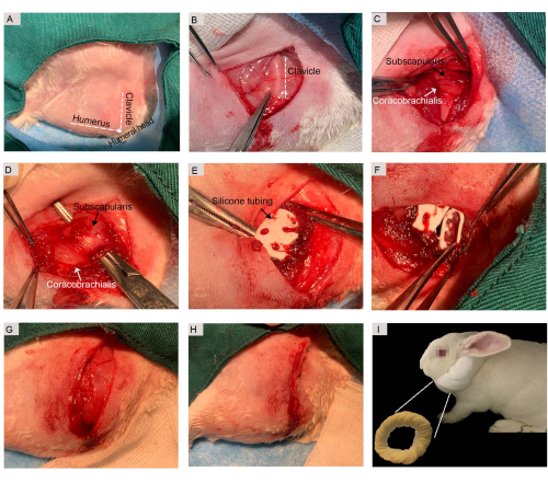
Figure 1: Surgical procedure for chronic-like SSC muscle-tendon injury model. (A) A surgical window was created and anatomical landmarks such as the humerus, humeral head, and clavicle were identified by palpation. (B) A 3.0 cm skin incision was made inferior to the clavicle. (C) The coracobrachialis muscle was split to expose the SSC muscle. (D) The SSC muscle-tendon unit was isolated. (E) A silicone-based penrose drain was used to wrap the SSC muscle-tendon tissue. (F) The SSC muscle-tendon was transected. (G) The coracobrachialis muscle was reapproximated using PGA sutures. (H) The skin incision was closed using nylon sutures. (I) Post-surgery, the rabbits were given a soft collar to wear. Abbreviations: SSC = subscapularis; PGA = poly glycolic acid. Please click here to view a larger version of this figure.

Figure 2: Gross morphology of representative SSC muscles. Black arrows represent white adipose tissues. Abbreviation: SSC = subscapularis. Please click here to view a larger version of this figure.

Figure 3: Histological analysis of chronic-like RC injury model at 4 weeks. (A) Representative H&E-stained histology images showed atrophic muscle fibers and accumulation of adipocytes. (B) Quantification of injured muscle fat accumulation percentage. n = 3 rabbits. Error bars indicate SEM. *, statistically significant (p≤ 0.05). Scale bars = 5,000 µm (A, left column), 600 µm (A, right column). Abbreviations: SSC = subscapularis; RC = rotator cuff; H&E = hematoxylin and eosin. Please click here to view a larger version of this figure.
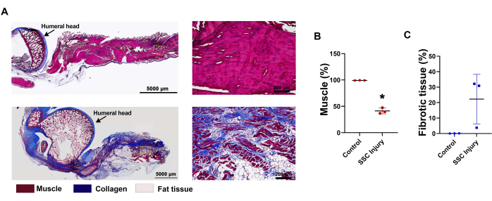
Figure 4: Histological analysis of chronic-like RC injury model at 4 weeks. (A) Masson's trichrome-stained images showed substantial fibrosis. Fibrous connective tissue are stained blue. (B) Quantification of the proportion of muscle and (C) fibrotic tissue. n = 3 rabbits. Error bars indicate SEM. *, statistically significant (p≤ 0.05). Scale bars = 5,000 µm (A, left column), 200 µm (A, right column). Abbreviations: SSC = subscapularis; RC = rotator cuff. Please click here to view a larger version of this figure.
Discussion
A reproducible and physiologically-relevant animal model provides the ability to advance the understanding of disease pathogenesis, evaluate the outcomes of clinical therapies, and improve and further develop surgical treatments35. In this study, a reliable and accurate rabbit SSC model that mimics aspects of human RC anatomy and pathophysiology was established. RC tears are related to progressive and likely irreversible muscular degenerative changes, resulting in a reduced healing potential. For example, Ko et al. showed that the reattachment of rabbit SSP at 6 weeks did not reverse muscle atrophy or FD in the following 6 weeks. Such FD-mediated muscle atrophy influences several important clinical parameters, including tendon-muscle strength and joint range of motion, which may affect the surgical outcomes36,37.
The protocol established here showed significant chronic-like attributes after the transection of SSC muscle-tendon units. Specifically, these changes include visibly decreased muscle mass and increased adipose content and fibrotic tissue (Figure 2, Figure 3, and Figure 4). These findings are consistent with degenerative changes reported in human RC tears38. In recent years, the rat has emerged as one of the most intensively studied animal models for RC disease and injury due to its high anatomical similarities with both human and rat SSPs traveling under the acromion38,39,40. However, it should be noted that the portion of rat SSP which passes under the acromial arch is muscular as opposed to tendinous, which is the case in humans41. Most importantly, Barton et al. recognized a lack of significant fat accumulation after SSP tendon detachment in rats23, which stands in contrast to the human condition42. As such, it is believed that the rabbit SSC complex may provide an appropriate model to mimic the chronic RC tear of humans.
To ensure the reproducibility of this model, two points are worth noting when performing this protocol. First, after the transection of muscle-tendon units, the free-end of the transected tendon may be at risk of forming adhesions, which can make tendon retrieval challenging for subsequent manipulations. To avoid this issue, a non-resorbable silicone tubing was used to wrap the free end of the muscle-tendon junction following transection to avoid spontaneous adhesion to surrounding tissues as well as spontaneous healing (Figure 1E). Further, the transected muscle-tendon unit during a second procedure for intervention (i.e., to perform a secure repair; data not shown) can be clearly identified by wrapping the end of injured tissues at the time of initial surgery. This technique is economical, effective, and can be easily implemented in surgery43. Second, rabbits are a highly sensitive species that may exhibit detrimental behavior following surgery. To avoid such issues, it is highly recommended that a soft collar is also applied to prevent undesired behavior, including self-mutilation, licking of surgical sites, and removal of sutures (Figure 1I). Compared to commercially conventional E-collars that are made of rigid plastic, the self-made soft collar did not cause any skin injury or other side effects that affected animal welfare or the quality of scientific inquiry. Together, such steps are critical to create an accurately reproducible rabbit RC injury model and provide the possibility for studying the regenerative repair strategies.
To study tendon pathophysiology and healing in an animal model, a distinct and reproducible injury must be created, and the study time points must be carefully selected. The vast majority of studies on tendon injury and healing have been performed on fully transected animal tendons44, as transection is a simple procedure that is highly reproducible and can adequately simulate the clinical scenario45,46. Huegel et al. showed that the injury of a partially transected tendon was less severe than that of a fully transected tendon, and immobilization had a detrimental effect on tendon mechanics, including increased joint stiffness47. To evaluate the atrophy and FD that is seen in the setting of massive RC tear, it is essential to define the experimentally observed characteristic time points. Gupta et al. have validated a RC injury model in the male rabbit and observed muscle atrophy at 2 and 6 week time points, with increased fat content at later time points (less than 5% fat content at 2 weeks vs. more than 10% fat content at 6 weeks), consistent with the pathological process observed in human RC tears11. In this study, a massive RC tear was created by transection of the SSC muscle-tendon unit in male and female rabbits for 4 weeks, which resulted in SSC muscle FD (36.5% fat content). Thus, a 4 week time point is appropriate for generating SSC muscle FD in male and female New Zealand white rabbits.
Several limitations to this study exist. These include: (i) steps associated with animal model generation, such as a relatively short time point and potentially inflammatory materials (silicone-based penrose tubing) for chronic-like injury generation; (ii) animal model characterization and analysis, such as lack of gait analysis and electromyography to assess joint kinematics and muscle contractile force generation; and (iii) animal model comparison, such as lack of comparison with other RC injury sites.
In terms of model generation, human RC injuries typically involve progressive atrophy and FD that may occur over the span of several years, which is relatively longer than the 4 week time point reported here. This is deemed to be acceptable, since an animal model that generates around 36.5% intramuscular fat in a relatively short time span will be logistically convenient and can be prolonged if deemed necessary. Moreover, the biocompatibility of silicone-based implants, such as penrose tubing, has been a source of long-standing controversy due to reports of cellular immune response and inflammation47; therefore, an alternative inert material, such as polyethylene glycol (PEG), may be substituted for wrapping the resected tendon if pursuing inflammation-associated RC studies.
In terms of animal model characterization and analysis, the lack of gait analysis49 and electromyograph studies50 may limit the study's findings to qualitative histological data. These aspects may be addressed in future studies by using video motion analysis51 and surface electromyography50 to generate quantitative data on shoulder kinematics and RC muscle performance.
In terms of model comparison, since the SSP and infraspinatus tendons in the rabbits have also been widely used for RC studies, comparing injury severity, including FD among these different injury sites in the future, will identify additional sites for model optimization.
In summary, this study has developed a protocol for modeling chronic-like RC injuries in male and female rabbits. This model is convenient for investigators owing to its simplicity (transection) and relatively short period to induce chronicity (4 weeks) while generating a large degree (36.5%) of intramuscular FD. As such, this protocol is expected to aid investigators in the study of RC pathophysiology, as well as facilitate the development of novel therapeutics for muscle-tendon repair and regeneration.
Disclosures
The authors have no competing interests to declare.
Acknowledgements
Dai Fei Elmer Ker's research is supported by funding from the Food and Health Bureau, Hong Kong SAR (Health Medical and Research Fund: 08190466), Innovation and Technology Commission, Hong Kong SAR (Tier 3 Award: ITS/090/18; Health@InnoHK program), Research Grants Council of Hong Kong, Hong Kong SAR (Early Career Scheme Award: 24201720 and General Research Fund: 14213922), and The Chinese University of Hong Kong (Faculty Innovation Award: FIA2018/A/01). Dan Wang's research is supported by funding from the Food and Health Bureau, Hong Kong SAR (Health Medical and Research Fund, 07180686), Innovation and Technology Commission, Hong Kong SAR (Tier 3 Award: ITS/333/18; Health@InnoHK program), and Research Grants Council of Hong Kong, Hong Kong SAR (General Research Fund: 14118620 and 14121121).
Materials
| Name | Company | Catalog Number | Comments |
|---|---|---|---|
| Surgical tools | |||
| 4-0 Poly glycolic acid (PGA) | e-Sutures | GBK884 | |
| Toothed Adson forceps | Taobao, China | ||
| Fine scissors | Taobao, China | ||
| Hemostatic forceps | Taobao, China | ||
| Needle holders | Taobao, China | ||
| Surgical scalpel with handle | Taobao, China | No. 11 blade | |
| Suture (4-0 Nylon) | Taobao, China | 19054 | Either nylon or silk sutures are acceptable for skin closure. Each suture has its own advantages and disadvantages and users are advised to choose one according to their preference. |
| Surgical accessories | |||
| Cotton balls | Taobao, China | ||
| Gauze | Taobao, China | ||
| Razor | Taobao, China | ||
| Surgical heating pad | Taobao, China | ||
| Surgical lamp | |||
| Syringe with needles | Taobao, China | 1 mL, 5 mL, 10 mL | |
| Drugs | |||
| Buprenorphine | LASEC, CUHK | 0.12 mg/kg | |
| Bupivacaine | Sigma-Aldrich | b5274-5g | 1-2 mg/kg |
| Cephalexin | Santa Cruz Biotechnology | sc-487556 | 20 mg/kg |
| Ketamine | LASEC, CUHK | 35 mg/kg | |
| Sodium pentobarbital | LASEC, CUHK | more than 60 mg/kg | |
| Xylazine | LASEC, CUHK | 5 mg/kg | |
| Equipment | |||
| Nikon Ni-U Eclipse Upright Microscope | Nikon Instruments Inc, USA | ||
| Software | |||
| Adobe Photoshop 20.01 | Adobe Inc, USA | ||
| Other reagents | |||
| Betadine | Taobao, China | 5% | |
| Ethanol | Taobao, China | 70% | |
| Ethylene diamine tetraacetic acid (EDTA) | Sigma-Aldrich | EDS-1KG | 10% |
| Paraformaldehyde (PFA) | Electron Microscopy Sciences | 15713 | 4% |
| Silicone tubing | Easy Thru, China | ISO13485 | |
| Saline | Taobao, China | ||
| Histological staining reagents | |||
| Eosin Stain Solution | Sigma-Aldrich | R03040 | 5% Aqueous |
| Hematoxylin Solution | Sigma-Aldrich | HHS32 | |
| Trichrome Stain (Masson) Kit | Sigma-Aldrich | HT15 |
References
- Goutallier, D., Postel, J. -M., Bernageau, J., Lavau, L., Voisin, M. -C. Fatty muscle degeneration in cuff ruptures. Pre-and postoperative evaluation by CT scan. Clinical Orthopaedics and Related Research. 304 (304), 78-83 (1994).
- Itoigawa, Y., Kishimoto, K. N., Sano, H., Kaneko, K., Itoi, E. Molecular mechanism of fatty degeneration in rotator cuff muscle with tendon rupture. Journal of Orthopaedic Research. 29 (6), 861-866 (2011).
- Mal Kim, H., et al. Relationship of tear size and location to fatty degeneration of the rotator cuff. The Journal of Bone and Joint Surgery. American Volume. 92 (4), 829-839 (2010).
- Melis, B., DeFranco, M. J., Chuinard, C., Walch, G. Natural history of fatty infiltration and atrophy of the supraspinatus muscle in rotator cuff tears. Clinical Orthopaedics and Related Research. 468 (6), 1498-1505 (2010).
- Li, K., Zhang, X., Wang, D., Tuan, R. S., Ker, D. F. E. Synergistic effects of growth factor-based serum-free medium and tendon-like substrate topography on tenogenesis of mesenchymal stem cells. Biomaterials Advances. , 146(2023).
- Derwin, K. A., Baker, A. R., Codsi, M. J., Iannotti, J. P. Assessment of the canine model of rotator cuff injury and repair. Journal of Shoulder and Elbow Surgery. 16, S140-S148 (2007).
- Grumet, R. C., Hadley, S., Diltz, M. V., Lee, T. Q., Gupta, R. Development of a new model for rotator cuff pathology: The rabbit subscapularis muscle. Acta Orthopaedica. 80 (1), 97-103 (2009).
- Renström, P., Johnson, R. J. Overuse injuries in sports. Sports Medicine. 2 (5), 316-333 (1985).
- Hertel, R., Lambert, S. M. Supraspinatus rupture at the musculotendinous junction. Journal of Shoulder and Elbow Surgery. 7 (4), 432-435 (1998).
- Oh, J. H., Chung, S. W., Kim, S. H., Chung, J. Y., Kim, J. Y. Neer Award: Effect of the adipose-derived stem cell for the improvement of fatty degeneration and rotator cuff healing in rabbit model. Journal of Shoulder and Elbow Surgery. 23 (4), 445-455 (2013).
- Gupta, R., Lee, T. Q. Contributions of the different rabbit models to our understanding of rotator cuff pathology. Journal of Shoulder and Elbow Surgery. 16, S149-S157 (2007).
- Otarodifard, K., Wong, J., Preston, C. F., Tibone, J. E., Lee, T. Q. Relative fixation strength of rabbit subscapularis repair is comparable to human supraspinatus repair at time 0. Clinical Orthopaedics and Related Research. 472 (8), 2440-2447 (2014).
- Liu, X., Manzano, G., Kim, H. T., Feeley, B. T. A rat model of massive rotator cuff tears. Journal of Orthopaedic Research. 29 (4), 588-595 (2011).
- Liu, X., et al. A mouse model of massive rotator cuff tears. The Journal of Bone and Joint Surgery. American Volume. 94 (7), 41(2012).
- Neer,, et al. Award 2007: Reversion of structural muscle changes caused by chronic rotator cuff tears using continuous musculotendinous traction. An experimental study in sheep. Journal of Shoulder and Elbow Surgery. 18 (2), 163-171 (2009).
- Warner, J. J., Parsons, I. M. Latissimus dorsi tendon transfer: A comparative analysis of primary and salvage reconstruction of massive, irreparable rotator cuff tears. Journal of Shoulder and Elbow Surgery. 10 (6), 514-521 (2001).
- Galatz, L. M., Ball, C. M., Teefey, S. A., Middleton, W. D., Yamaguchi, K. The outcome and repair integrity of completely arthroscopically repaired large and massive rotator cuff tears. The Journal of Bone and Joint Surgery. American. 86 (2), 219-224 (2004).
- Kim, H. M., Galatz, L. M., Lim, C., Havlioglu, N., Thomopoulos, S. The effect of tear size and nerve injury on rotator cuff muscle fatty degeneration in a rodent animal model. Journal of Shoulder and Elbow Surgery. 21 (7), 847-858 (2012).
- Carpenter, J. E., Thomopoulos, S., Flanagan, C. L., DeBano, C. M., Soslowsky, L. J. Rotator cuff defect healing: A biomechanical and histologic analysis in an animal model. Journal of Shoulder and Elbow Surgery. 7 (6), 599-605 (1998).
- Jal Soslowsky, L., et al. Rotator cuff tendinosis in an animal model: Role of extrinsic and overuse factors. Annals of Biomedical Engineering. 30 (8), 1057-1063 (2002).
- Thomopoulos, S., et al. The localized expression of extracellular matrix components in healing tendon insertion sites: An in situ hybridization study. Journal of Orthopaedic Research. 20 (3), 454-463 (2002).
- Su, W., et al. Effect of suture absorbability on rotator cuff healing in a rabbit rotator cuff repair model. The American Journal of Sports Medicine. 46 (11), 2743-2754 (2018).
- Barton, E. R., Gimbel, J. A., Williams, G. R., Soslowsky, L. J. Rat supraspinatus muscle atrophy after tendon detachment. Journal of Orthopaedic Research. 23 (2), 259-265 (2005).
- Gerber, C., Meyer, D. C., Schneeberger, A. G., Hoppeler, H., von Rechenberg, B. Effect of tendon release and delayed repair on the structure of the muscles of the rotator cuff: An experimental study in sheep. The Journal of Bone and Joint Surgery. American Volume. 86 (9), 1973-1982 (2004).
- Gerber, C., Schneeberger, A. G., Perren, S. M., Nyffeler, R. W. Experimental rotator cuff repair. A preliminary study. The Journal of Bone and Joint Surgery. American Volume. 81 (9), 1281-1290 (1999).
- Hal Coleman, S., et al. Chronic rotator cuff injury and repair model in sheep. The Journal of Bone and Joint Surgery. American Volume. 85 (12), 2391-2402 (2003).
- Turner, A. S. Experiences with sheep as an animal model for shoulder surgery: strengths and shortcomings. Journal of Shoulder and Elbow Surgery. 16, S158-S163 (2007).
- Turner, P. V., Brabb, T., Pekow, C., Vasbinder MA, Administration of substances to laboratory animals: routes of administration and factors to consider. J Am Assoc Lab Anim Sci. 50 (5), 600-613 (2011).
- Cooper, C. S., Metcalf-Pate, K. A., Barat, C. E., Cook, J. A., Scorpio, D. G. Comparison of side effects between buprenorphine and meloxicam used postoperatively in Dutch belted rabbits (Oryctolagus cuniculus). Journal of the American Association for Laboratory Animal Science. 48 (3), 279-285 (2009).
- Eal Ker, D. F., et al. Functionally graded, bone-and tendon-like polyurethane for rotator cuff repair. Advanced Functional Materials. 28 (20), 1707107(2018).
- Toumi, H., et al. Regional variations in human patellar trabecular architecture and the structure of the proximal patellar tendon enthesis. Journal of Anatomy. 208 (1), 47-57 (2006).
- Noor, R. A. M., Shah, N. S. M., Zin, A. A. M., Sulaiman, W. A. W., Halim, A. S. Disoriented collagen fibers and disorganized, fibrotic orbicularis oris muscle fiber with mitochondrial myopathy in non-syndromic cleft lip. Archives of Oral Biology. 140, 105448(2022).
- Wang, D., et al. Growth and differentiation factor-7 immobilized, mechanically strong quadrol-hexamethylene diisocyanate-methacrylic anhydride polyurethane polymer for tendon repair and regeneration. Acta Biomaterialia. 154, 108-122 (2022).
- Wang, D., et al. Combinatorial mechanical gradation and growth factor biopatterning strategy for spatially controlled bone-tendon-like cell differentiation and tissue formation. NPG Asia Materials. 13 (1), (2021).
- Kuyinu, E. L., Narayanan, G., Nair, L. S., Laurencin, C. T. Animal models of osteoarthritis: Classification, update, and measurement of outcomes. Journal of Orthopaedic Surgery and Research. 11, (2016).
- Safran, O., Derwin, K. A., Powell, K., Iannotti, J. P. Changes in rotator cuff muscle volume, fat content, and passive mechanics after chronic detachment in a canine model. The Journal of Bone and Joint Surgery. American. 87 (12), 2662-2670 (2005).
- Gerber, C., Fuchs, B., Hodler, J. The results of repair of massive tears of the rotator cuff. The Journal of Bone and Joint Surgery. American Volume. 82 (4), 505-515 (2000).
- Longo, U. G., Berton, A., Khan, W. S., Maffulli, N., Denaro, V. Histopathology of rotator cuff tears. Sports Medicine and Arthroscopy Review. 19 (3), 227-236 (2011).
- Schneeberger, A. G., Nyffeler, R. W., Gerber, C. Structural changes of the rotator cuff caused by experimental subacromial impingement in the rat. Journal of Shoulder and Elbow Surgery. 7 (4), 375-380 (1998).
- Soslowsky, L. J., Carpenter, J. E., DeBano, C. M., Banerji, I., Moalli, M. R. Development and use of an animal model for investigations on rotator cuff disease. Journal of Shoulder and Elbow Surgery. 5 (5), 383-392 (1996).
- Rowshan, K., et al. Development of fatty atrophy after neurologic and rotator cuff injuries in an animal model of rotator cuff pathology. The Journal of Bone and Joint Surgery. 92 (13), 2270-2778 (2010).
- Gladstone, J. N., Bishop, J. Y., Lo, I. K., Flatow, E. L. Fatty infiltration and atrophy of the rotator cuff do not improve after rotator cuff repair and correlate with poor functional outcome. The American Journal of Sports Medicine. 35 (5), 719-728 (2007).
- Chen, W. F., Kim, B. -S., Lin, Y. -T. Penrose drain interposition-A novel approach to preventing adhesion formation after tenolysis. The Journal of Hand Surgery. Asian-Pacific Volume. 27 (1), 174-177 (2022).
- Lui, P. P. Y. Stem cell technology for tendon regeneration: Current status, challenges, and future research directions. Stem Cells and Cloning: Advances and Applications. 8, 163-174 (2015).
- Howell, K., et al. Novel model of tendon regeneration reveals distinct cell mechanisms underlying regenerative and fibrotic tendon healing. Scientific Reports. 7, 45238(2017).
- Sharma, P., Maffulli, N. Tendinopathy and tendon injury: The future. Disability and Rehabilitation. 30 (20-22), 1733-1745 Forthcoming.
- Huegel, J., et al. Quantitative comparison of three rat models of Achilles tendon injury: A multidisciplinary approach. Journal of Biomechanics. 88, 194-200 (2019).
- Pal Heggers, J., et al. Biocompatibility of silicone implants. Annals of Plastic Surgery. 11 (1), 38-45 (1983).
- Liu, Y., et al. Evaluation of animal models and methods for assessing shoulder function after rotator cuff tear: A systematic review. Journal of Orthopaedic Translation. 26, 31-38 (2020).
- Disselhorst-Klug, C., Schmitz-Rode, T., Rau, G. Surface electromyography and muscle force: Limits in sEMG-force relationship and new approaches for applications. Clinical Biomechanics. 24 (3), 225-235 (2009).
- Kwon, D. R., Park, G. -Y., Moon, Y. S., Lee, S. C. Therapeutic effects of umbilical cord blood-derived mesenchymal stem cells combined with polydeoxyribonucleotides on full-thickness rotator cuff tendon tear in a rabbit model. Cell Transplantation. 27 (11), 1613-1622 (2018).
Reprints and Permissions
