This study shows a detailed protocol to perform ultrastructure expansion microscopy in three in vitro life cycle stages of Trypanosoma cruzi, the pathogen responsible for Chagas disease. We include the optimized technique for cytoskeletal proteins and pan-proteome labeling.
A subscription to JoVE is required to view this content. Sign in or start your free trial.
Method Article
Ultrastructural Expansion Microscopy in Three In Vitro Life Cycle Stages of Trypanosoma cruzi
* These authors contributed equally
In This Article
Summary
Abstract
We describe here the application of ultrastructure expansion microscopy (U-ExM) in Trypanosoma cruzi, a technique that allows increasing the spatial resolution of a cell or tissue for microscopic imaging. This is performed by physically expanding a sample with off-the-shelf chemicals and common lab equipment.
Chagas disease is a widespread and pressing public health concern caused by T. cruzi. The disease is prevalent in Latin America and has become a significant problem in non-endemic regions due to increased migration. The transmission of T. cruzi occurs through hematophagous insect vectors belonging to the Reduviidae and Hemiptera families. Following infection, T. cruzi amastigotes multiply within the mammalian host and differentiate into trypomastigotes, the non-replicative bloodstream form. In the insect vector, trypomastigotes transform into epimastigotes and proliferate through binary fission.The differentiation between the life cycle stages requires an extensive rearrangement of the cytoskeleton and can be recreated in the lab completely using different cell culture techniques.
We describe here a detailed protocol for the application of U-ExM in three in vitro life cycle stages of Trypanosoma cruzi, focusing on optimization of the immunolocalization of cytoskeletal proteins. We also optimized the use of N-Hydroxysuccinimide ester (NHS), a pan-proteome label that has enabled us to mark different parasite structures.
Introduction
Expansion microscopy (ExM) was described for the first time in 2015 by Boyden et al.1. It is an imaging protocol with which a conventional microscope can achieve a spatial resolution below the diffraction limit. This higher resolution is obtained because of a physical enlargement of the sample. To accomplish this, fluorescently labeled molecules are crosslinked to a hydrogel, which is subsequently expanded isotropically with water. As a result of this expansion, the signals are separated nearly isotropically in all three dimensions. This method employs low-cost chemicals and enables a spatial resolution of approximately 65 nm using conventional (confocal) microscopes, which is roughly four times better than the standard resolution of a confocal microscope (approximately 250 nm)1.
The next milestone, that has enabled the use of expansion microscopy in many biological fields, was the adaptation of immunofluorescence labeling with conventional antibodies2. Another adaptation from the initially published ExM protocol is the magnified analysis of the proteome (MAP)3. This method introduced the use of high concentrations of acrylamide and paraformaldehyde prior to sample-hydrogel immersion to prevent intra- and inter-protein crosslinking, which led to better preservation of the samples' protein content and subcellular architecture. This alternative protocol was optimized to obtain enhanced conservation of the overall ultrastructure of isolated organelles by utilizing lower concentrations of the fixative agents (formaldehyde/paraformaldehyde and acrylamide); this approach was termed ultrastructure expansion microscopy (U-ExM)4.
To gain even more resolution, the combination of ExM with super-resolution microscopy techniques, including stimulated emission depletion microscopy or single-molecule localization microscopy, has also been reported in order to reach resolutions below 20 nm5.
The use of ExM has been widely reported in the fields of neuroscience and cytoskeleton research6, but only a few studies have been conducted on parasitic protists. Our laboratory was the first to report the application of U-ExM in T. cruzi7. The foundation protocol is mainly based on the previous U-ExM reports in Toxoplasma gondii, Plasmodium ssp., and Trypanosoma brucei8,9,10,11.
One of the greatest advantages of ExM is its modular nature, which allows great flexibility to adapt to different biological samples. The protocol can be divided into steps (such as fixation, crosslinking prevention, or gelation) that can be easily adjusted by the user to meet their experimental requirements. Additionally, this pipeline can be modified to enhance compatibility with the model organism or to achieve a specific resolution. As a result, ExM offers tremendous potential for both advanced and non-advanced optical systems, ensuring wider applications in the future.
Chagas disease, also called American trypanosomiasis, is an endemic disease in Latin America caused by Trypanosoma cruzi, a protozoan parasite. The parasite's life cycle is complex and involves two developmental stages in mammals and two in the insect host (members of the Triatominidae family), which is the biological vector of this disease. Chagas disease belongs to the group of neglected tropical diseases listed by the World Health Organization and represents a significant economic and social problem in Latin America. Epidemiological studies estimate 8 million people around the world live with Chagas disease and over 10,000 deaths per year. These numbers exemplify the significance of Chagas disease as a public health issue worldwide. The geographic distribution of Chagas disease has changed in recent decades, with many infected individuals now residing in large urban areas globally due to increased migrations, as opposed to the primarily rural areas of Latin America where it was originally found12.
The developmental stages of T. cruzi differ throughout its life cycle, which can be replicated completely in vitro. Epimastigotes are replicative forms in the insect vector, and they have a spherical nucleus in the central region of the cell body and a bar-shaped kinetoplast (a mitochondrial DNA-containing structure unique to kinetoplastids) in the anterior region relative to the nucleus, with a free flagellum. Trypomastigotes are the infective, non-replicative form, and have an elongated nucleus, a rounded posterior kinetoplast, and a flagellum attached to the plasma membrane along the entire length of the parasite. Amastigotes are the intracellular replicative form; they have a nucleus in the central region, a rod-shaped kinetoplast in the anterior part of the cell body, and a reduced flagellum. The parasite's adaptability to different environments is a reflection of these morphological variations. It is also worth mentioning that this life cycle involves symmetrical division and different transitional developmental stages13. During differentiation, the trypanosomatids' cytoskeleton plays a critical role. This structure is formed by a corset of subpellicular microtubules arranged in an ordered array of stable microtubules below the plasma membrane. Also, a paraflagellar rod is present in these organisms, which is a lattice-like structure that runs parallel and is attached to the flagellar axoneme14. The precise cytoskeletal organization and nuclear structural changes along the cell cycle stages involve unique gene regulation mechanisms specific to trypanosomatids, making them interesting models for cell biology studies.
Given the small size of T. cruzi and other protozoan parasites, U-ExM presents an excellent tool for analyzing the structural features of these important pathogens. As mentioned earlier, the applicability of this technique on T. cruzi was validated for the first time by Dr. Alonso7. This report details a complete U-ExM protocol, with emphasis on the immunolocalization of cytoskeletal proteins during the different life cycle stages of T. cruzi. Also, we have optimized the use of N-Hydroxysuccinimide ester (NHS), a pan-proteome label that enables us to mark various parasite structures. In addition, an in vitro methodology to obtain the three stages of the parasite is described.
Access restricted. Please log in or start a trial to view this content.
Protocol
NOTE: Figure 1 illustrates the complete experimental design.
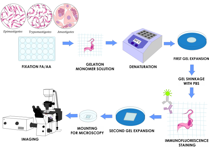
Figure 1: U-ExM workflow for three in vitro life cycle stages of T. cruzi. Please click here to view a larger version of this figure.
1. Preparation of the poly-D-lysine-coated coverslips
- Place a 10 cm x 10 cm square of sealing film in a Petri dish. Wash the coverslips by bathing them in absolute ethanol in a 35 mm glass Petri dish.
- Remove the coverslips with tweezers from the ethanol bath and drain the excess liquid with tissue paper. Place the coverslips over the sealing film.
- Absorb the rest of the ethanol with microscopy-grade paper. Add a 0.1% v/v solution of poly-D-lysine on the center of the coverslip and spread it with the tip to cover approximately 80% of its surface. Close the Petri dish and incubate for 1 h at 37 °C.
NOTE: For 22 mm2 coverslips, use 200 µL of poly-D-lysine solution; for 12 mm round coverslips, use 100 µL of poly-D-lysine solution. - Wash the coverslips with ultra-pure water three times. Use aspiration with a vacuum to remove the water between each wash. Keep at 4 °C for up to 1 week.
2. Solution preparation
- Prepare stock solutions of 38% (w/w) sodium acrylate (SA).
- Slowly add 19 g of SA to 31 mL of nuclease-free water while stirring.
NOTE: This solution is very viscous; pay attention when pipetting. - Once the SA is completely dissolved, store it in a sterile container. Keep it at 4 °C and change it every 6 months.
NOTE: SA sometimes shows signs of contamination with sodium polyacrylate depending on the brand, which is noticeable when preparing the stock solution as it turns yellowish and cloudy. Use it cautiously if this is the case.
- Slowly add 19 g of SA to 31 mL of nuclease-free water while stirring.
- Prepare a stock solution of 40% (w/v) acrylamide (AA) and a stock solution of 2% (w/v) N, N'-methylene bisacrylamide (BIS). Dissolve each compound in ultrapure water and filter with a 0.22 µm sterile syringe filter. Store in a sterile container at 4 °C.
CAUTION: AA and BIS are highly toxic substances. Work under a fume hood and use proper protection elements (gloves, protective clothing, mask, and safety glasses). - Prepare the protein crosslinking prevention (CP) solution by mixing 38 µL of a 37% formaldehyde (FA) solution with 50 µL of the 40% acrylamide stock solution (step 2.2) in 912 µL of phosphate-buffered saline (PBS; Table 1) to obtain a final concentration of 1.4% formaldehyde and 2% acrylamide.
NOTE: Always prepare the CP solution freshly and be extremely precise when pipetting. For example, use a P1000 pipette to take 900 µL of PBS and a P20 pipette to take the remaining 12 µL of PBS. - Prepare the monomeric solution
NOTE: Do not use the solution immediately after being prepared; store at -20 °C at least 24 h before being used.- Mix 500 µL of the 38% SA solution (step 2.1), 250 µL of the 40% AA solution, and 50 µL of the BIS solution (step 2.2), as well as 100 µL of 10x PBS (Table 1).
- Aliquot this final volume of 900 µL in 10x 1.5 mL microcentrifuge tubes with 90 µL of monomeric solution each. Store at -20 °C for up to 2 weeks.
NOTE: The SA solution may stay on the tip when pipetting because it is very viscous; be careful and dispense everything.
- Prepare the denaturing solution
- Mix 114.3 mL of a 350 mM sodium dodecyl sulfate (SDS) solution prepared in ultrapure water with 10 mL of a 4 M sodium chloride solution prepared in ultrapure water. Add 12 g of Tris while stirring in a 250 mL beaker.
CAUTION: SDS is highly toxic; use it under a fume hood wearing gloves, protective clothing, a mask, and safety glasses. - Adjust the pH to 9 with a concentrated solution of hydrochloric acid. Make up to 200 mL with ultrapure water and store in a sterile flask at 4 °C.
- Mix 114.3 mL of a 350 mM sodium dodecyl sulfate (SDS) solution prepared in ultrapure water with 10 mL of a 4 M sodium chloride solution prepared in ultrapure water. Add 12 g of Tris while stirring in a 250 mL beaker.
- Prepare a 10% ammonium persulfate (APS) solution and a 10% TEMED solution.
- Dissolve 0.1 g of APS in 1 mL of ultrapure water.
- Prepare 1 mL of a 10% TEMED solution in ultrapure water.
- Prepare 100 µL aliquots of both solutions in 1.5 mL microcentrifuge sterile tubes and store them at -20 °C for up to 1 month.
- Prepare the paraformaldehyde solution.
- Dissolve 2 g of paraformaldehyde in 40 mL of PBS while stirring at 60 °C. Add 1 M NaOH dropwise until the solution turns from white to colorless.
CAUTION: Formaldehyde is highly toxic; use it under a fume hood wearing gloves, protective clothing, a mask, and safety glasses. - Cool the solution at room temperature and adjust the pH with NaOH to 7.2 in a final volume of 50 mL. Filter with a 0.22 µm sterile syringe filter and store in a sterile container.
- Prepare aliquots of 1 mL in 1.5 mL sterile microcentrifuge tubes and store at -20 °C.
- Dissolve 2 g of paraformaldehyde in 40 mL of PBS while stirring at 60 °C. Add 1 M NaOH dropwise until the solution turns from white to colorless.
- Prepare the paraformaldehyde/glutaraldehyde solution.
- Add 0.2 g of paraformaldehyde in 3.5 mL of ultrapure water and 50 µL of 16 M NaOH solution. Heat the solution to 60 °C to dissolve the paraformaldehyde.
- Cool down and add 300 µL of 70% glutaraldehyde. Bring up the volume to 5 mL with ultrapure water and finally to 10 mL with PBS.
CAUTION: Paraformaldehyde and glutaraldehyde are highly toxic; use them under a fume hood wearing gloves, protective clothing, a mask, and safety glasses. - Prepare aliquots of 1 mL in 1.5 mL sterile microcentrifuge tubes and store at -20 °C.
3. Preparation of the parasite cultures
- Grow T. cruzi epimastigotes.
- Use an axenic culture in a T-25 flask (25 cm2 growth area) and maintain the cultures in the logarithmic phase by sub-culturing every 48-72 h in liver infusion tryptose (LIT) medium with 10% fetal calf serum (FCS; Table 1).
- Ensure the cap is securely closed and keep the culture flask vertically at 28 °C for incubation. Monitor the parasite growth by cell counting in a Neubauer chamber during each subculture.
NOTE: For the Dm28c strain used in this study, the concentration of epimastigotes in log-phase cultures is between 1-5 x 10⁷ parasites/mL. - Prepare a suspension of 2 x 10⁶ epimastigotes/mL from a log-phase culture in LIT medium supplemented with 10% FCS. Centrifuge the suspension at 5,000 x g for 10 min at room temperature (RT). Wash with PBS once or twice and resuspend in 200 µL of PBS.
- Adhere to the round 12 mm coverslip previously coated with poly-D-lysine (section 1). Incubate at RT for 15-20 min. Continue to the crosslinking prevention step (section 4).
NOTE: Alternatively, fix the epimastigotes with cold methanol for 7 min or paraformaldehyde/glutaraldehyde solution (step 2.8) for 10 min at RT prior to adherence of the parasites to the coverslip. It is possible to store the fixed parasites at 4 °C for up to 1 week.
- Obtain the amastigotes from infected Vero cells.
- Lay a sterile 12 mm round coverslip on the bottom of a 24-well tissue culture plate. Prepare a suspension of 2 x 10⁵ Vero cells/mL in Dulbecco's modified Eagle medium (DMEM) supplemented with 2% fetal calf serum (FCS). Seed 500 µL of the suspension per well.
NOTE: This study uses DMEM with 2% FCS to allow the cells to grow slower. - Incubate overnight (ON; 12-16 h) at 37 °C and 5% CO2 to ensure cell attachment.
- Following incubation, wash the cells twice using 500 µL of sterile PBS. Add T. cruzi trypomastigotes into the cells at a multiplicity of infection (MOI) of 10 in 100 µL of DMEM with 2% FCS per well, corresponding to one million trypomastigotes per well. Incubate at 37 °C and 5% CO2 for 6 h.
- After incubation, rinse the plates twice with PBS. Add 500 µL of DMEM supplemented with 2% FCS. At this point, continue to the crosslinking prevention step (section 4).
NOTE: Intracytoplasmic amastigotes will be visible through an inverted optical microscope 2 days post-infection (Supplementary Figure 1). Alternatively, fix the trypomastigotes with cold methanol for 7 min at -20 °C, or paraformaldehyde/glutaraldehyde for 10 min at RT, prior to adherence of the parasites to the coverslip.
- Lay a sterile 12 mm round coverslip on the bottom of a 24-well tissue culture plate. Prepare a suspension of 2 x 10⁵ Vero cells/mL in Dulbecco's modified Eagle medium (DMEM) supplemented with 2% fetal calf serum (FCS). Seed 500 µL of the suspension per well.
- Obtain trypomastigotes from infected Vero cells.
- Collect the supernatant of a Vero cell monolayer (30%-40% confluence) infected with trypomastigotes (MOI 1:10; ON incubation) 4 days post-infection.
NOTE: For a T-25 flask, the initial concentration of Vero cells used is 800,000 cells, and for a T-75 flask is two million cells. - Determine the trypomastigote concentration using a Neubauer chamber for cell counting. Centrifuge 4 x 10⁶ trypomastigotes at 7,000 x g at RT for 10 min.
- Rinse with PBS twice and resuspend in 200 µL of PBS. Apply to a round, 12 mm coverslip coated with poly-D-lysine (section 1). Incubate for 10-15 min at RT. Proceed with the crosslinking prevention step (section 4).
NOTE: Alternatively, fix the trypomastigotes with cold methanol for 7 min at -20 °C or paraformaldehyde/glutaraldehyde for 10 min at RT prior to adherence of the parasites to the coverslip.
- Collect the supernatant of a Vero cell monolayer (30%-40% confluence) infected with trypomastigotes (MOI 1:10; ON incubation) 4 days post-infection.
4. Performing crosslinking prevention (DAY 1)
- Submerge the 12 mm coverslip with the adhered parasites or infected cells (facing upward) in a 24-well plate with 0.5 mL of CP solution (step 2.3) in each well.
- Fill the empty wells with water to reduce evaporation. Seal the plate with a sealing film. Incubate for 5 h at 37 °C. This step can be extended up to an ON incubation at 4 °C.
NOTE: Always submerge the coverslip in the solution; do not pipette the fixative solution over the coverslips.
5. Performing gelation of the sample
- Assemble a humid chamber in a Petri dish with a sealing film on top of tissue paper (Figure 2A). Add water to the tissue paper and incubate at -20 °C for 20 min to cool down. Thaw a TEMED and an APS aliquot on ice for 20 min (step 2.6).
NOTE: Do not freeze-thaw the APS more than three times. - Lay the cool, humid chamber on ice (Figure 2A). Take the 24-well plate prepared in section 4 out of the incubator. Aspirate the CP solution with a 3 mL Pasteur pipette, leaving some solution, otherwise the coverslips will be hard to remove.
- Remove the 12 mm coverslips from the fixative solution with tweezers and lay them on tissue paper with the parasites facing up.
NOTE: It is helpful to use a sterile needle to lift the coverslip and then hold it with the tweezers. - To an aliquot of 90 µL of monomeric solution (step 2.4), add 5 µL of TEMED and 5 µL of APS previously thawed. Mix with a vortex mixer for no more than 2-3 s; it is not necessary to close the tube with the lid.
NOTE: Always add TEMED first and APS last. The addition of APS first and TEMED last to the monomeric solution makes the gelation process faster, giving no time to manipulate it. - Quickly make one drop of 35 µL over the sealing film of the humid chamber for each coverslip. Immediately pick up the coverslip with tweezers and lay over the drop (Figure 2B) with the parasites facing down.
NOTE: Make a maximum of two coverslips at a time; it is crucial not to delay this step because the solution polymerizes very quickly. - Incubate the humid chamber for 5 min on ice and then for 1 h at 37 °C. Turn on a heating block at 95 °C to ensure the correct temperature for the next step.
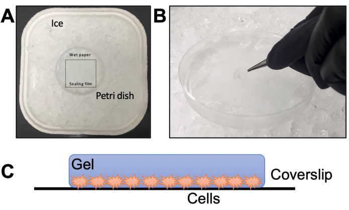
Figure 2: Gelation step details. (A) Assembly of the humid chamber. (B) Dropping the coverslips onto the monomer solution with TEMED and APS for gelation. (C) Schematic representation of the gel assembled for imaging. Please click here to view a larger version of this figure.
6. Denaturing the gelified samples and performing the isotropic expansion
- Remove the denaturation solution (step 2.5) from 4 °C. If it is precipitated, put it in a hot water bath until it dissolves completely.
- Add 2 mL of the denaturing solution to each well of a 6-well plate. Transfer the coverslips from step 5.3 to the plate with the denaturation solution. Incubate for 15 min at RT with gentle shaking so the gel detaches from the coverslip.
- Carefully transfer the gel (removing it from the 12 mm coverslip) with a metal spatula into a 1.5 mL sterile microcentrifuge tube with 1 mL of denaturing solution. Use cap locks to secure the tubes. Incubate for 1 h and 30 min at 95 °C in a heating block.
NOTE: Gels start to expand during this step; be gentle when transferring the gel to the 1.5 mL microcentrifuge tube.
CAUTION: After the incubation, the tube containing the gel is at 95 °C, which can be dangerous. Use protective gloves and let the tubes cool down before handling them to avoid burns and projections. - Perform the first round of expansion
- Aspirate the denaturing solution from the 1.5 mL microcentrifuge tube with the gel with a P1000 pipette. Transfer the gel from the 1.5 mL microcentrifuge tube to a Petri dish with 10 mL of ultrapure water for 30 min using a small spatula.
- Change the ultrapure water using a disposable 3 mL Pasteur pipette. Incubate ON at RT.
NOTE: Be gentle with the gels because, after the first 30 min of incubation, they became fragile. - Change the ultrapure water using a disposable 3 mL Pasteur pipette one more time.
NOTE: Three water incubations of 30 min each are sufficient, but for practicality, it is better to leave the second incubation ON.
7. Performing fluorescence labeling of the target proteins (DAY 2)
- Remove the water from the Petri dish with the gel with a disposable 3 mL Pasteur pipette. Measure the diameter of the gel with a caliper to calculate the expansion (between four and five times).
- Wash twice with 10 mL of PBS for 15 min. Cut the gel with a razor blade into squares of approximately 10 mm x 10 mm in the center of the circular gel. Use one square per condition to be tested.
NOTE: The gel shrinks after the PBS incubations. Gels can be stored in PBS at 4 °C for up to 1 week. - Transfer each square to a 12-well plate and incubate with 500 µL of primary antibody diluted in 2% PBS-bovine serum albumin (BSA) for 2 h and 30 min at 37 °C with shaking.
NOTE: Alternative antibody incubation can be performed ON at 4 °C. The minimum antibody volume that can be used is 300 µL in a 24-well plate. As a general rule, use twice the concentration of antibodies used for conventional immunolabeling. - Wash three times with 2 mL of PBS with 0.1% polysorbate 20 for 10 min while shaking in a 6-well plate.
- Transfer the gel to a 12-well plate and incubate with 500 µL of secondary antibody in PBS with DAPI and 10 µg/mL NHS-ester conjugated to the desired fluorophore for 2 h and 30 min at 37 °C with gentle shaking.
NOTE: As a general rule, use twice the concentration of antibodies used for conventional immunolabeling. Alternatively, this incubation can be performed ON at 4 °C. - Wash three times with 2 mL of PBS with 0.1% polysorbate 20 for 10 min while shaking in a 6-well plate.
- Transfer the gel to a Petri dish with ultrapure water. Incubate for 30 min. Change the water twice with a disposable 3 mL Pasteur pipette, as done in step 6.4.
8. Imaging and image processing (DAY 3)
- Remove the water from the Petri dishes with a disposable 3 mL Pasteur pipette and measure the gel diameter with a caliper to calculate the expansion factor.
- Cut a small piece of ~10 mm x 10 mm with a razor blade and place it on a 35 mm glass bottom dish.
NOTE: As an alternative, place the gel between a slide and a coverslip (without poly-D-lysine). The slide should have two smaller slide pieces cut with a diamond knife attached on the sides to form a chamber about the thickness of the gel. - Check the orientation at 10x or 20x magnification. In order to focus properly, ensure that the parasites are facing the coverslip; otherwise, one has to turn the gel around and check again (Figure 2C).
- Once the proper orientation is found, dry the remaining gel with microscopy paper. Cover the gel with a poly-D-lysine-coated coverslip.
- Add a small drop of ultrapure water to the gel to visualize with a confocal microscope.
NOTE: To ensure proper imaging, it is crucial to maintain the gel orientation throughout the process so that the side of the gel containing cells near its surface is facing the glass-bottom dish at the end. When using 35 mm dishes, adding poly-D-lysine to the coverslip placed in the bottom reduces the gel shifting during the image acquisition. - Image acquisition parameters
- Image the samples in a confocal microscope (Table of Materials) using a 63x oil immersion 1.4 numerical aperture (NA) objective.
- Acquire Z stacks, the width of each z step, and an exposure time per pixel, to be determined empirically depending on the sample, the signal intensity, and optimization of acquisition times. Use the scan zoom for effective magnification if desired.
- Open the Z stack using image processing software (Table of materials). For each channel, group the stacked images using the Group Z-project option. Select the maximum intensity projection.
- To merge images, use the Merge Channels tool and select the color of each channel. Add a scale bar using the Scale Bar tool in the processing software.
NOTE: Alternatively, one can color each channel as desired (see examples in the representative results).
Access restricted. Please log in or start a trial to view this content.
Results
If the protocol has been properly executed (Figure 1), samples will be visible as a planar and translucent gel that can be expanded up to a factor of 4-4.5x in water (Figure 3A). This expansion provided an effective resolution of about 70 nm, which may vary depending on the final expansion factor and imaging system employed. After the second expansion process and image acquisition in a confocal microscope, we were able to observe expansion factors of around 4.5....
Access restricted. Please log in or start a trial to view this content.
Discussion
Ultrastructural expansion microscopy is a technique that allows obtaining high-resolution images of biological samples by physically expanding them to several times their original size. The U-ExM protocol involves several critical steps that must be carefully executed to achieve optimal results4. First, the sample must be fixed with a CP agent and embedded in a swellable hydrogel matrix. The formaldehyde present in the CP solution interacts with the free covalent bonds of the acrylamide to prevent...
Access restricted. Please log in or start a trial to view this content.
Disclosures
The authors have no conflicts of interest to disclose.
Acknowledgements
We thank Dolores Campos for assisting with Vero cell culture and Romina Manarin for assisting with T. cruzi culture. This work was supported by Agencia Nacional de Promoción Científica y Tecnológica, Ministerio de Ciencia e Innovación Productiva from Argentina (PICT2019-0526), Consejo Nacional de Investigaciones Científicas y Técnicas (PIBAA 1242), and Research Council United Kingdom [MR/P027989/1].
Access restricted. Please log in or start a trial to view this content.
Materials
| Name | Company | Catalog Number | Comments |
|---|---|---|---|
| 0.22 micrometers sterile syringe filters PES | Membrane solutions | SFPES030022S | |
| 1 L beaker | Schott Duran | 10005227 | |
| 1.5-mL SPINWIN Micro Centrifuge Tube | Tarson | T38-500010 | |
| 10 mL disposable sterile serynge | NP | 66-32 | |
| 10 mL serological pipette sterile | Jet Biofil | GSP211010 | |
| 12-mm coverslips | Merienfeld GmbH | 01 115 20 | Round coverslips |
| 12-well plates | Jet Biofil | TCP011012 | |
| 22-mm coverslips | Corning | 2845-22 | Square coverslips |
| 24-well plates | Jet Biofil | TCP-011-024 | |
| 250 mL beaker | Schott Duran | C108.1 | |
| 3 mL Pasteur pipette | Deltalab | 200037 | |
| 35-mm glass bottom dishes | Matsunami glass ind | D11130H | |
| 4′,6-Diamidine-2′-phenylindole dihydrochloride | Sigma Aldrich | D9542 | DAPI |
| 5 ml serological pipette sterile | Jet Biofil | GSP010005 | |
| 6-well plates | Sarstedt | 83.3920 | |
| Acrilamide | BioRad | 1610101 | |
| Ammonium persulfate | Sigma Aldrich | A3678-25G | APS |
| ATTO 647 NHS ester | BOC Sciences | F10-0107 | For pan-proteome labelling |
| Biosafty Cabinet | Telstar | Bio II A/P | |
| Bovine Sodium Albumine | Sigma Aldrich | A7906 | BSA |
| CO2 Incubator | Sanyo | MCO-15A | |
| Confocal Microscope | Zeiss | LSM 880 | |
| Disposable Petridish | Tarsons | 460095 | 90 mm diameter |
| DMEM, High Glucose | Thermo Fisher Cientific | 12100046 | Powder |
| Electronic digital caliper | Radar | RADAR-SLIDE-CALIPER | |
| Ethanol Absolute | Supelco | 1,00,98,31,000 | |
| Fetal Calf Serum | Internegocios SA | FCS FRA 500 | Sterile and heat-inactivated |
| Fiji image processing package | ImageJ | doi:10.1038/nmeth.2019 | |
| Formaldehyde 37% | Sigma Aldrich | F8775 | FA |
| Glass Petridish | Marienfeld Superior | PM-3400300 | 60 mm diameter |
| Glucosa D(+) | Cicarelli | 716214 | |
| Glutaraldehyde 70% | Sigma Aldrich | G7776 | |
| Goat anti-Mouse IgG Secondary Antibody Alexa Fluor 555 | Invitrogen | A-21422 | |
| Goat anti-Rabbit IgG Secondary Antibody FICT | Jackson Immunoresearch | 115-095-003 | |
| Graduated cylinder | Nalgene | 3663-1000 | |
| Graduated glass flask | Glassco | GL-274.202.01 | 100 mL |
| Heating Block | IBR | Made in house | |
| Hemin | Frontier Scientific | H651-9 | |
| Hydrochloric acid 36.8-38.0% | Ciccarelli | 918110 | |
| Ice bucket | Corning | 1167U68 | |
| Incubator | Tecno Dalvo | TOC130 | |
| Liver Infusion | Difco | 226920 | |
| Magnetic stirrer and heater | Lab companion | HP-3000 | |
| Metal spatula | SALTTECH | 200MM | |
| Metal tweezers | Marienfeld Superior | PM-6633002 | |
| Methanol absolut | Cicarelli | 897110 | |
| Microcentrifuge tube 1.5 mL | Tarson | 500010-N | |
| Microscopy grade paper KimWipes | Kimtech Science | B0013HT2QW | |
| Milli-Q water sistem | Merk Millipore | IQ-7003 | |
| mouse anti- alpha tubulin clone DM1A | Sigma Aldrich | T9026 | |
| mouse anti-PFR | Purified antibodies | Donated by Dr. Ariel Silber (USP) | |
| N,N´-methylenbisacrilamide | ICN | 193997 | BIS |
| Na2HPO4 | Cicarelli | 834214 | |
| Neubauer chamber | Boeco | BOE 01 | |
| p1000 pipette | Gilson | PIPETMAN P1000 | |
| p1000 pipette tips | Tarson | TAR-521020B | |
| p20 pipette | Gilson | PIPETMAN P20 | |
| p20 pipette tips | Tarson | TAR-527108 | |
| p200 pipette | Gilson | PIPETMAN P200 | |
| p200 pipette tips | Tarson | TAR-521010Y | |
| Paraformaldehyde | Sigma Aldrich | P6148 | PFA |
| pH / ORP / °C meter | HANNA Instruments | HI 2211 | |
| Poly-D-Lysine 0.1% | Sigma Aldrich | P8920 | |
| Potassium Chloride | Cicarelli | 867212 | KCl |
| Razor blade | Printex | BS 2982:1992 | |
| Sealing FIlm "Parafilm M" | Bemis | PM996 | |
| Sodium Acrilate | Sigma Aldrich | 408220-25G | SA |
| Sodium Bicarbonate | Cicarelli | 929211 | NaHCO3 |
| Sodium Chloride | Cicarelli | 750214 | NaCl |
| Sodium Dodecyl Sulfate | BioRad | 1610302 | SDS |
| Sodium Hidroxide | Merk | 1-06498 | NaOH |
| Sorvall ST 16 Centrifuge | Thermo Fisher Scientific | 75004380 | |
| T-25 flasks | Corning | 430639 | |
| TEMED | Invitrogen | 15524-010 | |
| Tissue paper | Elite | ||
| Triptose | Merck | 1106760500 | |
| Tris | BioRad | 1610719 | |
| Tween-20 | Biopack | 2003-07 | Polysorbate 20 |
| Vaccum pump | Silfab | N33-A | |
| Vero cells | ATCC | CRL-1587 | |
| Vortex MIxer | Dragon Lab | MX-S |
References
- Chen, F., Tillberg, P. W., Boyden, E. S. Expansion microscopy. Science. 347 (6221), 543-548 (2015).
- Chozinski, T. J., et al. Expansion microscopy with conventional antibodies and fluorescent proteins. Nature Methods. 13 (6), 485-488 (2016).
- Ku, T., et al. Multiplexed and scalable super-resolution imaging of three-dimensional protein localization in size-adjustable tissues. Nature Biotechnology. 34 (9), 973-981 (2016).
- Gambarotto, D., et al. Imaging cellular ultrastructures using expansion microscopy (U-ExM). Nature Methods. 16 (1), 71-74 (2018).
- Tillberg, P. W., Chen, F. Expansion microscopy: Scalable and convenient super-resolution microscopy. Annual Review of Cell and Developmental Biology. 35, 683-701 (2019).
- Jurriens, D., van Batenburg, V., Katrukha, E. A., Kapitein, L. C. Mapping the neuronal cytoskeleton using expansion microscopy. Methods in Cell Biology. 161, 105-124 (2021).
- Alonso, V. L. Ultrastructure expansion microscopy (U-ExM) in Trypanosoma cruzi: localization of tubulin isoforms and isotypes. Parasitology Research. 121 (10), 3019-3024 (2022).
- Kalichava, A., Ochsenreiter, T. Ultrastructure expansion microscopy in Trypanosoma brucei. Open Biology. 11 (10), 210132(2021).
- Amodeo, S., et al. Characterization of the novel mitochondrial genome segregation factor TAP110 in Trypanosoma brucei. Journal of Cell Science. 134 (5), (2021).
- Liffner, B., Absalon, S. Expansion microscopy reveals plasmodium falciparum blood-stage parasites undergo anaphase with a chromatin bridge in the absence of mini-chromosome maintenance complex binding protein. Microorganisms. 9 (11), 2306(2021).
- Dos Santos Pacheco, N., Soldati-Favre, D. Coupling auxin-inducible degron system with ultrastructure expansion microscopy to accelerate the discovery of gene function in Toxoplasma gondii. Methods in Molecular Biology. 2369, 121-137 (2021).
- Lidani, K. C. F., et al. Chagas disease: From discovery to a worldwide health problem. Frontiers in Public Health. 7, 166(2019).
- Carrea, A., Diambra, L. Systems biology approach to model the life cycle of Trypanosoma cruzi. PloS One. 11 (1), e0146947(2016).
- Vidal, J. C., De Souza, W. Morphological and Functional Aspects of Cytoskeleton of Trypanosomatids. Cytoskeleton - Structure, Dynamics, Function and Disease. , InTech. (2017).
- Kunz, T. C., Götz, R., Gao, S., Sauer, M., Kozjak-Pavlovic, V. Using expansion microscopy to visualize and characterize the morphology of mitochondrial cristae. Frontiers in Cell and Developmental Biology. 8, 617(2020).
- Laporte, M. H., Klena, N., Hamel, V., Guichard, P. Visualizing the native cellular organization by coupling cryofixation with expansion microscopy (Cryo-ExM). Nature Methods. 19 (2), 216-222 (2022).
- Sahabandu, N., et al. Expansion microscopy for the analysis of centrioles and cilia. Journal of Microscopy. 276 (3), 145-159 (2019).
- Wen, G., Leen, V., Rohand, T., Sauer, M., Hofkens, J. Current progress in expansion microscopy: chemical strategies and applications. Chemical Reviews. 123 (6), 3299-3323 (2023).
- Trinks, N., et al. Subdiffraction-resolution fluorescence imaging of immunological synapse formation between NK cells and A. fumigatus by expansion microscopy. Communications Biology. 4 (1), 1151(2021).
- White, B. M., Kumar, P., Conwell, A. N., Wu, K., Baskin, J. M. Lipid expansion microscopy. Journal of the American Chemical Society. 144 (40), 18212-18217 (2022).
- Steib, E., et al. TissUExM enables quantitative ultrastructural analysis in whole vertebrate embryos by expansion microscopy. Cell Reports Methods. 2 (10), 100311(2022).
- Shaib, A. H., et al. Visualizing proteins by expansion microscopy. bioRxiv. , (2023).
- M'Saad, O., et al. All-optical visualization of specific molecules in the ultrastructural context of brain tissue. bioRxiv. , (2022).
Access restricted. Please log in or start a trial to view this content.
Reprints and Permissions
