This protocol describes a setup for the crystallization of the sterol transporter ABCG5/G8. ABCG5/G8 is reconstituted into bicelles for hanging-drop crystallization. The protocol does not require specialized materials or substrates, making it accessible and easy to adapt in any laboratory for determining the protein structure through X-ray crystallography.
Method Article
ABCG5/G8 Crystallization in a Lipidic Bicelle Environment for X-Ray Crystallography
In This Article
Summary
Abstract
ATP-binding cassette (ABC) transporters constitute lipid-embedded membrane proteins. Extracting these membrane proteins from the lipid bilayer to an aqueous environment is typically achieved by employing detergents. These detergents disintegrate the lipid bilayer and solubilize the proteins. The intrinsic habitat of membrane proteins within the lipid bilayer poses a challenge in maintaining their stability and uniformity in solution for structural characterization. Bicelles, which comprise a blend of long and short-chain phospholipids and detergents, replicate the natural lipid structure. The utilization of lipid bicelles and detergents serves as a suitable model system for obtaining high-quality diffraction crystals, specifically to determine the high-resolution structure of membrane proteins. Through these synthetic microenvironments, membrane proteins preserve their native conformation and functionality, facilitating the formation of three-dimensional crystals. In this approach, the detergent-solubilized heterodimeric ABCG5/G8 was reintegrated into DMPC/CHAPSO bicelles, supplemented with cholesterol. This setup was employed in the vapor diffusion experimental procedure for protein crystallization.
Introduction
ATP-binding cassette (ABC) transporters constitute a superfamily of membrane proteins responsible for diverse ATP-dependent transport processes across biological membranes1,2,3,4,5. These transporter proteins are implicated in cardiovascular diseases and play a significant role in facilitating cholesterol efflux to the bile for subsequent excretion in the liver. Consequently, cholesterol metabolism and balance have garnered considerable interest over the years6. A specific mechanism involved in the elimination of cholesterol and other sterols from the body involves members of the human ABCG subfamily, notably the heterodimeric ABCG5/G87,8,9,10. Mutations in either of these genes disrupt the heterodimer, leading to loss of function and causing sitosterolemia, a disorder affecting sterol trafficking11,12,13. Given the disease's relevance and their role in promoting cholesterol efflux, sterol transporters have attracted significant attention. Nevertheless, the intricate details of their molecular mechanism and substrate selectivity remain largely undisclosed. Thus, the elucidation of the crystal structure of ABCG5/G8 is a crucial stride toward comprehending the mechanisms and downstream functions in cholesterol transport.
Membrane proteins require anchoring within membranes to fold and function correctly. Consequently, extracting membrane proteins from their natural environment often results in protein instability, misfolding, and loss of function14,15. These challenges underscore the primary hurdles faced in membrane protein crystallization. However, the reconstitution of proteins into synthetic detergent bilayers, like bicelles, has emerged as a solution to this predicament, enabling the maintenance of membrane proteins within a native-like bilayer milieu16. Bicelles are assemblies of synthetic phospholipids and detergents suspended and solubilized in water. Notably, they adopt a bilayer structure that mimics biological membranes16,17,18. Bicelles can transition between liquid and gel phases based on temperature and viscosity. Bicelle crystallization capitalizes on the small bilayer discs and low viscosity at reduced temperatures, facilitating thorough mixing of proteins and bicelle solutions. The size of the bicelles depends on the detergent-to-lipid ratio during preparation19,20. The prevalent detergents for bicelle formation include 3-[(3-cholamidopropyl)dimethylammonio]-2-hydroxy-1-propanesulfonate (CHAPSO), along with 3-[(3-cholamidopropyl)dimethylammonio]-1-propanesulfonate (CHAPS) and 1,2-ditridecanoyl-sn-glycerol-3-phosphocholine (DHPC)21. These detergents are used in conjunction with lipids such as di-myristoyl-phosphatidylcholine (DMPC) and 1-palmitoyl-2-oleoyl-phosphatidylcholine (POPC). Furthermore, recent studies have demonstrated the full functionality of membrane proteins within bicelles under physiological conditions. For example, Lee and colleagues successfully crystallized and reported the crystal structure of ABCG5/ABCG8 based on a lipid bilayer22,23. In the crystallization process, protein-bicelle mixtures can be accommodated using standard equipment, including high-throughput crystallization robots24. The feasibility of utilizing bicelles, however, hinges on the proteins' thermostability due to the crystallization conditions at higher temperatures. Nevertheless, when compared to other techniques, the requisite crystallization conditions for membrane proteins generally remain mild, involving low concentrations of precipitant, salt, and buffer. This renders both protein-bicelle mixtures and vapor diffusion effective and easily implementable tools for structural studies of membrane proteins.
This protocol outlines essential steps in protein preparation and bicelle crystallization for determining the X-ray crystal structure of ABCG5/G8 at high resolution (Figure 1).
Protocol
1. Cloning and protein expression
- Clone the human ABCG5/G8 gene into Pichia pastoris yeast following previous protocols25,26. Briefly, derive pSGP18 and pLIC expression vectors from pPICZB. Add a tag that encodes a rhinovirus 3C protease site followed by a calmodulin binding peptide (CBP) to the C-terminus of ABCG8 cDNA (pSGP18-G8-3C-CBP).
- Add a six-histidine tag separated by a glycine (His6GlyHis6) to the C-terminus of ABCG5 cDNA (pLIC-G5-H12). Co-transform the plasmids into Pichia strain KM71H using electroporation.
NOTE: Please see the Table of Materials for the details of the plasmids, media and buffers used. - Grow transformed yeast cells on MD agar plates at 28 °C.
- Add a six-histidine tag separated by a glycine (His6GlyHis6) to the C-terminus of ABCG5 cDNA (pLIC-G5-H12). Co-transform the plasmids into Pichia strain KM71H using electroporation.
- After 1-2 days, select 10-12 colonies and inoculate them into 10 mL of minimal glycerol yeast nitrogen base (MGY) media using 50 mL centrifuge tubes for small-scale culture.
- Make three small holes in the centrifuge tube lid for better aeration. Allow cells to grow at 28 °C with constant shaking at 250 rpm until the optical density at 600 nm (OD600) reaches 10, usually taking 1-2 overnights.
NOTE: Cell growth usually takes between 12-24 h.
- Make three small holes in the centrifuge tube lid for better aeration. Allow cells to grow at 28 °C with constant shaking at 250 rpm until the optical density at 600 nm (OD600) reaches 10, usually taking 1-2 overnights.
- On the next day, take 1 L of sterile MGY media and inoculate it with the primary culture in a 2.4 L flask. Incubate the flask at 28 °C in a shaker incubator at 250 rpm for 24 h.
- To maintain pH between 5-6, add 10% ammonium hydroxide (NH4OH) until the pH stabilizes.
- Adjust the pH and induce protein expression by adding 1 mL of pure methanol per 1 L culture (0.1 % (v/v) methanol).
NOTE: Feed cells subsequently with 5 mL pure methanol per liter culture (0.5 % (v/v) methanol) every 12 h for a total duration of 36-48 h. - Harvest cells by centrifuging at 15,000 x g for 30 min at 4 °C.
- Collect cell pellets and resuspend them in lysis buffer (0.33 M sucrose, 0.3 M Tris-Cl pH 7.5, 0.1 M aminohexanoic acid, 1 mM EDTA, and 1 mM EGTA) to a concentration of 0.5 g/mL. Store the suspension at -80 °C. Typically, one can recover 30 ± 5 g of cell mass from 1 L of cultured cells.
NOTE: Store cell pellets directly in the freezer or perform immediate resuspension in lysis buffer for membrane preparations.
2. Preparation of microsomal membrane
- Thaw the cells and add protease inhibitors (2 µg/mL leupeptin, 2 µg/mL pepstatin A, 2 mM PMSF, see Table of Materials).
- To further lyse cells, use an ice-chilled emulsifier or microfluidizer (see Table of Materials) at 25,000-30,000 psi. Repeat this process 3-4 times.
- Centrifuge to remove cell debris: spin at 3,500-4,000 x g for 15 min, followed by a second spin at 15,000 x g for 30 min. Maintain both spins at 4 °C.
- To isolate microsomal membrane vesicles, transfer the supernatant to ultracentrifuge tubes and subject it to ultracentrifugation at 2,00,000 x g for 1.5 h at 4 °C.
- Resuspend the membrane pellet in 50 mL of Buffer A (50 mM Tris-Cl pH 8.0, 100 mM NaCl, and 10% glycerol) using a dounce homogenizer. Store the suspension at -80 °C.
3. Protein preparation-purification of heterodimers
- Thaw the frozen microsomal membranes, and adjust the concentration to 4-6 mg/mL using solubilization buffer. The buffer should contain 50 mM Tris-HCl, pH 8.0, 100 mM NaCl, 10% glycerol, 1% (w/v) β-dodecyl maltoside (β-DDM), 0.5% (w/v) cholate, 0.1 % (w/v) cholesteryl hemisuccinate (CHS), 5 mM imidazole, 5 mM β-mercaptoethanol (β-ME), 2 µg/mL leupeptin, 2 µg/mL pepstatin A, and 2 mM PMSF (see Table of Materials).
NOTE: One can mix equal volumes of the membrane preparation and the solubilization buffer, or use 2x solubilization buffer without protease inhibitors and reducing agents. Short boiling of the buffer helps dissolve CHS efficiently. For purification, use only the 4 °C-cooled buffer.- Stir the mixture at medium speed for 1 h at 4 °C. Follow this with additional stirring at room temperature (RT) for 20-30 min.
- Centrifuge the mixture at 1,00,000 x g for 30 min at 4 °C to remove insoluble membranes. Collect the solubilized supernatant and add 20 mM imidazole and 0.1 mM TCEP.
- Perform affinity column chromatography26: bind the solubilized supernatant to pre-equilibrated Ni-NTA beads (10-15 mL) (see Table of Materials) in Buffer A (step 2.2.1) overnight.
NOTE: Avoid using glycerol in running buffers from this point onward.- Wash the column twice with 10 column volumes of Buffer B (50 mM HEPES, pH 7.5, 100 mM NaCl, 0.1 % (w/v) β-DDM, 0.05 % (w/v) cholate, 0.01 % (w/v) CHS, 0.1 mM TCEP) containing 25 mM imidazole.
- Wash the column with 10 column volumes of Buffer B containing 50 mM imidazole.
- Elute the protein using Buffer C (Buffer B with 200 mM imidazole).
- Add 1 mM TCEP (see Table of Materials) and 10 mM MgCl2 to the eluted proteins.
- Validate the eluted fractions on an SDS-PAGE gel to confirm the correct protein size26.
NOTE: Typical protein yield (1st Ni-NTA): 10-20 mg protein per 6 L culture. Use DDM at 10x or 5x of its critical micelle concentration (CMC), approximately 0.01%. This protocol employs 0.1% DDM. - Dilute the peak fractions from Ni-NTA elution with an equal volume of Buffer D1 (Buffer B with 1 mM CaCl2, 1 mM MgCl2), mix, and load the protein fractions onto a CBP column (3-5 mL) (see Table of Materials) that has been pre-equilibrated with CBP wash Buffer D1.
- Perform sequential washes on the CBP column to exchange detergents using Buffer D1 and D2 (Buffer B with 1 mM CaCl2, 1 mM MgCl2, 0.1% (w/v) decyl-maltose neopentyl glycol (DMNG), without β-DDM): first, wash with 3 column volumes of D1; second, wash with 3 column volumes of D1:D2 (3:1, v/v); third, wash with 3 column volumes of D1:D2 (1:1, v/v); fourth step, wash with 3 column volumes of D1:D2 (1:3, v/v), followed by 6-10 column volumes of D2.
- Elute the protein using CBP wash Buffer D2 with 300 mM NaCl in 1 mL fractions from the CBP column (total 10 mL). Concentrate the eluted fractions to 1-2 mL.
NOTE: The typical protein yield (1st CBP) is 5-15 mg protein per 6 L culture. Maltose neopentyl glycol (MNG) detergents enhance purified protein storage at 4 °C. Both DMNG and Lauryl MNG (LMNG) were used, with DMNG yielding better X-ray diffracting crystals. Use DMNG at 10-20x of its critical micelle concentration (CMC), approximately 0.003%. This protocol used 0.1% DMNG. A fraction of the CBP eluate can be further purified by gel-filtration chromatography (step 4.4.) to analyze proteins' ATPase activity or assess mono-dispersity through transmission electron microscopy (TEM).
4. Protein preparation-pre-crystallization treatment
- Cleave the N-linked glycans and CBP tags using endoglycosidase H (Endo H, ~0.2 mg per 10-15 mg purified protein) and HRV-3C protease (~2 mg per 10-15 mg purified protein), respectively (see Table of Materials). Incubate overnight at 4 °C.
- During the Endo H and 3C protease incubation, perform reductive alkylation on the pooled proteins. Begin by incubating with 20 mM iodoacetamide (see Table of Materials) overnight at 4 °C. Follow this with a 1 h incubation with an additional 2 mM iodoacetamide on ice.
NOTE: This step further stabilizes protein storage for up to one month at 4 °C. - Employ a second CBP column (1-2 mL) to separate the cleaved CBP tag. Use Buffer D2 for this process.
NOTE: The typical protein yield (2nd CBP) is 5-10 mg protein per 6 L culture. - Purify the CBP tag-free protein using gel filtration chromatography. The buffer should contain 10 mM HEPES, pH 7.5, 100 mM NaCl, 0.1% (w/v) DMNG, 0.05 % (w/v) cholate, and 0.01% (w/v) CHS.
NOTE: The typical protein yield (gel-filtration) is 2-8 mg protein per 6 L culture. During this step, the absence of a DDM peak (~65 kD) during gel filtration indicates successful detergent exchange to DMNG. - Modify the pooled protein fractions through reductive methylation: add 20 mM dimethylamine borane (DMAB, see Table of Materials) and 40 mM formaldehyde to the protein. Incubate for 2 h at 4 °C on an oscillatory shaker. Add 10 mM DMAB.
- Repeat step 4.5, including the addition of 10 mM DMAB, and incubate overnight (12-18 h) at 4 °C.
- Halt the reaction by adding 100 mM Tris-Cl, pH 7.5.
- Load the methylated protein onto a 2 mL Ni-NTA column pre-equilibrated with 100 mM Tris-Cl, pH 8.0, and 100 mM NaCl.
- Wash the column using 10 column volumes of wash buffer (10 mM HEPES, pH 7.5, 100 mM NaCl, with 0.5 mg/mL DOPC: DOPE (3:1, w/w), 0.1% (w/v) DMNG, 0.05% (w/v) cholate, 0.01% (w/v) CHS).
- Elute the relipidated protein using elution buffer (10 mM HEPES, pH 7.5, 100 mM NaCl, 200 mM imidazole, 0.5 mg/mL DOPC: DOPE (1:1, w/w), 0.1% (w/v) DMNG, 0.05% (w/v) cholate, 0.01% (w/v) CHS).
NOTE: The typical protein yield (2nd Ni-NTA) is 1-5 mg protein per 6 L culture. - Pass the protein eluates through a PD-10 desalting column pre-equilibrated with the buffer used in step 4.4.
- Incubate the desalted and relipidated protein overnight with cholesterol (prepared in isopropanol or ethanol) to a final concentration of ~20 µM.
- The next morning, remove the precipitant by ultracentrifugation at 1,50,000 x g for 10 min at 4 °C. Collect the supernatant.
- Concentrate the protein to a final concentration of 30-50 mg/mL using a 100 kDa cutoff centrifugal concentrator.
- Remove the precipitant using a benchtop refrigerated centrifuge at the top speed for 30 min at 4 °C.
- Keep the supernatant on ice at 4 °C and establish crystallization conditions in the bicelles.
NOTE: The concentrated proteins should be used for crystal growth within a week. Do not freeze the proteins.
5. Protein crystallization in bicelles
- Prepare a 10% bicelle stock solution with DMPC lipids and CHAPSO detergent in a 3:1 (w/w) ratio (see Table of Materials).
NOTE: Use CHAPSO at 5x of its critical micelle concentration (CMC), approximately 0.5%. This maintains detergent concentration around its CMC in the protein-bicelle mixture (step 5.2).- Add deionized H2O-dissolved detergent (CHAPSO) to pre-dried lipids (mixture of 5 mol% cholesterol and 95 mol% DMPC).
NOTE: Prepare various lipid compositions in chloroform, and dry them in a glass test tube using a nitrogen gas stream at RT. Eliminate residual solvents by placing them in a vacuum chamber overnight, forming a thin lipid layer. - Resuspend lipids and detergent using a water-bath sonicator.
- Sonicate the bicelle mixture in ice-chilled water using continuous power until the solution becomes transparent.
NOTE: Use hearing protection and ensure a sufficient ice supply to keep the mixture in a liquid phase. - Remove undissolved components with a 0.2 µm centrifugal filter (see Table of Materials).
NOTE: Store the aliquoted bicelle solution at -80 °C.
- Add deionized H2O-dissolved detergent (CHAPSO) to pre-dried lipids (mixture of 5 mol% cholesterol and 95 mol% DMPC).
- Create a protein/bicelle mixture on ice by gently combining 10% bicelles (step 5.1.4) and proteins (step 4.7.4) in a 1:4 (v/v) ratio, achieving a final protein concentration between 5-10 mg/mL.
- Incubate the protein and bicelle mixture on ice for 30 min.
- Set up crystallization conditions in a hanging drop vapor diffusion format using 48-well plates.
- Mix equal volumes (0.5 or 1 µL) of protein/bicelle mixture and crystallization reservoir solution containing 1.6 M-2.0 M ammonium sulfate, 100 mM MES (pH 6.5), 0%-4% PEG 400, and 1 mM TCEP (see Table of Materials).
NOTE: Create a matrix of the reservoir solution prior to each experiment, adjusting ammonium sulfate (1.6-2.0 M) and PEG 400 (0%-4%). - Incubate for crystallization at 20 °C.
- Check the crystallization trays the next day to ensure proper cover glass sealing.
- Monitor crystal growth at least once daily. High-quality crystals usually appear within 1-2 weeks, measuring 50-150 µm x 20-50 µm x 2-5 µm.
NOTE: Crystals may take longer to form at lower protein concentrations. Mature crystals should be harvested within one month. - Soak protein crystals in 0.2 M sodium malonate and flash freeze them in liquid nitrogen using 50 or 100 µm cryo-loops.
NOTE: If an X-ray diffractometer is available, test a few crystals with a 15-30 min X-ray beam exposure to reveal diffraction up to 5 Å. Higher-resolution diffraction requires a synchrotron light source. Using 0.2 M sodium malonate as a cryo-protectant, a crystal measuring 100 µm x 50 µm x 2 µm can provide around 90 diffraction image frames with synchrotron X-ray.
- Mix equal volumes (0.5 or 1 µL) of protein/bicelle mixture and crystallization reservoir solution containing 1.6 M-2.0 M ammonium sulfate, 100 mM MES (pH 6.5), 0%-4% PEG 400, and 1 mM TCEP (see Table of Materials).
Results
Recombinant ABC half-transporters, human ABCG5, and ABCG8, are co-expressed in Pichia pastoris yeast. The yeast membrane fraction is then fractionated through centrifugation. As outlined in this protocol, the heterodimeric proteins are extracted using tandem column chromatography. Subsequently, chemically pre-treated proteins are crystallized by incubating them with phospholipid/cholesterol bicelles. Schematic overviews of the purification and crystallization processes are provided in Figure 1.
To assess the monodispersity of the purified proteins, samples containing 0.01-0.05 mg/mL of proteins are stained with 1%-2% uranyl acetate. These samples are then examined using negative-stain TEM (Figure 2A). In order to evaluate protein stability without undergoing freeze-thaw cycles, analytical gel filtration chromatography is employed. This analysis involves monitoring the time-course storage of purified proteins through the use of small, equal-volume aliquots of the proteins (Figure 2B). There might be a slight loss of proteins at the peak fractions after a week of incubation at 4 °C, possibly due to residual soluble protein aggregates. Nonetheless, the overall protein yield remains sufficient for crystal growth. The use of negative stain TEM and analytical gel filtration chromatography is a standard practice to assess the suitability of proteins for crystallization, particularly from different engineered constructs.
For the assessment of protein quality at each step of the column chromatography process, as well as after the pre-crystallization chemical treatment, aliquots of fractions corresponding to two Ni-NTA columns, two CBP columns, one gel filtration, and reductive alkylation are loaded onto a 10% SDS-PAGE gel (Figure 3). Additionally, the same reaction environment utilized for alkylation can be applied for mercury labeling with ethyl mercury (EMTS), although this is beyond the scope of the current study.
The growth of crystals is monitored daily using a tabletop stereo microscope equipped with a polarizer. Crystals that are mature and suitable for data collection generally attain dimensions of 50 µm x 100 µm x 2 µm (Figure 4). During the crystal harvesting process, smaller crystals or clusters are deliberately avoided.
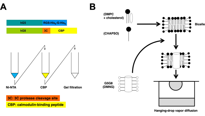
Figure 1: Schematic overviews for purification (A) and bicelle crystallization (B) of heterodimeric ABCG5/G8. Constructs of recombinant human ABCG5 (hG5) and ABCG8 (hG8) carry RGS-H6-G-H6 and 3C-CBP tags, respectively (A, top). Tandem affinity column chromatography, followed by gel filtration chromatography to achieve heterodimeric purification (A, bottom). Please click here to view a larger version of this figure.

Figure 2: Evaluation of mono-dispersity (A) and stability (B) of purified proteins. (A) Electron micrograph of negatively stained ABCG5/G8 (G5G8) heterodimers using TEM. Representative particles are highlighted in solid white circles. Scale bar = 100 nm. (B) Alkylated proteins stored at 4 °C analyzed by analytical gel filtration chromatography over the course of a month with a slight loss of proteins after a week. Please click here to view a larger version of this figure.

Figure 3: SDS-PAGE analysis of protein eluates of column chromatography and reductive alkylation. Various volumes (1-10 µL) of protein fractions were loaded onto a 10% Tris/Glycine gel and ran for 45 min at a constant voltage of 200 V. The gel was stained with Coomassie blue, destained, air-dried, and scanned by a tabletop scanner. 1° & 2° Ni: first and second Ni-NTA columns; 1° & 2° CBP: first and second CBP columns; Peak Fractions solid line: pooled fractions for crystallization; Peak Fractions dashed line: shoulder fractions; EMTS: ethyl mercury thiosalicynate; IA: iodoacedamide. Please click here to view a larger version of this figure.
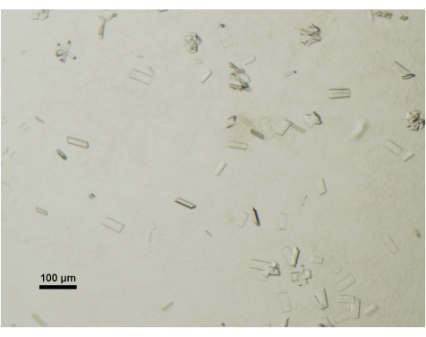
Figure 4: Assessment of protein crystal maturation by light microscopy. Mature crystals of ABCG5/G8 from a crystallization drop were visualized under a tabletop and polarizer-equipped stereo microscope. Scale bar = 100 µm. Please click here to view a larger version of this figure.
Discussion
The challenges associated with crystallizing membrane proteins have prompted the development of lipid-bilayer-driven crystallization methods, such as the bicelle27 or lipid cubic phase (LCP)14 approaches. However, achieving successful crystallization of membrane proteins still hinges on the critical and sometimes bottlenecked step of protein preparation. Notably, ABC transporters present a formidable hurdle in growing crystals suitable for X-ray crystallography. This protocol provides comprehensive hands-on guidance for streamlining the preparation of human ABCG5/G8 sterol transporter and fostering crystal growth through the bicelle crystallization approach.
A key consideration in devising this protocol was the imperative for a substantial protein yield in the initial phases of protein purification, allowing for a certain degree of protein loss during pre-crystallization treatment (Figure 3). Common strategies for addressing this challenge involve extensive protein engineering, utilization of diverse expression hosts, and exploration of orthologs or homologs, among other approaches. Nevertheless, with this seemingly intricate procedure, a number of pivotal steps have been identified that underpin the protocol's success and also provide insights into potential limitations that may arise when studying other ABC transporters or membrane proteins in general.
Firstly, this protocol employs thorough centrifugation at each step to minimize protein aggregation. Additionally, continuous monitoring of the thermostability of the purified proteins is crucial. Electron microscopy is utilized to verify protein monodispersity, while analytical gel filtration tracks protein stability over time (Figure 2). Alternative techniques like circular dichroism (CD) or differential scanning calorimetry (DSC) could also be incorporated. Furthermore, the incorporation of lipids at specific stages is essential to maximize both the activity and crystallogenesis of the purified ABCG5/G8. For instance, cholate and CHS are necessary to exhibit measurable ATP hydrolysis; phospholipids are indispensable for maintaining the stability of methylated proteins; and cholesterol is a requisite component of the bicelle solution, fostering crystal growth suitable for high-resolution X-ray diffraction (Figure 4).
In essence, the entire procedure can be accomplished within a week's worth of effort. In contrast to LCP, the retrieval of crystals from hanging-drop crystallization trays is straightforward. Looking ahead, with a substantial protein yield (approximately 10 mg), this protocol is readily adaptable for developing crystallographic investigations involving ABCG5/G8 mutants or other transporter proteins. This is particularly pertinent for cases that currently evade visualization through electron microscopy.
Disclosures
The authors do not have anything to disclose.
Acknowledgements
This work is supported by a Natural Sciences and Engineering Research Council Discovery Grant (RGPIN 2018-04070) and a Canadian Institutes of Health Research Project Grant (PJT-180640) to JYL. This protocol is based on the original reports in ABCG5/G8 crystal structures reported earlier by Farhat et al.22 and Lee et al.23.
Materials
| Name | Company | Catalog Number | Comments |
|---|---|---|---|
| ABCG5 | National Institute of Health collection | NCBI accession number NM_022436 | |
| ABCG8 | National Institute of Health collection | NCBI accession number NM_022437 | |
| ÄKTA FPLC system | Cytiva (formerly GE Healthcare Life Sciences) | ||
| CaCl2 | Wisent | 600-024-CG | Anhydrous |
| CBP | Agilent | 214303 | Calmodulin binding peptide affinity resin |
| Centrifugal concentrators (Vivaspin) | Sartorius | ||
| CHAPSO | Anatrace | C317 | Anagrade |
| Cholesterol | Anatrace | CH200 | |
| CHS | Steraloids | C6823-000 | |
| DMAB | MilliporeSigma | 180238 | 97% |
| DMNG | Anatrace | NG322 | |
| DMPC | Anatrace | D514 | |
| DOPC | Avanti | 850375 | |
| DOPC | Anatrace | D518 | |
| DOPE | Avanti | 850725 | |
| DTT | Fisher | BP172 | |
| Dual Thickness MicroLoops | MiTeGen | ||
| EDTA | BioShop | EDT003 | Disodium salt, dihydrate |
| EGTA | MilliporeSigma | 324626 | |
| Emulsifier (EmulsiFex-C3) | Avestin | ||
| Endo H | New England Biolabs | P0702 | |
| Ethanol | Greenfield | P016EAAN | Ethyl Alcohol Anhydrous |
| Formaldehyde | MilliporeSigma | 252549 | ACS Reagent |
| Glycerol | BioShop | GLY004 | |
| HEPES | BioShop | HEP001 | |
| HRV-3C protease | Homemade | ||
| Imidazole | BioShop | IMD510 | Reagent grade |
| Iodoacetamide | MilliporeSigma | I1149 | BioUltra |
| Isopropanol | Fisher | BP2618212 | |
| Leupeptin | BioShop | LEU001 | |
| MES | MilliporeSigma | 69892 | BioUltra |
| Methanol | Fisher | A412P | |
| MgCl2 | Wisent | 800-070-CG | Hydrated |
| microfluidizer (LM 20) | Microfluidics | ||
| NaCl | BioShop | SOD002 | |
| NH4OH | Fisher | A669-212 | ACS Reagent |
| Ni-NTA superflow | Qiagen | 30430 | Nickel-charged resins |
| PEG 400 | MilliporeSigma | 202398 | |
| Pepstatin | BioShop | PEP605 | |
| PMSF | MilliporeSigma | P7626 | |
| pSGP18 and pLIC | Homemade (derived from pPICZ, Invitrogen) | ||
| SDS | BioShop | SDS003 | |
| Sodium cholate | Fisher | 229101 | |
| Sodium malonate | MilliporeSigma | 63409 | |
| Sucrose | Wisent | 800-081-WG | Ultra pure |
| Superdex 200 30/100 GL | Cytiva (formerly GE Healthcare Life Sciences) | 28990944 | Prepacked gel-filtration column |
| TCEP | |||
| TEM | FEI, Technai | ||
| Tris Base | Fisher | BP152 | |
| β-DDM | Anatrace | D310S | Sol Grade |
| β-mercaptoethanol | MilliporeSigma | ||
| ε-aminocaproic acid | Fisher | AAA1471936 |
References
- Hamada, H., Tsuruo, T. Purification of the 170- to 180-kilodalton membrane glycoprotein associated with multidrug resistance. 170- to 180-kilodalton membrane glycoprotein is an ATPase. Journal of Biological Chemistry. 263 (3), 1454-1458 (1988).
- Higgins, F., Hiles, D., Whalley, K., Jamieson, J. Nucleotide binding by membrane components of bacterial periplasmic binding protein-dependent transport systems. The EMBO Journal. 4 (4), 1033-1039 (1985).
- Higgins, F., et al. A family of related ATP-binding subunits coupled to many distinct biological processes in bacteria. Nature. 323 (6087), 448-450 (1986).
- Horio, M., Gottesman, M., Pastan, I. ATP-dependent transport of vinblastine in vesicles from human multidrug-resistant cells. Proceedings of the National Academy of Sciences of the United States of America. 85 (10), 3580-3584 (1988).
- Mimmack, L., et al. Energy coupling to periplasmic binding protein-dependent transport systems: stoichiometry of ATP hydrolysis during transport in vivo. Proceedings of the National Academy of Sciences of the United States of America. 86 (21), 8257-8261 (1989).
- Grundy, M. Absorption and Metabolism of Dietary Cholesterol. Annual Review of Nutrition. 3 (1), 71-96 (1983).
- Berge, E., et al. Accumulation of dietary cholesterol in sitosterolemia caused by mutations in adjacent ABC transporters. Science. 290 (5497), 1771-1775 (2000).
- Repa, J., et al. Regulation of ATP-binding cassette sterol transporters ABCG5 and ABCG8 by the Liver X receptors α and β. Journal of Biological Chemistry. 277 (21), 18793-18800 (2002).
- Yu, L., et al. Stimulation of cholesterol excretion by the Liver X receptor agonist requires ATP-binding cassette transporters G5 and G8. Journal of Biological Chemistry. 278 (18), 15565-15570 (2003).
- Yu, L., et al. Expression of ABCG5 and ABCG8 is required for regulation of biliary cholesterol secretion. Journal of Biological Chemistry. 280 (10), 8742-8747 (2005).
- Lütjohann, D., Björkhem, I., Beil, F., von Bergmann, K. Sterol absorption and sterol balance in phytosterolemia evaluated by deuterium-labeled sterols: effect of sitostanol treatment. Journal of Lipid Research. 36 (8), 1763-1773 (1995).
- Miettinen, A. Phytosterolaemia, xanthomatosis and premature atherosclerotic arterial disease: a case with high plant sterol absorption, impaired sterol elimination and low cholesterol synthesis. European Journal of Clinical Investigation. 10 (1), 27-35 (1980).
- Salen, G., et al. Sitosterolemia. Journal of Lipid Research. 33 (7), 945-955 (1992).
- Caffrey, M. Membrane protein crystallization. Journal of Structural Biology. 142 (1), 108-132 (2003).
- Michel, H. Crystallization of membrane proteins. Trends in Biochemical Sciences. 8 (2), 56-59 (1983).
- Dürr, N., Gildenberg, M., Ramamoorthy, A. The magic of bicelleslights up membrane protein structure. Chemical Reviews. 112 (11), 6054(2012).
- Dürr, N., Soong, R., Ramamoorthy, A. When detergent meets bilayer: Birth and coming of age of lipid bicelles. Progress in nuclear magnetic resonance spectroscopy. 69 (1), 1-22 (2013).
- Dufourc, J. Bicelles and nanodiscs for biophysical chemistry. Biochimica et Biophysica Acta (BBA) - Biomembranes. 1863 (1), 183478(2021).
- Beaugrand, M., et al. Lipid concentration and molar ratio boundaries for the use of isotropic bicelles. Langmuir. 30 (21), 6162-6170 (2014).
- Sanders, R., Schwonek, P. Characterization of magnetically orientable bilayers in mixtures of dihexanoylphosphatidylcholine and dimyristoylphosphatidylcholine by solid-state NMR. Biochemistry. 31 (37), 8898-8905 (1992).
- Seddon, M., Curnow, P., Booth, J. Membrane proteins, lipids and detergents: not just a soap opera. Biochimica et Biophysica Acta (BBA) - Biomembranes. 1666 (1-2), 105-117 (2004).
- Farhat, D., et al. Structural analysis of cholesterol binding and sterol selectivity by ABCG5/G8. Journal of Molecular Biology. 434 (20), 167795(2022).
- Lee, J. Y., et al. Crystal structure of the human sterol transporter ABCG5/ABCG8. Nature. 533 (7604), 561-564 (2016).
- Ujwal, R., Bowie, U. Crystallizing membrane proteins using lipidic bicelles. Methods. 55 (4), 337-341 (2011).
- Johnson, H., Lee, J. Y., Pickert, A., Urbatsch, L. Bile acids stimulate ATP hydrolysis in the purified cholesterol transporter ABCG5/G8. Biochemistry. 49 (16), 3403-3411 (2010).
- Wang, Z., et al. Purification and ATP hydrolysis of the putative cholesterol transporters ABCG5 and ABCG8. Biochemistry. 45 (32), 9929-9939 (2006).
- Faham, S. Crystallization of bacteriorhodopsin from bicelle formulations at room temperature. Protein Science. 14 (3), 836-840 (2005).
Reprints and Permissions
