Endothelial/vascular aging and atherogenesis are key mechanisms that promote the development of cardiovascular diseases. The present protocol describes methods to evaluate arterial stiffness, endothelial dysfunction, and atherogenesis in patients with related risk factors, which are highly valuable in the cardiovascular research field.
Method Article
Pulse-Wave Velocity, Flow-Mediated Dilation, and Carotid Intima-Media Thickness to Assess Cardiovascular Risk in Population with Metabolic Syndrome
In This Article
Summary
Abstract
Pulse-wave velocity (PWV), flow-mediated dilation (FMD), and carotid intima-media thickness (CIMT) are established methods used in research and clinical settings to assess arterial stiffness, endothelial function, and subclinical atherogenesis. These measurements may reflect vascular disease and atherosclerotic progression, which are major causes of adverse cardiovascular events. These methods are particularly valuable in determining cardiovascular dysfunction among populations with different risk factors, such as diabetes mellitus, hypertension, and other metabolic dysfunction-related conditions. They provide a non-invasive and reliable source of information that complements clinical practice. Early detection, risk assessment, and therapeutic decisions regarding cardiovascular disease can be achieved, ultimately contributing to improved patient outcomes. Traditional tools for evaluating cardiovascular disease do not reveal whether metabolic syndrome affects early subclinical cardiovascular disease in patients with obesity. Recent research has highlighted the importance of including arterial stiffness and endothelial function in a comprehensive cardiovascular assessment. Therefore, the aim of the present study is to describe methods that provide information on early subclinical vascular aging, endothelial dysfunction, and atherogenic disease, enabling vascular-targeted risk stratification among populations with obesity and different metabolic profiles.
Introduction
Obesity is a major health problem worldwide due to its associated complications, such as hypertension, dyslipidemia, liver disease, atherosclerosis, insulin resistance, and type 2 diabetes mellitus (T2DM), as well as an increased risk of cardiovascular diseases (CVD)1.
The constellation of these conditions, known as Metabolic Syndrome (MS), has been reported to be a major cause of CVD pathogenesis, which is one of the leading causes of death, accounting for up to 30% of all deaths worldwide2. Obese individuals have a higher requirement for oxygen and nutrients throughout the body due to an increased blood supply demand, leading to significant hemodynamic changes. These changes can result in decreased nitric oxide (NO) availability, increased oxidative stress, and vascular endothelial dysfunction3,4,5.
Atherosclerotic diseases are major cardiovascular conditions and represent the leading cause of death worldwide. This is a clinical manifestation of multiple possible factors, including genetic and environmental factors6. People with metabolic abnormalities, such as insulin resistance or prediabetes, have been shown to have a significantly higher prevalence and incidence of coronary atherosclerosis than healthy people. Furthermore, congested blood vessels with highly lipidic plaque have been found even before the appearance of clinical manifestations of metabolic dysfunction7,8,9,10.
Arterial stiffness, endothelial dysfunction, and atherogenesis have been described as important factors in the development of cardiovascular diseases. These processes are related to vascular aging and atherogenic plaque formation in critical vessels like coronary, carotid, or limb arteries. Translational research has evidenced that arterial stiffness, endothelial dysfunction, and atherogenesis are related to common vascular damage induced by chronic inflammation, lower NO production, and oxidative stress11,12.
Measurement of Carotid-femoral pulse-wave velocity (cfPWV) represents the gold standard method to measure arterial stiffness. cfPWV can be measured using a carotid tonometer simultaneously with a leg cuff to capture blood pressure waveforms at the carotid and femoral sites. Then, a software can perform velocity calculation by computing D/Δt, where D is the transit distance between carotid and femoral pulse sites, and Δt is the time delay from the peak ECG R-wave to the foot of the corresponding pressure waveform between the carotid and femoral waveforms. Increased stiffness of central arteries, like the aorta, causes a higher speed of the ejected pulse from the left ventricle through the arteries, as well as a faster return of the reflected pressure, with a consequent elevation of pressure during left ventricular ejection, which potentially decreases coronary artery perfusion. Therefore, cfPWV may be useful as a marker of coronary artery disease, stroke, and cardiovascular diseases13,14.
Likewise, Pulse Wave Analysis (PWA) is a non-invasive vascular parameter that assesses central pressure wave characteristics, where aortic systolic and diastolic blood pressures are the main variables. By measuring arterial stiffness and elastic compliance, PWA reflects arterial distensibility, which is closely related to cardiovascular risk. This method allows for measuring parameters like the Augmentation Index, which has the ability to predict the severity of cardiovascular and coronary artery diseases. The Augmentation Index may be described as follows: an early incident arterial wave is produced after left ventricular ejection, with a subsequent reflected wave originating from the periphery. The velocity of these waves increases according to arterial stiffness, and if the reflected wave arrives at the central aorta early, aortic systolic pressure will increase. This is known as Augmented Pressure (AP), whereas its percentage relative to Pulse Pressure is known as the Augmentation Index. PWA can be measured through the applanation tonometry method, involving a slight compression of the brachial artery so that its transmural pressure is zero. At this point, Mean Arterial Pressure can be measured. After scaling the arterial pressure waveform, the systolic part of the AP waveform is analyzed, also considering biometric and demographic data15,16,17. Particularly, the applanation tonometry method (SphygmoCor) has shown acceptable repeatability and significant correlation with invasive aortic catheterization in determining aortic PWV, as well as good agreement with the Artery Society Guidelines18,19,20.
Other vascular tests like flow-mediated dilation (FMD) and carotid intima-media thickness (CIMT) represent non-invasive techniques performed by ultrasonography with linear transducers. These assessment procedures are useful for evaluating vascular health, specifically endothelial dysfunction and subclinical atherogenesis, respectively. Both have shown prognostic ability for cardiovascular events. FMD is commonly considered a reflection of endothelium-dependent arterial function, primarily mediated by nitric oxide. It serves as a surrogate marker for vascular health and has been utilized non-invasively to compare subject groups and assess the effects of interventions on individuals21.
The aim of the present study is to describe the use of methods that yield the determination of markers reflecting early subclinical vascular aging, endothelial dysfunction, and atherogenic disease. Such information allows risk stratification among populations with obesity and different metabolic profiles. These methods might be useful to determine cardiovascular damage and prognosis, as well as to evaluate vascular and atherogenic responses to pharmacologic and non-pharmacologic interventions, particularly among populations with metabolic risk factors.
Protocol
The institutional research ethics committee from the National Medical Center "20 de Noviembre" ISSSTE approved this protocol (ID No. 386.2013). All enrolled patients provided written informed consent. The details of the equipment and software used in this study are listed in the Table of Materials.
Patient inclusion/exclusion criteria:
Eligible patients were older than 18 years and diagnosed with morbid obesity (Body Mass Index [BMI] >40 kg/m² or BMI >35 kg/m² with obesity-related health conditions, such as diabetes mellitus, hypertension, or obstructive sleep apnea/hypopnea) and candidates for bariatric surgery. Patients were excluded if they had used weight-reducing therapy during the 6 months prior to enrollment, had significant inflammatory diseases, severe renal and/or hepatic disease, active malignancy, pregnancy, or evidence of cardiovascular disease (either self-reported or diagnosed with ischemic heart disease, coronary artery disease, myocardial structural abnormalities, cardiac interventions, or being under treatment for any of these conditions).
1. Evaluation of cardiometabolic profile
NOTE: The study sample used for this experiment comprised 21 Metabolically Healthy Obese (MHO) and 25 Metabolically Unhealthy Obese (MUO) patients, determined by the absence or presence of metabolic syndrome, respectively. The participants were aged 43 ± 9 years old, with a BMI of 45 ± 7.8 kg/m², and 78% were female. The most prevalent co-morbidities were type 2 diabetes mellitus, systemic arterial hypertension, and/or dyslipidemia. The sample was intended to be age-matched.
- Evaluate cardiometabolic profile
- Obtain demographic and anthropometric characteristics, such as age, gender, height, weight, chronic diseases (t2DM, hypertension, etc.), and medications consumed. Calculate BMI by dividing weight by the square of height. Obtain waist circumference by measuring between the lower point of the last rib and the iliac crest.
- Perform routine clinical biochemical tests, including glucose, insulin, liver function tests, and lipid profile22.
- Designate patients as MUO or MHO, as determined by the presence or absence of metabolic syndrome, respectively.
NOTE: Metabolic Syndrome is diagnosed according to the NCEP/ATP III criteria23. A patient is diagnosed with metabolic syndrome if they present at least three of the following five risk factors: - Abdominal obesity (waist circumference >102 cm in men or >88 cm in women). (2) Serum triglyceride level ≥150 mg/dL (1.7 mmol/L). (3) HDL cholesterol level <40 mg/dL (1.0 mmol/L) in men or <50 mg/dL (1.3 mmol/L) in women. (4) Systolic blood pressure ≥130 mmHg or diastolic blood pressure ≥85 mmHg. (5) Fasting plasma glucose level ≥100 mg/dL (5.6 mmol/L).
2. Vascular aging (arterial stiffness)
NOTE: Vascular aging may be evaluated in terms of aortic stiffness, which is determined by the central aortic pulse pressure and carotid-femoral pulse wave velocity (cfPWV). Nowadays, cfPWV is the gold standard for determining arterial stiffness13.
- Aortic pressure assessment
NOTE: Aortic pressure assessment is performed using a device for measuring arterial wave reflection and Pulse Wave Analysis (PWA), on which central aortic pressure waveform parameters are determined.- Create a profile of the patient in the device software and introduce data such as patient ID, name, birth date, gender, and height.
- Place the patient in a supine position for at least 5 min before beginning the assessment.
- Place a brachial cuff and secure it around the patient's arm, centered on the brachial artery, ensuring that the center of the cuff and the heart are at the same level.
- The device automatically performs PWA. Press the start button. The cuff will automatically inflate for the first time to determine brachial systolic and diastolic pressure. Then, the cuff will deflate and inflate again to capture the PWA waveform.
- Obtain PWA by applanation tonometry, which allows recording peripheral pulse and generation of central aortic pressure waveforms. Peripheral locations for applanation tonometry include brachial or radial arteries.
- After scaling the arterial pressure waveform, analyze the systolic part of the waveform, also considering biometric and demographic data.
NOTE: The software calculates based on the formula K·Psa·(1 + Ts/Td), where Psa is the area under the systolic part of the curve above end-diastolic pressure, Ts and Td are the durations of systole and diastole, respectively, and K is a constant related to stroke volume24,25. - Calculate the Augmentation Index following the NOTE below.
NOTE: An early incident arterial wave is produced after left ventricular ejection, with a subsequent reflected wave originating from the periphery. The velocity of these waves increases according to arterial stiffness, and if the reflected wave arrives at the central aorta early, aortic systolic pressure will increase. This is known as Augmented Pressure, whereas its percentage relative to the Pulse Pressure is known as the Augmentation Index (Aix). - Obtain an automatic report of the test, containing aortic parameters, including average central pressure waveform: SP (aortic Systolic Pressure), DP (aortic Diastolic Pressure), PP (Aortic Pulse Pressure), MAP (Mean Arterial Pressure), and HR (Heart Rate); clinical parameters which are represented on a bar graph; as well as Aix.
- Perform cfPWV determination through applanation tonometry method (SphygmoCor)
- Place a femoral cuff around the patient's thigh, as high as possible, ensuring the tube is centered at the top of the leg.
- Find the carotid pulse on the patient's neck below the jaw. Ask the patient to slightly rotate their head aside; if required, place a pillow below the neck to provide support. Once the carotid pulse is found at the strongest perceived site, place an indicator mark on the patient's skin.
- Obtain three measurements. First, the distance between the carotid pulse and the suprasternal notch; second, the distance between the suprasternal notch and the femoral cuff; and finally, the distance between the femoral artery by palpating the pulse on the patient's groin and the femoral cuff.
NOTE: All distances shall be taken in straight lines (avoiding the patient's body curves) and must be submitted in millimeters.- Then, introduce information on the three distances into the cfPWV software.
- Place the tip of the tonometer on the site where the carotid pulse was previously located. Then press the START button. The sensor will automatically detect the carotid pulse and will record the shape of the pulse once a regular pattern is registered. The device will sync both pulses (carotid and femoral) to determine the wave pulses and estimate the cfPWV and Pulse Transit Time (Figure 1).
- Ensure that the software automatically performs velocity calculation by computing D/Δt, where D is the transit distance between carotid and femoral pulse sites, and Δt is the time delay from the peak ECG R-wave to the foot of the corresponding pressure waveform between the carotid and femoral waveforms.
- Evaluate a quality control test, as reported by the device, to determine whether the measurements are acceptable.
NOTE: Hold the tonometer tip like a pencil to ensure maximum stability. Pressure and position adjustments on the tonometer should be delicate and soft to obtain accurate measurements, which will be indicated by a green or yellow color of an upper line on the screen and waveforms; otherwise, the color will turn red, indicating a need for a reduction in tonometer pressure (if the indicator of pressure level goes up) or an increase in tonometer pressure (if the indicator goes down). The operator must ensure that there is a well-defined upstroke on the carotid waveforms, as it is an important feature that will be used to determine the cfPWV. It is important to locate the screen of the computer in a convenient place for the operator to view prior to the determination. If the screen is in a place where the operator finds it difficult to view, it can make the data capture challenging. The device will need a minimum of 10 s of consistent simultaneous waveforms of the carotid and femoral pulses and will automatically capture the waveforms; nevertheless, some circumstances will require the operator to capture the pulse waves manually (Figure 2).
3. Endothelial dysfunction (flow-mediated dilation [FMD])
NOTE: The Flow Mediated Dilation (FMD) test is a non-invasive technique to evaluate vascular health; it is specifically useful for assessing endothelial function and has been described as a useful tool to predict future cardiovascular events21. It is performed using ultrasonography with a linear transducer.
- Perform the FMD protocol according to international recommendations26. Use a sphygmomanometer and place the cuff around the right forearm.
- Place a high-resolution linear transducer in B-mode coupled to the compatible analysis software on the brachial artery.
- Scan longitudinally 5-10 cm above the elbow. Acquire the clearest B-mode image of the anterior and posterior intimal interfaces and determine the baseline artery diameter.
- Hold the transducer at the same point to ensure consistency of the site of measurement.
- Occlude for 5 min using the blood pressure cuff, reaching a pressure of 30-50 mmHg above the determined systolic pressure.
- After cuff deflation, perform a continuous record of the longitudinal image of the brachial artery for 3 min, and determine the diameter again.
- Calculate FMD as the percentage change relative to the vessel diameter before cuff inflation, as follows: (peakdiameter − baselinediameter)/baselinediameter (peak diameter − baseline diameter) / baseline diameter (peakdiameter − baselinediameter)/ baselinediameter x 100 (Figure 3). FMD% measures the ability of the arteries to respond with endothelial nitric oxide release during reactive hyperemia (flow-mediated)27.
- Further evaluate endothelial dysfunction by determining plasma NO (Nitric Oxide assay kit, commercially available) as measured by enzyme-linked immunosorbent assay.
- Prepare the standard curve and samples, then add them to wells in the assay plate.
- Add nitrate reductase and enzyme cofactor and incubate for 60 min at room temperature to convert nitrate to nitrite.
- Add enhancers and Griess reagents; then develop at room temperature for 10 min, allowing nitrite to turn into a deep purple azochromophore compound, which accurately reflects NO amounts.
- Analyze in a microplate reader by measuring at an optical density of 540 nm.
4. Subclinical atherogenesis (carotid intima-media thickness [CIMT])
NOTE: Patients must be placed in a supine position comfortably, with the head rotated to expose the jugular vein and the carotid artery; a rolled-up towel or pillow under the neck can be used to better expose the carotid.
- Perform the carotid intima-media thickness (CIMT) measurement protocol according to expert Consensus28,29 as follows: apply a 4.0 MHz ultrasonographic probe to identify neck vascular structures, such as the carotid artery and jugular vein, as well as the thyroid gland, in a transverse orientation at the base of the neck.
- Locate the right carotid artery using a transverse orientation of the transducer, moving in a cephalic direction until the carotid bulb and the bifurcation of the internal and external carotid artery are identified. Then, rotate the transducer 90° to achieve a longitudinal view of the carotid bulb.
- Determine carotid intima-media thickness by measuring the distance between the intima-lumen and media-adventitia interfaces along a 1 cm range distal from the carotid bulb (Figure 4).
NOTE: The protocol does not include follow-up, medications, or interventions.
Results
Subjects were classified as MHO and MUO based on their cardiometabolic profiles. The MUO group exhibited a higher prevalence of chronic diseases, such as systemic arterial hypertension, type 2 diabetes mellitus (t2DM), and dyslipidemia. Similarly, the MUO phenotype showed elevated levels of glucose and HbA1c, as well as differences in triglycerides and total cholesterol (Table 1).
Vascular aging was then assessed, reflecting arterial stiffness and endothelial dysfunction, determined through aortic hemodynamic parameters, FMD, and Nitric Oxide (NO), respectively. Additionally, subclinical atherosclerosis was evaluated by CIMT measurement (Table 2).
The MUO group exhibited lower values of FMD (4.87% ± 1.80% vs. 7.32% ± 2.90%, p = 0.001; Table 2) and plasma NO (154.3 µM/L ± 6.15 µM/L vs. 170.6 µM/L ± 4.97 µM/L, p = 0.04; Table 2 and Figure 5) compared to the MHO group. However, no statistical differences were found in aortic hemodynamic parameters and CIMT.
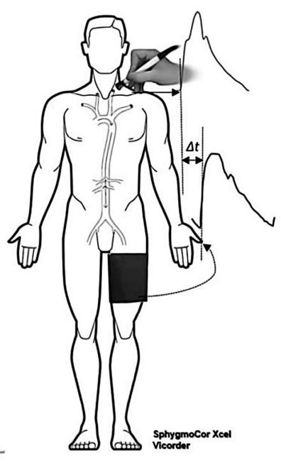
Figure 1: Schematic representation of carotid-femoral pulse wave velocity assessment. This figure illustrates the methodology for assessing carotid-femoral pulse wave velocity (PWV), a key measure of arterial stiffness and cardiovascular health. Carotid tonometry and femoral pulse determination are depicted as integral steps in the arterial PWV measurement system. Please click here to view a larger version of this figure.

Figure 2: Carotid and femoral pulse waveforms determined to assess the carotid-femoral pulse wave velocity. Time (x-axis) and amplitude of the waveform (y-axis) are shown. Please click here to view a larger version of this figure.
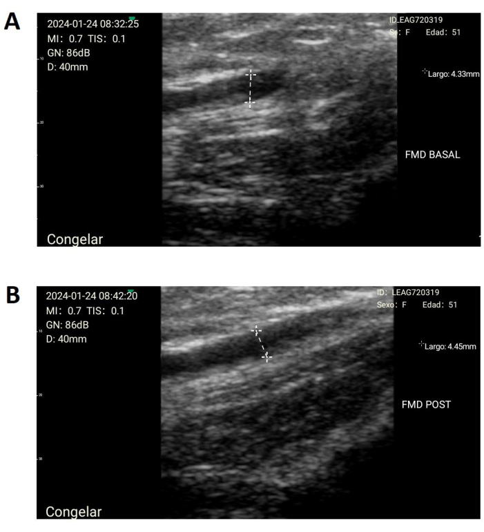
Figure 3: Flow-mediated dilation (FMD). Diameter assessment of the brachial artery, either (A) before or (B) after artery occlusion, to determine the endothelial function through the calculation of the dilation. Please click here to view a larger version of this figure.
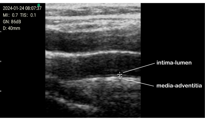
Figure 4: Carotid ultrasonography to determine the intima-media thickness (CIMT). It is important to make three measurements from anterior, lateral, and posterior angles. CIMT determination is performed on both sides of the patient's neck. Please click here to view a larger version of this figure.
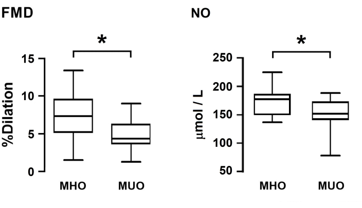
Figure 5: Endothelial function tests. Comparison of flow-mediate dilation (FMD, left) and plasma Nitric Oxide (NO, right) between MHO and MUO patients. Metabolic Syndrome significantly lowers NO bioavailability; therefore, these patient's arteries have a reduced ability to dilate after stress during the FMD test. *p < 0.05. Please click here to view a larger version of this figure.
| Metabolically Healthy Obese (MHO) | Metabolically Unealthy Obese (MUO) | p value | |
| n = 21 | n = 25 | ||
| Age | 43.3 ± 7.63 | 46.7 ± 7.75 | 0.0695 |
| Male sex | 5 (23.8) | 9 (36) | 0.5223 |
| Systemic Arterial Hypertension | 7 (33.3) | 19 (76) | 0.0067* |
| Type 2 Diabetes Mellitus | 1 (4.8) | 17 (68) | <0.0001* |
| Dyslipidemia | 3 (14.3) | 16 (64) | 0.0009* |
| BMI | 46.27 ± 4.86 | 47.61 ± 6.90 | 0.2288 |
| Glucose | 90.59 ± 2.46 | 100.4 ± 3.38 | 0.0273* |
| HbA1c | 5.514 ± 0.10 | 6.186 ± 0.19 | 0.0053* |
| Insulin | 26.68 ± 3.6 | 46.64 ± 9.57 | 0.2723 |
| Triglycerides | 109.5 ± 7.82 | 197.1 ± 37.15 | 0.0005* |
| HDLc | 49.86 ±5.38 | 40 ± 1.72 | 0.1163 |
| LDLc | 115.9 ± 7.095 | 115.3 ± 8.23 | 0.7131 |
| Total Cholesterol | 186.7 ± 6.47 | 167.7 ± 11.84 | 0.0392* |
| Categorical variables were compared by Fisher exact test; whereas quantitative variables were compared by 2-way, independent U-mann Withney or T-test, according to normality assessment. Statistical significance was considered if p<0.05. Abbreviatures: BMI; Body Mass Index, HbA1c, Glycated Hemoglobin A1c | |||
Table 1: Baseline characteristics of the patients. The table compares baseline characteristics between subjects with MUO (metabolically unhealthy obesity) and MHO (metabolically healthy obesity).
| Metabolically Healthy Obese (MHO) | Metabolically Unhealthy Obese (MUO) | p | |
| n = 21 | n = 25 | ||
| Aortic Systolic Pressure (mmHg) | 121.1 ± 3.77 | 119.7 ± 2.93 | 0.9608 |
| Aortic Diastolic Pressure (mmHg) | 79.9 ± 2.56 | 75.96 ± 1.79 | 0.515 |
| AIX (%) | 26 ± 3.16 | 30.08 ± 2.19 | 0.2823 |
| Pulse Wave Velocity (m/s) | 6.089 ± 0.32 | 6.854 ± 0.26 | 0.3154 |
| FMD (%) & | 7.32 ± 2.90 | 4.87 ± 1.80 | 0.0011* |
| CIMT (mm) | 0.69 ± 0.02 | 0.704 ± 0.03 | 0.933 |
| Nitric Oxide (uM/L) | 170.6 ± 4.97 | 154.3 ±6.15 | 0.0443* |
| Quantitative variables were compared by 2-way, independent U-mann Withney or T-test, according to normality assessment. Statistical significance was considered if p<0.05. Abbreviatures: AIX – Aortic Augmentation index; FMD – Flow-Mediated Dilation; CIMT – Cartid Intima-Media Thickness. (&) Due to limited physical area in our lab, FMD data shown were obtained in a sitting position, which may alter FMD value, since FMD measurement is recommended in supine position. | |||
Table 2: Cardiovascular risk evaluation. The table compares cardiovascular risk factors between subjects with MUO (metabolically unhealthy obesity) and MHO (metabolically healthy obesity).
Discussion
Addressing vascular health and understanding and managing cardiovascular risk are essential for the prevention, early intervention, and reduction of the global burden of cardiovascular diseases. In this regard, the combined use of methods to assess the elasticity and compliance of the arterial wall (including aortic hemodynamic parameters, cfPWV for arterial stiffness, and Augmentation Index), endothelial nitric oxide production, and atherosclerosis provide a more comprehensive evaluation. These methods are highly useful for translational studies, characterization of cardiovascular risk factors, and cardiovascular assessments in clinical trials, thereby enhancing our understanding of vascular health and the progression of cardiovascular and coronary artery diseases30,31.
For instance, Flow Mediated Dilation (FMD) has been employed to better understand endothelial and vascular consequences induced by COVID-19, to characterize endothelial dysfunction in immune system disorders such as Systemic Lupus Erythematosus, and to explore whether endothelial damage contributes to hypertension and cardiovascular risk in hyperuricemia32,33,34.
Furthermore, other studies have investigated the impact of lifestyle modifications, such as exercise and dietary changes, on arterial stiffness, endothelial function, and CIMT. By utilizing these tools, researchers can objectively measure the efficacy of interventions and monitor changes in arterial health over time35.
In the present study, markers reflecting vascular aging in populations with cardiometabolic risk were compared. Interestingly, the group with higher cardiometabolic risk showed significantly impaired endothelial function, as determined by FMD and NO levels, whereas no difference was observed in the progression of atherosclerosis according to CIMT.
Here, the results of FMD are consistent with findings from other studies36,37, supporting the impact of metabolic risk on vascular dilation response. The lack of association between FMD and CIMT may be attributed to the time lag between endothelial dysfunction and the subsequent development of vascular lesions. Furthermore, endothelial dysfunction is likely associated with inflammatory responses, whereas morphological changes in vascular structure may be linked to extracellular remodeling38. This suggests that individuals with metabolic syndrome may exhibit different responses to therapies targeting vascular function, while atherosclerosis progression may be influenced by other factors. This underscores the importance of using multiple biomarkers to comprehensively evaluate overall vascular health.
Some challenges may arise during measurements of arterial stiffness, FMD, and CIMT if the equipment is inadequate or the environment is not suitable. For validation, evaluating inter-observer and intra-observer variability is recommended. Likewise, some limitations of these techniques include: (1) results obtained may apply to specific vascular functions and anatomical regions, thus not reflecting overall vascular health; (2) the accuracy of measures can depend on the operator, and results may vary within the same individual over time due to factors such as hydration status, body position, and recent meals; (3) while tests can indicate the presence of arterial stiffness, endothelial dysfunction, or accelerated atherogenesis, they may not provide insights into underlying causes. Furthermore, vascular health is dynamic and can change over time, and measurements may not capture the complete cardiovascular risk profile of an individual. Ongoing research is improving our understanding of these measurements, and refinements in measurement techniques and interpretation are still evolving.
In conclusion, aortic hemodynamic parameters, FMD, and CIMT represent useful, non-invasive diagnostic tools for evaluating cardiovascular health and disease.
Disclosures
The authors have nothing to disclose.
Acknowledgements
The authors thank the support of Institutional Program E015.
Materials
| Name | Company | Catalog Number | Comments |
|---|---|---|---|
| Device for measuring arterial wave reflection and Pulse Wave Analysis | ATCOR | SphygmoCor | Analyzer of pulse wave for central pressure. It contains a brachial cuff and a femoral cuff |
| Microplate reader for absorbance, Sunrise | Tecan | 30190079 | Detection Mode: Absorbance; Wavelength Range: 340 nm - 750 nm; Filter Wavelength: 405 nm, 450 nm, 492 nm, 620 nm; Plate Format 96 well plates |
| Nitric oxide assay kit | Abcam | ab65328 | Nitric Oxide Assay Kit, Colorimetric, Abcam Cat. ab65328 for 96-well plates |
| Portatil ultrasound to measure FMD | Sonolife | MED 36-13 | Ultrasonography linear transducer |
| Software for FMD WirelessUSG | SonoStarMed Technologies | WirelessUSG v. 3.6.52 | Software used to measure artery diameter for FMD |
| Software used to calculate vascular parameters from Waveform Analysis | ATCOR | SphygmoCor XCEL | Software used to integrate patient profile, waveform analysis, calculation of PWA, PWV and other vascular parameters |
| Sphygmomanometer | Homecare | ANEROIDE 1000 | 100% cotton self-adjustable bracelet with hook, Adult artery indicator cuff. |
| Ultrasound to measure CIMT | Philips | EPIQ7L12-3 Broadband Linear Array Transducer | Linear transducer (Broadband Linear Array Transducer) |
References
- Csige, I., et al. The impact of obesity on the cardiovascular system. J Diabetes Res. 2018, 3407306(2018).
- McAloon, C. J., et al. The changing face of cardiovascular disease 2000-2012: An analysis of the World Health Organization Global Health Estimates data. Int J Cardiol. 224, 256-264 (2016).
- Virdis, A. Endothelial dysfunction in obesity: Role of inflammation. High Blood Press Cardiovasc Prev. 23 (2), 83-85 (2016).
- Medina-Leyte, D. J., et al. Endothelial dysfunction, inflammation and coronary artery disease: Potential biomarkers and promising therapeutical approaches. Int J Mol Sci. 22 (8), 3850(2021).
- Mikael, L. R., et al. Vascular aging and arterial stiffness. Arq Bras Cardiol. 109 (3), 253-258 (2017).
- Liang, Y., et al. The Mechanisms of the development of atherosclerosis in prediabetes. Arq Bras Cardiol. 22 (8), 4108(2021).
- Açar, B., et al. Association of prediabetes with higher coronary atherosclerotic burden among patients with first diagnosed acute coronary syndrome. Angiology. 70, 174-180 (2019).
- Amano, T., et al. Abnormal glucose regulation is associated with lipid-rich coronary plaque: Relationship to insulin resistance. JACC Cardiovasc Imaging. 1, 39-45 (2008).
- Fan, J., Watanabe, T. Atherosclerosis: Known and unknown. Pathol Int. 72 (3), 151-160 (2022).
- Coutinho, M. S. S. A. Abdominal adiposity and intima-media carotid thickness: An Association. Arq Bras Cardiol. 112 (3), 228-229 (2019).
- Zanoli, L., et al. Arterial stiffness in the heart disease of CKD. J Am Soc Nephrol. 30 (6), 918-928 (2019).
- Boutouyrie, P., et al. Arterial stiffness and cardiovascular risk in hypertension. Circ Res. 128 (7), 864-886 (2021).
- Ben-Shlomo, Y., et al. Aortic pulse wave velocity improves cardiovascular event prediction: An individual participant meta-analysis of prospective observational data from 17,635 subjects. J Am Coll Cardiol. 63 (7), 636-646 (2014).
- Butlin, M., Ahmad, Q. Large artery stiffness assessment using SphygmoCor technology. Pulse. 4 (4), 180-192 (2017).
- Choi, J., Kim, S. Y., Joo, S. J., Kim, K. S. Augmentation index is associated with coronary revascularization in patients with high Framingham risk scores: A hospital-based observational study. BMC Cardiovasc Disord. 19 (15), 131(2015).
- Doupis, J., Papanas, N., Cohen, A., McFarlan, L., Horton, E. Pulse wave analysis by applanation tonometry for the measurement of arterial stiffness. Open Cardiovasc Med J. 10, 188-195 (2016).
- Saugel, B., et al. Cardiac output estimation using pulse wave analysis-physiology, algorithms, and technologies: A narrative review. Br J Anaesth. 126 (1), 67-76 (2021).
- Grillo, A., et al. Short-term repeatability of non-invasive aortic pulse wave velocity assessment: comparison between methods and devices. Am J Hypertens. 31 (1), 80-88 (2017).
- Salvi, P., et al. Non-invasive estimation of aortic stiffness through different approaches. Hypertension. 74 (1), 117-129 (2019).
- Milan, A., et al. Current assessment of pulse wave velocity: Comprehensive review of validation studies. J Hypertens. 37 (8), 1547-1557 (2019).
- Thijssen, D. H., et al. Assessment of flow-mediated dilation in humans: A methodological and physiological guideline. Am J Physiol Heart Circ Physiol. 300 (1), H2-H12 (2011).
- Costa Pereira, L. M., et al. Assessment of cardiometabolic risk factors, physical activity levels, and quality of life in stratified groups up to 10 years after bariatric surgery. Int J Environ Res Public Health. 16 (11), 1975(2019).
- Grundy, S. M., Hansen, B., Smith, S. C. Jr, Cleeman, J. I., Kahn, R. A. Clinical management of metabolic syndrome: Report of the American Heart Association/National Heart, Lung, and Blood Institute/American Diabetes Association conference on scientific issues related to management. Arterioscler Thromb Vasc Biol. 24 (2), e19-e24 (2004).
- Williams, R. R., Wray, R. B., Tsagaris, T. J., Kuida, H. Computer estimation of stroke volume from aortic pulse contour in dogs and humans. Cardiology. 59 (6), 350-366 (1974).
- Kouchoukos, N. T., Sheppard, L. C., McDonald, D. A. Estimation of stroke volume in the dog by a pulse contour method. Circ Res. 26 (5), 611-623 (1970).
- Corretti, M. C., et al. Guidelines for the ultrasound assessment of endothelial-dependent flow-mediated vasodilation of the brachial artery: A report of the International Brachial Artery Reactivity Task Force. J Am Coll Cardiol. 39 (2), 257-265 (2002).
- Atkinson, G., Batterham, A. M. The percentage flow-mediated dilation index: A large-sample investigation of its appropriateness, potential for bias and causal nexus in vascular medicine. Vasc Med. 18 (6), 354-365 (2013).
- Touboul, P. J., et al. Mannheim carotid intima-media thickness consensus (2004-2006). An update on behalf of the Advisory Board of the 3rd and 4th Watching the Risk Symposium, 13th and 15th European Stroke Conferences, Mannheim, Germany, 2004, and Brussels, Belgium, 2006. Cerebrovasc Dis. 23, 75-80 (2007).
- Touboul, P. J., et al. Mannheim carotid intima-media thickness and plaque consensus (2004-2006-2011). An update on behalf of the advisory board of the 3rd, 4th, and 5th watching the risk symposia at the 13th, 15th and 20th European Stroke Conferences, Mannheim, Germany, 2004, Brussels, Belgium, 2006, and Hamburg, Germany, 2011. Cerebrovasc Dis. 34, 290-296 (2012).
- Thijssen, D. H. J., et al. Expert consensus and evidence-based recommendations for the assessment of flow-mediated dilation in humans. Eur Heart J. 40 (30), 2534-2547 (2019).
- Lucas-Herald, A. K., Christian, D. Carotid intima-media thickness is associated with obesity and hypertension in young people. Hypertension. 79 (6), 1177-1179 (2022).
- Mansiroglu, A. K., Seymen, H., Sincer, I., Gunes, Y. Evaluation of endothelial dysfunction in COVID-19 with flow-mediated dilatation. Arq Bras Cardiol. 119 (2), 319-325 (2022).
- Sincer, I., et al. Association between serum total antioxidant status and flow-mediated dilation in patients with systemic lupus erythematosus: an observational study. Anatol J Cardiol. 15 (11), 913-918 (2015).
- Sincer, I., et al. Significant correlation between uric acid levels and flow-mediated dilatation in patients with masked hypertension. Clin Exp Hypertens. 36 (5), 315-320 (2014).
- Nakanishi, K., et al. Carotid intima-media thickness and subclinical left heart dysfunction in the general population. Atherosclerosis. 305, 42-49 (2020).
- Klobučar, I., et al. Associations between endothelial lipase, high-density lipoprotein, and endothelial function differ in healthy volunteers and metabolic syndrome patients. Int J Mol Sci. 24 (3), 2073(2023).
- Sandoo, A., et al. The association between functional and morphological assessments of endothelial function in patients with rheumatoid arthritis: a cross-sectional study. Arthritis Res Ther. 15 (5), R107(2013).
- Toyoda, S., et al. Relationship between brachial flow-mediated dilation and carotid intima-media thickness in patients with coronary artery disease. Int Angiol. 39 (5), 433-442 (2020).
Reprints and Permissions
