Here, we describe the implementation of phase-resolved functional lung MRI as a contrast-agent-free proton MR technique for the assessment of pulmonary ventilation and perfusion dynamics. Validated and applicable across different field strengths and age groups, it could enhance clinical decision-making in the future by aiding in disease quantification and therapy monitoring.
Method Article
Phase-Resolved Functional Lung MRI for Pulmonary Ventilation and Perfusion (V/Q) Assessment
In This Article
Summary
Abstract
Fourier decomposition is a contrast agent-free 1H MRI method for lung perfusion (Q) and ventilation (V) assessment. After image registration, the time series of each voxel is analyzed with regard to the cardiac and breathing frequency components.
Using a standard 2D spoiled gradient-echo sequence with a temporal resolution of ~300 ms, an image-sorting algorithm was developed to produce phase-resolved functional lung imaging (PREFUL) with an increased temporal resolution. Thus, it is feasible to evaluate regional flow volume loops (FVL) during tidal volume breathing and depict the propagation of the pulse wave during the cardiac cycle. This method can be applied at 1.5T or 3T with standard MR hardware without the necessity for sequence programming, as the described protocol can be implemented with the default SPGRE sequence on most systems.
PREFUL ventilation MRI has been validated using 129Xe and 19F gas imaging with good regional agreement. Perfusion-weighted PREFUL MRI has been validated using SPECT as well as dynamic contrast enhanced (DCE) MRI. PREFUL has been tested in a dual center dual vendor setting and is currently applied in several ongoing multicenter trials. Furthermore, it is feasible across a range of field strengths (0.55T-3T) and different age groups, including newborns.
Quantitative V/Q PREFUL MRI has been used in patients with cystic fibrosis, chronic obstructive pulmonary disease, chronic thromboembolic pulmonary hypertension, and corona virus disease-2019 to quantify disease and monitor treatment change after therapy. Furthermore, PREFUL V/Q imaging has been shown to predict transplant loss due to chronic lung allograft dysfunction in patients after lung transplantation. In summary, PREFUL MRI is a validated technique for quantitative ventilation and pulmonary pulse wave/perfusion imaging for regional pulmonary disease detection, quantification, and treatment monitoring with potential added value to the current clinical routine.
Introduction
The respiratory system, with its intricate mechanisms, is vulnerable to various diseases. Prominently, chronic respiratory conditions such as chronic obstructive pulmonary disease (COPD), cystic fibrosis (CF), and chronic thromboembolic pulmonary hypertension (CTEPH) considerably reduce life expectancy1. As a result, early diagnosis, monitoring, and therapeutic response assessment have become paramount.
Pulmonary function tests (PFTs) can derive global lung function parameters like the Tiffeneau-Pinelli index, defined as the ratio of the forced expiratory volume in one second (FEV1) and forced vital capacity (FVC)2. Such parameters are well established in the clinical routine but lack regional information and require a high level of patient compliance. In this regard, imaging can offer additional insights and possibilities for more sensitive parameters. Computed tomography (CT) offers high-resolution imaging of parenchymal morphology, and recent techniques like parametric-response mapping also retrieve functional information3. Nevertheless, single photon emission computed tomography (SPECT) remains the current gold standard for depicting ventilation and perfusion (V/Q) in the lung4. Common to all, the mentioned imaging modalities require exposure to ionizing radiation, which needs special consideration in cases of monitoring and vulnerable groups. Consequently, there is an ongoing effort to promote MRI as an alternative modality.
Inherently, the lung is a challenging organ for MRI due to its low proton density and fast signal decay5. Among the multitude of approaches, the most widespread solutions include the use of hyperpolarized gas (e.g., 129Xe MRI) for ventilation6 and intravenous gadolinium-based contrast agent application for perfusion depiction7. These methods offer a high signal-to-noise (SNR) ratio and are widely considered gold standard methods in the MR community. A more recent approach avoids the application of any contrast agent and is feasible with conventional proton MR in free breathing with a total acquisition time of ~1 min/slice. Thus, potential adverse events and recently debated long-term effects of contrast agents are avoided and easier dissemination without the requirement for additional hyperpolarization and multi-nuclear hardware is enabled. Additionally, the problem of finding an adequate inflation state, which can affect the derived ventilation defect values8 is avoided by the free-breathing acquisition.
This indirect MR signal-based approach was first introduced by Zapke et al. who utilized the reciprocal relationship of proton-weighted signal S and lung volume V: S~1/V.9 It is based on the process of transforming images acquired in free-breathing to one common inflation state (typically in an intermediate position between end-expiration and end-inspiration), thereby compensating for motion and allowing to analyze the signal time series in each voxel. Thereafter, a ventilation measurement can be derived from these so-called registered images by using equation (1) by Klimeš et al.10:
 (1)
(1)
With the volumes/signals in inspiration (Insp), expiration (Exp), and registered state (Reg). Thereafter, the method was expanded by introducing Fourier Decomposition to differentiate between signal modulations associated with breathing frequency (ventilation) and pulse frequency (perfusion) and therefore, derive a perfectly spatially matched V/Q map from one acquisition11. This is made possible by the typical gap between breathing and heart frequencies, so that both components which are on top of each other in the time domain are effectively discriminated in the frequency domain by Fourier analysis. After the transition from low-field (0.35T) to 1.5T with an optimized balanced steady-state free precession sequence (bSSFP)12, this method started to gain more attention with several follow-up studies13,14,15.
Since breathing and pulse are subject to variability and commercially available bSSFP (gradient compensated) imaging at 1.5T can result in substantial banding artifacts (clear lines of signal void), a related method was proposed with spoiled gradient echo sequence (SPGRE) in combination with broad low-pass and high-pass filtering16,17. This captures the more complex spectrum of real breathing- and pulse-related modulations. The following calculation of the amplitude in the time domain avoids the necessity to select one specific frequency peak. Further optimization was achieved by splitting the typical one-step registration towards one reference state into two separate steps. Thereby the fact that during free-breathing a range of different respiration phases are acquired between end-inspiration and end-expiration with varying degrees of required deformation towards a fixed state is utilized. After choosing several groups and identifying the group of the individual images the following procedure is performed: 1) Registration inside the respective respiration state group, 2) Step-by-Step inter-group registration from one adjacent group to the next (e.g., 1->2, 2->3,…) to the group representing the reference group. This approach was further expanded by phase estimation for each image to establish a higher apparent temporal resolution to facilitate the analysis of ventilation and perfusion dynamics, leading to the phase-resolved functional lung (PREFUL) MR terminology to differentiate this branch from other related techniques18. Follow-up studies made use of the additional information provided by the full respiration and cardiac cycles and showed potentially increased sensitivity of such parameters19,20,21.
Validation with the gold standard SPECT revealed a dice coefficient of ≥67% for defect regions22, and more direct ventilation measurement with 129Xe showed ventilation defect percentages correlation ≥62% in a mixed COPD/CF/healthy cohort23 and 84% in a CF multi-center, multi-vendor cohort24, which also demonstrated a similar correlation to lung clearance index of PREFUL and 129Xe (r = 0.82 and r = 0.91). The perfusion analysis of the same study showed that there were no significant differences in spatial overlap with DCE among the evaluated centers25. Concordance with DCE and agreement of PREFUL results between centers was also reported for a prospective sub-study including nine centers26. A reproducibility analysis in COPD patients resulted in a coefficient of variation below 15% for all parameters27. Current studies suggest that the FVL parameter has higher predictive power and sensitivity to detect treatment changes compared to the "static" ventilation parameter, which only takes the end-inspiratory and end-expiratory phases into account. Responsiveness to treatment with regional flow-volume loop (FVL) measurements was demonstrated after inhaler treatment with indacaterol-glycopyrronium (IND/GLY) in COPD28. In concordance, the FVL parameter predicted graft loss in double-lung transplant patients, whereas spirometry could not (P = .02 vs. P = 0.33)29. First feasibility studies show that functional pulmonary imaging with PREFUL can be realized in free-breathing infants and neonates with standard clinical MRI hardware30,31. Glandorf et al. compared PREFUL parameters at 1.5T and 3T (SPGRE sequence) and found no significant differences for most parameters, which were highly reproducible despite the difference in field strength32. This might be an important advantage, as not every site has access to 1.5T or lower field strength scanners. Recently, the feasibility and detection of persistent symptoms after COVID-19 infection at 0.55T was demonstrated by evaluating bSSFP data with PREFUL33.
In summary, despite being a relatively novel technique, PREFUL has been extensively studied. Important criteria such as validation with more direct and established measurements, reproducibility, sensitivity for pathology, and responsiveness for treatment and progression changes were assessed. Nevertheless, still, only a few specialized centers are using this technique despite the low technological requirements. Therefore, the aim of this work is to summarize the latest methodology of PREFUL MR in written and visual form. This information can be used to establish this technique in more centers and thus, in the long-term, lead to a more mature technique.
Protocol
This study received approval from the ethics committee of Hannover Medical School, ensuring that it adhered to rigorous ethical standards throughout the research process. The investigation strictly followed the guidelines outlined in the Declaration of Helsinki, emphasizing the ethical conduct of medical research. Additionally, informed consent was diligently obtained from all study participants (or their parent or legal guardian) before their participation in the MRI examination. See Figure 1 for a simplified overview of the core protocol steps, consisting of acquisition, registration, filtering, and sorting, and finally, cardiac and respiratory cycle synthesis. In the following sections, all involved steps are described in full detail.
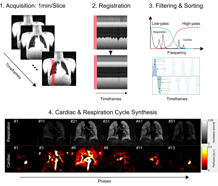
Figure 1: Schematic overview of the core components of PREFUL. 1) Acquisition in free-breathing, which necessitates 2) motion compensation via registration as demonstrated with the profile plots and enables a voxel-by-voxel analysis of the Fourier components as illustrated in step 3) filtering and sorting. After low-pass (ventilation) and high-pass (perfusion) filtering, 4) the estimated phase is used to sort the images to a higher apparent temporal resolution and synthesize one full cardiac and respiratory cycle. Note that this is a simplified outline and full details are described in the manuscript. Further steps, which are omitted from this figure, include parameter quantification and report generation. Abbreviation: PREFUL = phase-resolved functional lung. Please click here to view a larger version of this figure.
1. Recruitment
- Recruit adult patients or minors in a PREFUL MRI trial for patients and healthy controls based on their diagnosis of a pulmonary disorder based on spirometry examination and/or previous imaging (e.g., X-ray or CT scan); ability to give informed consent; ability to lie flat and remain still for the duration of the MRI scan; and no known contraindications to MRI (e.g., metallic implants, claustrophobia).
- Exclude patients if they are pregnant, have had prior lung surgery within the last 6 months, have severe respiratory distress or requirement for continuous oxygen supplementation, or have had previous adverse reactions to gadolinium-based contrast agents if contrast-enhanced MRI is used in addition to PREFUL.
- Recruit healthy controls if there is no known history of lung disease based on clinical examination and self-report; lung function tests are normal; they are able to give informed consent; they are able to lie flat and remain still for the duration of the MRI scan; and have no known contraindications to MRI.
- Exclude individuals as healthy controls according to current or past history of smoking, exposure to known lung toxins or occupational hazards, family history of hereditary lung diseases, any known chronic disease that might influence lung function, and pregnancy.
- Acquire consent forms. Continue with the protocol if a written informed consent form is obtained from the participant containing the purpose and procedures of the research, any potential risks and benefits, assurances of confidentiality, duration of the study, and the right to withdraw without consequence.
- Apply the following additional steps when recruitment includes minors.
- Obtain written informed consent from at least one parent or guardian in addition to the assent of the minor.
- Present the whole process in an age-appropriate manner that is understandable to the minor.
- For younger minors, use visual aids, storybooks, or simplified explanations.
- Ensure that the study is relevant to the age group and that minors are not being included unnecessarily.
- Allow minors to be accompanied by a trusted adult (e.g., parent, guardian) during all trial-related procedures unless this interferes with the trial's integrity.
NOTE: The described steps assume the trial was reviewed and approved by the Ethics Committee.
- Apply the following additional steps when recruitment includes minors.
2. Acquisition
- Conduct the prescan safety procedure.
- Prior to an MRI, conduct a detailed prescreening of patients to identify potential contraindications such as previous surgeries, implanted devices, tattoos, or exposure to metal fragments.
- Upon arrival, educate the patients about the procedure's magnetic properties and risks.
- Instruct the patients to remove all personal metallic items, including jewelry, watches, and certain clothing components, and provide them with a gown if necessary.
- Have a trained professional visually inspect patients for overlooked metallic objects.
- Vet all medical devices or implants for MRI compatibility.
- Ensure emergency protocols are in place for both staff and patient safety.
- Position participant and scan
- Orient the patient head-first and lay them in a supine position on a 0.55T, 1.5T, or 3T system.
- Provide hearing protection, an emergency bell, padding, and a blanket for safety and comfort.
- Position a multi-channel flex coil just beneath the chin to ensure optimal coil sensitivity across all lung areas.
- Secure the coil's placement to maintain stability without hindering the patient's breathing.
- Instruct the patient to close their eyes, then mark the center of the lung using the MR laser.
- Align the lung center at the isocenter and allow the patient to open their eyes again.
- Scan the initial localizers to establish a general orientation, followed by a transversal morphological scan to identify the tracheal bifurcation.
- Anchor the first coronal slice at the tracheal bifurcation as a consistent landmark to enhance reproducibility.
- Depending on the scan protocol, either capture three slices spaced with a slice-to-slice distance (measured from edge to edge) of 100% (of slice thickness) or acquire multiple slices spanning the entire lung with a distance of 20% or 33%.
- Acquire each slice completely separately and not interleaved.
- Upload the reconstructed images to the picture archiving and communication system (PACS) for subsequent access and analysis. Alternatively, for adherence to privacy and data protection standards, manually export images to a designated network drive or a similar storage solution.
NOTE: For detailed information on sequence protocol and parameters, refer to Table 1 and Table 2. For a visual representation of slice positioning, see Figure 2.

Figure 2: A typical slice positioning for a PREFUL experiment visualized with a 3D gradient echo in transversal orientation. Note that the first slice is positioned at tracheal bifurcation as a reproducible landmark. The 2nd and 3rd slices are positioned with a 100% slice gap in the anterior and posterior directions. Exemplary histograms show value distributions with proper and inadequate (low) scaling. The latter leads to a low dynamic range and loss of accuracy. An inadequate high scaling, which leads to clipping (not shown here), should be avoided as well. Abbreviation: PREFUL = phase-resolved functional lung. Please click here to view a larger version of this figure.
Table 1: A typical outline of a PREFUL protocol. After a localizer, a 3D volume of the lung is acquired in transversal orientation. This acquisition is used to plan the following PREFUL acquisitions (see also Table 2 for sequence settings and Figure 2 for slice positioning). Other sequences can be added depending on the study. Abbreviation: PREFUL = phase-resolved functional lung. Please click here to download this Table.
Table 2: The sequence parameters summary for PREFUL acquisition with spoiled gradient echo sequence. Abbreviations: PREFUL = phase-resolved functional lung; SPGRE = spoiled gradient echo sequence. Please click here to download this Table.
3. Postprocessing
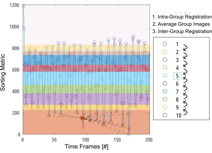
Figure 3: Schematic presentation of the group-oriented registration to minimize the required deformation for motion compensation. After dividing the images (represented by circles) into 10 groups based on a sorting metric (e.g., segmented lung area), the images are registered inside each group to an intermediate position (illustrated by dashed lines for group 1). Then, the registered images are averaged and used for the final step of Inter-Group registration in a step-by-step fashion toward the intermediate group. Abbreviation: GOREG = group-oriented registration. Please click here to view a larger version of this figure.

Figure 4: Illustration of the sorting algorithm for perfusion and ventilation. For perfusion (left), a piece-wise fitting (upper row) is performed to estimate the phase and resort the acquisitions (lower row). For ventilation (right), outliers are excluded (upper row) and sorted according to a cosine model (lower row) based on amplitude and amplitude differential to distinguish between expiration and inspiration phases. Please click here to view a larger version of this figure.

Figure 5: Exemplary flow-volume loops and the corresponding FVL-Correlation Metric of a 43-year-old female patient with COPD. Note that with changing FVL, the FVL-CM decreases. Abbreviations: FVL = flow-volume loop; FVL-CM = FVL-correlation metric; COPD = chronic obstructive pulmonary disease. Please click here to view a larger version of this figure.
Table 3: Application of thresholds to the parameter maps and combination of defect maps. Please click here to download this Table.
- Registration
NOTE: In the following section, the procedure for non-rigid registration to a reference volume (preferably the mid-level respiration level) using a group-oriented (GOREG) scheme for compensation of breathing and cardiac motion is described.- Retrieve the images.
- Initially, perform lung segmentation on all unregistered images, with a trained U-Net and apply a low-pass filter with a cut-off of 0.7 Hz to obtain an estimate for the respiration phases.
NOTE: This will initiate the fully automated processing pipeline, which will perform the subsequent steps in the background. The demonstrated and described processing steps are not limited to a specific app or language and therefore, can be implemented in a custom app with many programming languages. - Classify high amount of segmented voxels as inspiration and low amount as expiration.
- Group the images by dividing them into 10th-percentiles, ensuring each group of the resulting 10 groups contains an equal number of images.
- Select ANTs34 (BSplineSyN with cross-correlation metric) or Forsberg35,36 (polynomial expansion with elastic and fluid regularization) as the registration algorithm.
- Conduct Intra-registration for each group towards the intermediate lung position of the respective group.
- Average the group results to obtain one image for each group.
- Conduct Inter-registration going from each group image towards the next neighbor in the direction of the 5th group.
- Apply the chain of deformations to the original images, as necessary to reach the respiratory position represented by the averaged image in group 5. For example, for Image 36, which belongs to group 3, the following deformation fields are applied: 36->Image 57 (~Intermediate Lung Position in group 3) -> Step 3->4 -> Step 4->5.
NOTE: For a detailed description of the GOREG procedure, refer to Figure 3. Perform registration with parallel computing to reduce processing time. GOREG registration is performed to minimize the deformation amount required for each registration step and hence ensure stable algorithm convergence. Nevertheless, registration can also be performed with just one deformation step toward the intermediate lung position. While all published 2D PREFUL studies used ANTs, Forsberg yields results up to 6x faster with results being of comparable quality as reported by a 3D PREFUL study37. Exemplary results in this report were generated with Forsberg registration. Refer to Figure 1 to see an illustration of the registration effect on diaphragm movement.
- General filtering
- Denoise the registered images using image-guided filtering38, employing the temporally averaged registered image as the guiding image. Apply the following settings: NeighborhoodSize = [10, 10], DegreeOfSmoothing = 1.
- For ventilation and perfusion analysis, use a low-pass or high-pass filter with a cut-off at 0.7 Hz to suppress the respective other component. Exclude the first 20 images from all further processing steps, except for quantified perfusion calculation, to ensure steady state in the included time series.
NOTE: A change of the cut-off might be necessary if subjects have a respiration rate above ~40 breaths/min.
- Segmentation
NOTE: Final segmentation is performed using the registered images in intermediate lung position in a two-step procedure as outline in the following.- Perform lung boundary (lung ROI) segmentation on the temporally averaged registrated images with a trained U-Net or manually.
- Then, exclude large central vessels to refine lung boundary segmentation and obtain a region of interest or ROI for lung parenchyma.
- Perfusion
NOTE: The following steps are required to precisely estimate the cardiac phase for each image in the acquired series, which are sampled at a relatively low frequency (~3-5 images/s), especially in comparison to the heart rate (typically 40-90 bpm). Rearrangement of the data according to the determined cardiac phases is used to obtain the complete cardiac cycle with enhanced temporal resolution, surpassing the data acquisition's sampling rate18 (refer to Figure 4 for an illustration of the sorting procedure). For phase estimation, a search ROI with a strong perfusion-weighted signal is required. Employ an iterative search algorithm as follows22.- Connect the lung boundary ROI to include the mediastinum in the search ROI.
- Generate a simple perfusion-weighted map by calculating the standard deviation across the image sequence.
- Identify regions corresponding to the 98th percentile of this map within the search ROI as seed ROIs for subsequent steps.
- Perform the piece-wise fitting, increasing the size of the seed points as long as the fitting performance improves.
- Rank the expanded seed ROIs according to their fitting performance.
- Iteratively combine the best-expanded seed ROIs with the second-best, third-best, etc., until either the combination doesn't improve the metric or all seed ROIs are considered. Consider the final ROI to be the vessel ROI used for cardiac phase estimation.
- Spatially average the signal inside the optimized phase estimation to produce one signal-time series for phase estimation.
- Perform the piece-wise estimation by segmenting the signal into smaller portions using the local maxima of the signal, followed by a piecewise sinusoidal fit considering parameters like amplitude, phase offset, and frequency (Figure 4).
- Phase-sort the images to represent one cardiac cycle.
- Employ Nadaraya-Watson kernel regression with a Gaussian kernel (sigma = 0.1) to interpolate 15 phases onto a uniformly spaced time grid encompassing a single cardiac cycle.
NOTE: Refer to Figure 1 for a subset of an exemplary synthesized full cardiac cycle of a healthy volunteer, starting at diastole, transitioning to systole, and returning to diastole.
- Ventilation
NOTE: For perfusion analysis, note that the heart frequency remains relatively stable with negligible amplitude variations. In contrast, ventilation tends to experience more variations in tidal volume and frequency, leading to different respiratory states with identical respiratory phases not always having the same amplitude. Inspired by Fischer et al.'s self-gating approach, it is essential to categorize ventilation based on signal amplitude.- Exclude extreme outliers using empirical rules (data below the 5th or above the 97th percentile).
- Derive the amplitude range R and offset C from the signal-time series created for registration grouping.
- Define a model function A(t) with an arbitrarily selected frequency fRespiration (here 0.3 Hz):

- Classify the data into inspiration and expiration states based on the slope.
- Achieve more refined phase determination according to the model function:


- Subsequently, align samples based on their phase and apply the Nadaraya-Watson kernel regression to compute ventilation at uniformly spaced intervals during the respiratory cycle.
- Calculate the regional ventilation (RVent) for each phase in analogy to equation 1, substituting the inspiration phase with the respective phase.

NOTE: Refer to Figure 1 to see a subset of an exemplary synthesized full respiratory cycle of a healthy volunteer, beginning at expiration, transitioning to inspiration, and then returning to expiration. For an illustration of the sorting algorithm, see Figure 4.
- Parameter calculation
NOTE: Using the synthesized full respiration and cardiac cycles, one can derive further parameters. A selection of the most important parameters is described in the following. - Regional ventilation (RVent)
- Using the inspiratory phase derive RVent according to:

- Using the inspiratory phase derive RVent according to:
- Flow-volume-loop correlation metric (FVL-CM)
NOTE: To assess all respiration phases, a series of steps is performed to generate an MRI equivalent to FVL analysis in analogy to pulmonary function testing.- Calculate the slope of regional ventilation (RVent) as a surrogate for flow using the first time derivative of RVent. Employ a symmetric difference quotient with step length h:

- Optionally, display regional or averaged RVent slopes as a function of the respective RVent, thereby generating a PREFUL equivalent to FVL analysis.
- Determine a reference ROI by identifying the largest connected region with RVent values in the 80th to 90th percentile range in the lung parenchyma ROI.
- Average the flow-volume loops inside the determined reference ROI.
- To determine the similarity of each lung FVC to the reference, cross-correlate each FVC in the lung parenchyma ROI to the reference with zero lag:
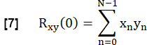
- Normalize it according to:
![Static equilibrium equation; Rxy,norm(0)=1/√[Rxx(0)Ryy(0)]Rxy(0); mathematical expression.](/files/ftp_upload/66380/66380eq09v5.jpg "figure-protocol-13")
Here, x and y represent the reference and the respective RVent flow curve.
NOTE: Note that zero-lag is employed so that delayed ventilation results in a lower correlation.
NOTE: Refer to Figure 5 for an illustration of the FVL calculation and the derived FVL-CM metric.
- Calculate the slope of regional ventilation (RVent) as a surrogate for flow using the first time derivative of RVent. Employ a symmetric difference quotient with step length h:
- Quantified perfusion
NOTE: Quantification is performed according to Glandorf et al. using the first images acquired during the transient state36.- Normalize the first four registered images to the mid-inspiration level using the lung voxel amount A as calculated in step 3.1.2. This reduction of modulation caused by varying proton density is expressed as:

- Perform an exponential fit to estimate the signal associated with maximal magnetization using the model:

- Determine a map Q related to parenchymal perfusion using the cardiac cycle phase which shows the most maximal signals in the lung parenchyma ROI.
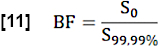
- For the estimation of regional blood fraction (BF), normalize the S0 value by averaging the values above the 99.99th percentile in the search ROI (full blood voxel):
- Estimate the exchange fraction (EF) during a cardiac cycle by considering the ratio of the maximal median signal difference between steady state (SS) and initial state as determined by S0 and the flow-related signal difference Q:

- Determine the heart frequency fHeart in 1/s from the vessel ROI using Fourier analysis (frequency corresponding to the largest peak).
- Calculate the final quantified Perfusion (QQ) in mL∙min-1∙100 mL-1 as follows:

NOTE: Here, the voxel volume (VV) is canceled [mL/mL], and the conversion factor 60 s/min and convention factor 100/100 are used to display the final result in [mL∙min-1∙100 mL-1]. EF and BF are dimensionless ratios.
- Normalize the first four registered images to the mid-inspiration level using the lung voxel amount A as calculated in step 3.1.2. This reduction of modulation caused by varying proton density is expressed as:
- Thresholding and statistics
- Statistically describe the aforementioned parameters for the middle slice and all slices with mean value (all values/middle slice values) and standard Deviation (all values / only middle slice values).
- Additionally, normalize the standard deviation to the coefficient of variation to obtain a relative account of dispersion. Select the mean value and coefficient of variation as final statistical outputs.
- Apply thresholds to the parameter maps to generate defect maps and derive defect percentage values (see Table 3).
- Classify values below the thresholds as ventilation or perfusion defect (VD/QD). Combine these maps further to quantify the overlap of defects and normal regions (V/Q classes), including the following combinations as shown in the four-fold table in Table 3:
- Calculate the defect percentage of Ventilation Defect (VD), Perfusion Defect (QD), and Ventilation/Perfusion (V/Q) classes as the number of voxels with the respective class in relation to the total lung parenchyma voxels:
Defect Percentage = #DefectVoxels/#LungParenchyma - Calculate this defect percentage for each slice and the compound coronal slices. For this study, choose the combined approach, where a ventilation defect is determined by an OR-operation: VD = VD(RVent) OR VD(FVL-CM).
NOTE: Described analysis was performed with a commercial software app (see Table of Materials) using the Forsberg registration toolbox.
| Parameter | Threshold | Comment |
| RVent | 90th Percentile * 0.4 | Adaptive Threshold |
| FVL-CM | 90% | Fixed threshold |
| Q | 90th Percentile * 0.15 | Adaptive Threshold |
| V/Q | NO QD | QD |
| NO VD | Normal | VQ Mismatch (Exclusive QD) |
| VD | VQ Mismatch (Exclusive VD) | VQ Defect-Match |
Results
The lower part of Figure 2 illustrates the consequence of proper and inadequate scaling with a corresponding effect on the dynamic range. Figure 6 shows the inhomogeneous signal distribution, which is representative of scans without and with coil normalization. Avoiding a low dynamic range and images without coil normalization is recommended.
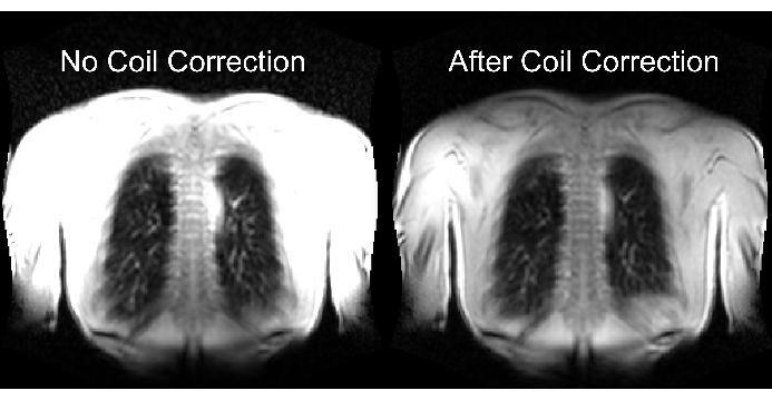
Figure 6: Exemplary images after acquisition without coil correction (incorrect) and with coil correction (correct). Note the artificial signal enhancement at the body boundaries near the coil elements. Please click here to view a larger version of this figure.
Figure 7 illustrates successful and failed automatic segmentation. Note that the failed segmentation does not include all lung voxels, which will falsify further analysis and statistics. Special care is required for cases with infiltrates as such voxels can be misclassified as vessels due to their high signal by AI models or not segmented at all.
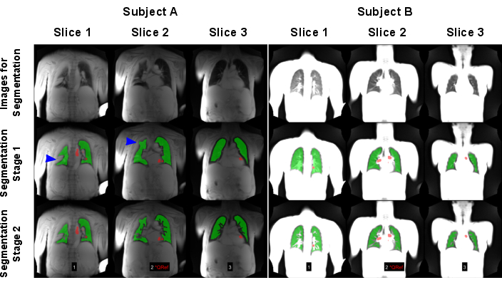
Figure 7: An example of automated segmentation resulting in a failed (Subject A: 83-year-old male with COPD) and a successful result (Subject B: 30-year-old female healthy control). The first row shows the images, which were used as input for the AI models. The second row shows the results of the first segmentation stage consisting of finding the lung boundary. The third row shows the final result after the exclusion of vessels. As shown by the blue arrows, the algorithm was challenged by the high-signal lung variances causing wrong lung boundary detection. Note that the images were normalized by maximal signal, which led to different results due to missing coil normalization of the scan performed on subject A. Red regions show the ROIs, which were automatically detected for perfusion-phase sorting. Abbreviations: COPD = chronic obstructive pulmonary disease; ROIs = regions of interest. Please click here to view a larger version of this figure.
Figure 8 and Figure 9 show representative parameter maps for a healthy control (age 30, female) and a COPD patient (age 60, male). Note that the healthy control shows a more homogeneous ventilation and perfusion and thus, fewer defect voxels. The corresponding reports of the ROI statistics can be found in Table 4 and Table 5.

Figure 8: PREFUL parameter maps of a 30-year-old female healthy control. The perfusion (1st row), regional ventilation (2nd row), flow-volume loop correlation metric (3rd row), and thresholded V/Q maps (4th row). Note the homogeneous distribution of the parenchymal values and low defect percentages. Abbreviations: PREFUL = phase-resolved functional lung; V = ventilation; Q = perfusion. Please click here to view a larger version of this figure.

Figure 9: PREFUL parameter maps of a 60-year-old male COPD patient. The perfusion (1st row), regional ventilation (2nd row), flow-volume loop correlation metric (3rd row), and thresholded V/Q maps (4th row). Note the heterogeneous distribution of the parenchymal values and high defect percentages. Abbreviations: COPD = chronic obstructive pulmonary disease; PREFUL = phase-resolved functional lung; V = ventilation; Q = perfusion. Please click here to view a larger version of this figure.
Table 4: Exemplary report of PREFUL parameters obtained for a healthy control (30-year-old female). Note the low coefficient-of-variation and defect values, which are in line with the parameter maps presented in Figure 8 for the same subject. See also Table 5 and Figure 9. Abbreviation: PREFUL = phase-resolved functional lung. Please click here to download this Table.
Table 5: Exemplary report of PREFUL parameters obtained for a COPD patient (60-year-old male). Note the high coefficient-of-variation and defect values, which are in line with the parameter maps presented in Figure 9 for the same subject. See also Table 4 and Figure 8. Please click here to download this Table.
Supplementary Material: Animated Explanation of the PREFUL Algorithm. Please click here to download this file.
Discussion
Critical steps
One of the most common pitfalls during acquisition is inadequate signal scaling, which causes a loss of information during DICOM conversion by reduced precision of digital data representation. Consequently, this can lead to problems during the postprocessing stage. Another even more critical pitfall is the acquisition of multiple slices in an interleaved fashion. Thereby, the effective temporal resolution of the individual slices is critically reduced. Additionally, depending on the distance of the slices, this can have an impact on the perfusion contrast and quantification since the inflow relies on fresh spins without magnetization history. Special care is required during protocol setup, especially regarding gradient strength, asymmetric echo, bandwidth, and parallel imaging. Deviations from the suggested settings for even just one of these parameters can lead to inadequate TE and temporal resolution.
The postprocessing consists of multiple steps, which should be followed in the described order. For example, a registration after low-pass filtering is not meaningful. Consequently, failure at one step leads to a breakdown during the next steps. This makes the registration stage especially important. Since there is no single registration algorithm, depending on the respective implementation, parameters must be set empirically. Without finetuning of these parameters, a false registration will prevent the generation of any meaningful result. Another possibly time-consuming and critical step during postprocessing is segmentation. False segmentations can lead to completely wrong parameter calculations (e.g., by including non-lung regions) in the final report. Such mis-segmentations are more likely to occur with deep learning algorithms, which are accustomed to certain image appearances and are applied to images from another vendor/machine with a slightly different appearance. A visual quality check of segmentation accuracy, with potential manual correction, is therefore mandatory.
Troubleshooting
The typical troubleshooting procedure is to follow all steps one by one and check the plausibility of the intermediate results. The procedure for the main steps is as follows: Check that the images are acquired in free breathing with the correct sequence and settings. Next, check that the dynamic range of the signals is appropriate (~50 AU in the lung parenchyma). If raw data are still available, repeat the reconstruction of the images with an appropriate scaling factor avoiding the need for a new acquisition of data. Check that the registration was performed without major artifacts and remaining motion. Next, check if small ROIs show a time series with expected ventilation- and perfusion-related modulations. Then, check if the applied filters alter the images in the expected manner (e.g., no high-frequency modulations in low-pass filtered data). Check if the synthesized respiratory and cardiac cycles are physiologic and don't show sudden jumps. Check the segmentation accuracy. Note that a search on a finer resolution level might be necessary as soon as the main step, during which the problem occurs, was identified.
Limitations
Although the presented protocol is known to produce reproducible and sensitive results, the numbers of involved steps and parameters during acquisition and post-processing allow for nearly endless optimization and are intertwined. Therefore, a bottom-up approach should be followed by first addressing optimizations of the sequence protocol (e.g., regarding SNR and functional contrast-to-noise ratio). For the following postprocessing optimizations, a predefined ground truth in the form of a digital lung model might be useful40. As presented, this model mimics a free-breathing acquisition and includes several classes to simulate ventilation/perfusion defects. Including a known deformation due to movement, registration algorithms can also be tested directly. Despite these advantages, each model is inherently limited by the accuracy of mapping complex reality to a finite and simplified model.
The thresholds presented in this protocol were found to show reasonable results for healthy volunteers and across different patient cohorts by empirical analysis. Nevertheless, as outlined before, adjustment is likely required depending on the sequence, field strength, and cohort.
A general limitation of PREFUL is the extensive post-processing, which is not readily available as a medical product yet, although first work-in-progress versions from Siemens Healthineers and BioVisioneers are available for scientific purposes in a scientific collaboration/commercial setting. Calculations typically involve parallel processing, which poses especially high demands on CPU and RAM and might require modern workstations or server solutions to effectively process large amounts of data. Further, the time-consuming postprocessing steps currently impede an instant presentation of the results, which would be desirable for the clinical workflow.
Comparison to other methods
There are a multitude of similar approaches like PREFUL, including the predecessor Fourier Decomposition and its other derivates such as Matrix Pencil Decomposition41 and the slightly different approach Self-gated Non-Contrast-enhanced Functional Lung MRI (SENCEFUL MRI)42. While Fourier Decomposition and similar methods operate in the frequency domain, PREFUL uses less strict Fourier-filtering and subsequent calculation of amplitudes in the time domain. Therefore, there is no requirement to select specific peaks corresponding to ventilation/perfusion. This can result in less susceptibility to respiration variability, which is known to occur in human subjects.
While PREFUL performs image sorting, SENCEFUL uses sorting of k-space lines, leading to more flexibility. Nevertheless, SENCEFUL requires sequences with self-gating capabilities, while PREFUL can be performed with a conventional spoiled gradient echo sequence. Similarly, bSSFP commonly used in Fourier Decomposition-based approaches is known for better SNR and blood flow contrast but typically requires more optimization for lung acquisition especially at 3T43. Nevertheless, other than that there is no reason to not combine PREFUL with bSSFP acquisition44.
All these signal-based approaches assume that certain unwanted signal influences, including T1, T2/T2*, diffusion, through-plane motion, and non-orthogonally perfused voxels, are negligible. While the progressed validation of PREFUL indirectly suggests that indeed such influences are not critical, Triphan et al. showed that there is a dependence on the effective T1 and TE, which is explained by the different weighting of the blood and parenchymal components depending on the TE45. In this light, the initial advantage of bSSFP to visualize blood due to T2/T1 contrast might pose an additional challenge to establish an accurate quantification in comparison to the simpler contrast mechanics of an SPGRE. Nevertheless, further studies that directly address the influence of various MR-variables, for example, as performed by Glandorf et al. for contrast media46,47, are desirable as they can directly quantify the effect on PREFUL.
Importance
Being a free-breathing, contrast-media-free method, PREFUL shares many advantages with the previously mentioned related methods: 1) No ionizing radiation and contrast agent application, 2) No requirement for additional hardware or personnel, 3) acquisition, which depends only on minimal patient compliance. These advantages make PREFUL a convenient monitoring tool, especially for vulnerable groups such as children with chronic pulmonary disease. Although SNR is low with SPGRE sequence, the availability, and a lack of requirement for additional sequence programming/sharing further promote the dissemination of this approach.
As discussed in the introduction section, the number of studies showing good validation, reproducibility, sensitivity results, and monitoring capabilities show that the importance of this technique and corresponding dynamic parameters is on a rising trajectory and will be further supported by wide dissemination.
Disclosures
Filip Klimeš, Andreas Voskrebenzev, and Jens Vogel-Claussen are shareholders of BioVisioneers GmbH, a company, which has an interest in pulmonary magnetic resonance imaging methods.
Acknowledgements
This work was funded by the German Center for Lung Research (DZL). The authors would like to express a deep gratitude towards all, who contributed and supported the further development of PREFUL, in particular: Marcel Gutberlet, Till F. Kaireit, Lea Behrendt, Julian Glandorf, Sonja Lüdiger, Tawfik Moher Alsady, Katharina Bünemann, Marius Wernz, Robin Müller, Maximilian Zubke, Gesa Pöhler, Agilo Kern, Cristian Crisosto, Milan Speth, Arnd Obert, Julienne Scheller, Jim Wild, Edwin van Beek, Helen Marshall, Jens Gottlieb, Martha Dohna, Diane Renz, Anna-Maria Dittrich, Tobias Welte, Jens Hohlfeld, Patrick Zardo, Giles Santyr, Franz Wolfgang Hirsch, Robert Grimm, Bastian Bier, Bassem Ismail, André Fischer, Berthold Kiefer, Gregor Thoermer and Rebecca Ramb. Furthermore, the authors would also like to thank the radiographers and study participants. In particular, we thank Frank Schröder and Sven Thiele from the Department of Radiology (Hannover Medical School) for outstanding technical assistance in performing the MRI examinations.
Materials
| Name | Company | Catalog Number | Comments |
|---|---|---|---|
| Advanced Normalization Tools (ANTs) | - | Image registration toolbox (https://stnava.github.io/ANTs/; https://github.com/fordanic/image-registration) | |
| Forsberg | - | Image registration toolbox | |
| MRI | Siemens Healthineers AG, Munich, Germany | 0.55T / 1.5T / 3T Scanner | |
| PREFUL App | BioVisioneers GmbH, Laatzen, Germany | PREFUL analysis, Figures and reports |
References
- Vogelmeier, C. F., et al. Global strategy for the diagnosis, management, and prevention of chronic obstructive lung disease 2017 Report: GOLD Executive Summary. Eur Respir J. 49 (3), 1700214(2017).
- Ranu, H., Wilde, M., Madden, B. Pulmonary function tests. Ulster Med J. 80 (2), 84-90 (2011).
- Galbán, C. J., et al. Computed tomography-based biomarker provides unique signature for diagnosis of COPD phenotypes and disease progression. Nat Med. 18 (11), 1711-1715 (2012).
- Roach, P. J., Schembri, G. P., Bailey, D. L. V/Q Scanning using SPECT and SPECT/CT. J Nucl Med. 54 (9), 1588-1596 (2013).
- Wild, J. M., et al. MRI of the lung (1/3): methods. Insights Imaging. 3 (4), 345-353 (2012).
- Albert, M. S., et al. Biological magnetic resonance imaging using laser-polarized 129Xe. Nature. 370 (6486), 199-201 (1994).
- Berthezène, Y., et al. Contrast-enhanced MR imaging of the lung: assessments of ventilation and perfusion. Radiology. 183 (3), 667-672 (1992).
- Hughes, P. J. C., et al. Assessment of the influence of lung inflation state on the quantitative parameters derived from hyperpolarized gas lung ventilation MRI in healthy volunteers. J Appl Physiol. 126 (1), 183-192 (2019).
- Zapke, M., et al. Magnetic resonance lung function - a breakthrough for lung imaging and functional assessment? A phantom study and clinical trial. Respir Res. 7 (1), 106(2006).
- Klimeš, F., et al. Free-breathing quantification of regional ventilation derived by phase-resolved functional lung (PREFUL) MRI. NMR Biomed. 32 (6), e4088(2019).
- Deimling, M., Jellus, V., Geiger, B., Chefd'hotel, C. Time resolved lung ventilation imaging by Fourier decomposition. Proc. Intl. Soc. Mag. Reson. Med. 16, 2639(2008).
- Bauman, G., et al. Non-contrast-enhanced perfusion and ventilation assessment of the human lung by means of fourier decomposition in proton MRI. Magn Reson Med. 62 (3), 656-664 (2009).
- Bauman, G., et al. Lung ventilation- and perfusion-weighted Fourier decomposition magnetic resonance imaging: In vivo validation with hyperpolarized 3He and dynamic contrast-enhanced MRI. Magn Reson Med. 69 (1), 229-237 (2013).
- Bauman, G., et al. Validation of Fourier decomposition MRI with dynamic contrast-enhanced MRI using visual and automated scoring of pulmonary perfusion in young cystic fibrosis patients. Eur J Radiol. 82 (12), 2371-2377 (2013).
- Sommer, G., et al. Non-contrast-enhanced preoperative assessment of lung perfusion in patients with non-small-cell lung cancer using Fourier decomposition magnetic resonance imaging. Eur J Radiol. 82 (12), e879-e887 (2013).
- Voskrebenzev, A., Gutberlet, M., Becker, L., Wacker, F., Vogel-Claussen, J. Reproducibility of fractional ventilation derived by Fourier decomposition after adjusting for tidal volume with and without an MRI compatible spirometer. Magn Reson Med. 76 (5), 1542-1550 (2016).
- Voskrebenzev, A., Gutberlet, M., Kaireit, T. F., Wacker, F., Vogel-Claussen, J. Low-pass imaging of dynamic acquisitions (LIDA) with a group-oriented registration (GOREG) for proton MR imaging of lung ventilation. Magn Reson Med. 78 (4), 1496-1505 (2017).
- Voskrebenzev, A., et al. Feasibility of quantitative regional ventilation and perfusion mapping with phase-resolved functional lung (PREFUL) MRI in healthy volunteers and COPD, CTEPH, and CF patients. Magn Reson Med. 79 (4), 2306-2314 (2018).
- Voskrebenzev, A., et al. Imaging-based spirometry in chronic obstructive pulmonary disease (COPD) patients using phase resolved functional lung imaging (PREFUL). Proc. Intl. Soc. Mag. Reson. Med. 26, 1079(2018).
- Alsady, T. M., et al. MRI-derived regional flow-volume loop parameters detect early-stage chronic lung allograft dysfunction. J Magn Reson Imaging. 50 (6), 1873-1882 (2019).
- Pöhler, G. H., et al. Chronic thromboembolic pulmonary hypertension perioperative monitoring using phase-resolved functional lung (PREFUL)-MRI. J Magn Reson Imaging JMRI. 52 (2), 610-619 (2020).
- Behrendt, L., et al. Validation of automated perfusion-weighted phase-resolved functional lung (PREFUL)-MRI in patients with pulmonary diseases. J Magn Reson Imaging. 52 (1), 103-114 (2020).
- Kaireit, T. F., et al. Flow volume loop and regional ventilation assessment using phase-resolved functional lung (PREFUL) MRI: comparison with 129 xenon ventilation MRI and lung function testing. J Magn Reson Imaging JMRI. 53 (4), 1092-1105 (2021).
- Marshall, H., et al. 129Xe and free-breathing 1H ventilation MRI in patients with cystic fibrosis: a dual-center study. J Magn Reson Imaging. 57 (6), 1908-1921 (2023).
- Behrendt, L., et al. A dual center and dual vendor comparison study of automated perfusion-weighted phase-resolved functional lung magnetic resonance imaging with dynamic contrast-enhanced magnetic resonance imaging in patients with cystic fibrosis. Pulm Circ. 12 (2), e12054(2022).
- Moher Alsady, T., et al. Multicenter standardization of phase-resolved functional lung MRI in patients with suspected chronic thromboembolic pulmonary hypertension. J Magn Reson Imaging. , (2023).
- Pöhler, G. H., et al. Repeatability of phase-resolved functional lung (PREFUL)-MRI ventilation and perfusion parameters in healthy subjects and COPD patients. J Magn Reson Imaging. 53 (3), 915-927 (2021).
- Voskrebenzev, A., et al. PREFUL MRI depicts dual bronchodilator changes in COPD: a retrospective analysis of a randomized controlled trial. Radiol Cardiothorac Imaging. 4 (2), e210147(2022).
- Vogel-Claussen, J., et al. Phase-resolved functional lung (PREFUL) MRI-derived ventilation and perfusion parameters predict future lung transplant loss. Radiology. 307 (4), e221958(2023).
- Zanette, B., et al. Clinical feasibility of structural and functional MRI in free-breathing neonates and infants. J Magn Reson Imaging. 55 (6), 1696-1707 (2022).
- Dyke, J. P., et al. Assessment of lung ventilation of premature infants with bronchopulmonary dysplasia at 1.5 Tesla using phase-resolved functional lung magnetic resonance imaging. Pediatr Radiol. 53 (6), 1076-1084 (2023).
- Glandorf, J., et al. Comparison of phase-resolved functional lung (PREFUL) MRI derived perfusion and ventilation parameters at 1.5T and 3T in healthy volunteers. PLOS ONE. 15 (12), e0244638(2020).
- Lévy, S., et al. Free-breathing low-field MRI of the lungs detects functional alterations associated with persistent symptoms after COVID-19 infection. Invest Radiol. 57 (11), 742-751 (2022).
- Avants, B. B., et al. A reproducible evaluation of ANTs similarity metric performance in brain image registration. NeuroImage. 54 (3), 2033-2044 (2011).
- Forsberg, D., Andersson, M., Knutsson, H. Extending image registration using polynomial expansion to diffeomorphic deformations. SSBA Symposium on Image Analysis. , Stockholm, Sweden. (2012).
- Forsberg, D. fordanic/image-registration. , Available from: https://github.com/fordanic/image-registration (2022).
- Klimeš, F., et al. Evaluation of image registration algorithms for 3D phase-resolved functional lung ventilation magnetic resonance imaging in healthy volunteers and chronic obstructive pulmonary disease patients. NMR Biomed. 36 (3), e4860(2023).
- He, K., Sun, J., Tang, X. Guided image filtering. IEEE Trans Pattern Anal Mach Intell. 35 (6), 1397-1409 (2013).
- Glandorf, J., et al. Perfusion quantification using voxel-wise proton density and median signal decay in PREFUL MRI. Magn Reson Med. 86 (3), 1482-1493 (2021).
- Voskrebenzev, A., Gutberlet, M., Klimeš, F., Wacker, F., Vogel-Claussen, J. Introduction of a Digital Lung Model for Validation and Refinement of Functional Lung Imaging Methods. Proc. Intl. Soc. Mag. Reson. Med. 31, 4820(2023).
- Bauman, G., Bieri, O. Matrix pencil decomposition of time-resolved proton MRI for robust and improved assessment of pulmonary ventilation and perfusion. Magn Reson Med. 77 (1), 336-342 (2017).
- Fischer, A., et al. SElf-gated Non-Contrast-Enhanced FUnctional Lung imaging (SENCEFUL) using a quasi-random fast low-angle shot (FLASH) sequence and proton MRI. NMR Biomed. 27 (8), 907-917 (2014).
- Bauman, G., Pusterla, O., Bieri, O. Functional lung imaging with transient spoiled gradient echo. Magn Reson Med. 81 (3), 1915-1923 (2019).
- Rotärmel, A., et al. GRE bSSFP vs. FLASH based Fourier decomposition lung MRI at 1.5T: evaluation of image quality, fractional ventilation and lung perfusion in healthy volunteers. Prog Intl Soc Mag Reson. 26, 2-4 (2018).
- Triphan, S. M. F., et al. Echo time-dependent observed lung T1 in patients with chronic obstructive pulmonary disease in correlation with quantitative imaging and clinical indices. J Magn Reson Imaging. 54 (5), 1562-1571 (2021).
- Glandorf, J., et al. Effect of intravenously injected gadolinium-based contrast agents on functional lung parameters derived by PREFUL MRI. Magnetic Resonance in Medicine. 83 (3), 1045-1054 (2020).
- Glandorf, J., et al. Influence of gadolinium, field-strength and sequence type on quantified perfusion values in phase-resolved functional lung MRI. PloS One. 18 (8), e0288744(2023).
Reprints and Permissions
