This protocol involves a laparoscopic anatomical resection of liver segment VII with liver parenchymal transection following a priority approach. This technique is unaffected by anatomical variations.
Method Article
Laparoscopic Anatomical Liver Segment VII Resection with Liver Parenchymal Transection Following a Priority Approach
In This Article
Summary
Abstract
Laparoscopic liver resection procedures have rapidly developed, including surgical techniques ranging from early local resection of the liver margin and superficial lesions to semi-hepatectomy and even complex liver segment resection. Segment VII of the liver is located at the base of the subphrenic space, making it difficult to access and resulting in a lengthy and challenging resection procedure. This liver segment resection involves high volumes of blood loss throughout the procedure. To reduce bleeding caused by the anatomy of the hepatic porta, our center uses a surgical technique that is guided by the right hepatic vein. This technique is unaffected by anatomical variations, such as the dorsal branch of the hepatic pedicle crossing the right hepatic vein to innervate hepatic segment VII. Here, we provide a method and approach for the anatomical resection of segment VII of the liver with anatomical variations of the portal vein, which can be recommended for clinical practice.
Introduction
Hepatocellular carcinoma (HCC) globally ranks among the most prevalent cancers1. In 2022, China reported 367,700 new liver cancer cases, marking the disease as the fourth most commonly diagnosed cancer in the country2. This high incidence rate exerts substantial pressure on China's healthcare system, posing a considerable public health challenge. Anatomical hepatectomy can usually completely remove the tumor-bearing portal vein basin, ensuring the preservation of the liver parenchyma to avoid postoperative liver failure3. Therefore, anatomical liver segmentectomy has become one of the main surgical methods for treating HCC4. Laparoscopic liver resection is widely used because of its minimal trauma, low incidence of complications, quick postoperative recovery, and ability to remove tumors to the same degree as open surgery5,6. The safety and effectiveness of laparoscopic liver resection for treating HCC have been widely recognized worldwide7,8. However, laparoscopic liver segment VII resection surgery has several challenges, including liver segment VII exposure, manipulation, and dissection, as well as approaching the liver pedicle9. The operative field for liver segment VII resection is situated at the base of the subphrenic space, and laparoscopic visualization is susceptible to being obstructed in the supine position10. Thus, limited visibility and restricted maneuvering space during hepatic parenchymal dissection can result in uncontrollable intraoperative hemorrhage. In addition, excessive manipulation and compression of the liver compromise the "no-touch principle" critical to oncological management, thereby increasing the risk of tumor cell dissemination11. Therefore, laparoscopic anatomical liver segmentectomy presents challenges for the resection of liver segment VII9. Moreover, the plane separating the liver segments VII and VIII is difficult to identify12. One of the primary challenges in liver dissection is determining the correct anatomical plane. There are two key considerations. First, the dorsal branch of the hepatic pedicle in liver segment VIII often branches across the area where the right hepatic vein supplies segment VII of the liver. Second, the ischemic border between segments VII and VIII may not adequately encompass the tumor area or provide sufficient margins. As a result, it is currently impossible to establish a clear boundary between segments VII and VIII by solely dissecting the hepatic pedicle in segment VII.
Our center has implemented the liver parenchyma priority method after years of experience, prioritizing liver parenchyma dissection over liver pedicle disconnection13. While performing these surgeries, the entire hepatic blood supply is blocked as the liver parenchyma is intermittently detached, all without extending the surgical scope. The only modification made is to the order of the surgical processes, making it easier to block the incoming hepatic blood flow and decreasing the chance of bleeding from hilar dissection. The liver parenchyma priority method is not influenced by anatomical variations, such as hepatic segment VII innervations by the dorsal branch of the hepatic pedicle crossing the right hepatic vein, which is advantageous for advancing and using anatomical liver resection surgery.
Here, we present the case of a 54-year-old male patient admitted to our hospital in September 2022 whose liver mass had been present for one week. His medical history revealed hepatitis B. Abdominal contrast-enhanced computed tomography revealed a mass with heterogeneous enhancement located at segment VII of the liver (size: 28 mm × 25 mm × 23 mm) (Figure 1). The measured value of alpha-fetoprotein in this patient was 559.6 ng/mL. The Child-Pugh score indicated liver function grade A, and the Indocyanine Green (ICG) clearance rate R15 was 6.2% (<10%). The cancer was classified as stage A according to the BCLC algorithm and stage Ia according to the CNLC algorithm. Further, 3D reconstruction revealed that the dorsal branch of the VIII segment of the liver pedicle crosses the right hepatic vein and supplies the segment VII area of the liver. If the traditional preferential dissection approach is used to reach the segment VII liver pedicle to reveal the ischemic line or if fluorescence staining is used, the resection range will not completely cover the entire segment VII area. Therefore, we adopted the liver parenchyma priority approach for the anatomical resection of segment VII, thereby avoiding errors caused by this specific variation in the resection range.
This surgery applies to all VII segment resections containing special portal vein variations.
Protocol
All methods described in this study were approved by the Ethics Committee of Guangdong Provincial Hospital of Traditional Chinese Medicine (Ethics Approval No: ZE2024-226-01). Surgical indications and contraindications were based on the CSCO primary liver cancer diagnosis and treatment guidelines.
1. Inclusion/exclusion criteria
- Inclusion criteria:
- Confirm that the mass is located in segment VII of the liver and identify it as a primary or secondary malignant tumor or benign liver disease.
- Verify that the surgical procedure planned is the resection of segment VII of the liver using the parenchyma priority method.
- Ensure that the preoperative Child-Pugh grade is A.
- Check that the future liver remnant (FLR) represents more than 40% of the standard liver volume (SLV).
- Exclusion criteria:
- Exclude cases where tumors invade the portal or hepatic veins.
- Exclude cases with intrahepatic or distant metastases of the tumor.
- Exclude patients with vital organ pathology that prevents anesthesia administration and surgical intervention tolerance.
2. Preoperative preparation
- Ensure the availability of laparoscopic imaging systems, pneumoperitoneum images, and ultrasound scalpels.
- Ensure the patient undergoes preoperative bowel preparation and preoperative risk assessment before anesthesia administration. For patients who take aspirin, aspirin should be discontinued before the surgery.
- Intravenously administer antibiotics 30 min before the surgery. Dilute cefuroxime sodium (1.5 g) in 100 mL of 0.9% sodium chloride solution.
- Perform tracheal intubation under general anesthesia. Place an arterial catheter and a central venous catheter. Ensure the central venous pressure at 2-3 mmHg14.
- Surgical position: Place the patient in a supine split-leg position. Raise the right waist by 15°, and ensure the chief surgeon stands between the patient's legs (Figure 2).
3. Surgery
- Preparation phase
- Ensure the surgeon washes their hands before the procedure. Disinfect the skin at the surgical site, extending 15 cm twice with 5% tincture of iodine and three times with 75% alcohol.
- Place sterile towels at the nipple line, the left mid-clavicular line, the right mid-axillary line, and the bilateral anterior superior iliac spines, exposing the surgical area.
- Trocar placement
- Make a 2-cm incision to the right of the umbilicus as a laparoscopic hole (Figure 3).
- Insert a pneumoperitoneum needle following the Veress method to establish a pneumoperitoneum. Maintain pneumoperitoneum pressure at 12-14 mmHg11 (Figure 4).
- Place a 12-mm Trocar at 4 cm below the midline of the right clavicle from the rib margin. Place a 5-mm trocar in the right anterior axillary line 4 cm below the rib margin. Subsequently, place 5-mm and 10-mm Trocars in the front of the midline of the sternum and 2 cm from the lower edge of the xiphoid process, respectively (Figure 5).
- Adjust the angle of the operating table so that the patient's head is positioned higher than their feet (15-20°), high on the right, and low on the left (15-20°) (Figure 6).
NOTE: This angle results in the intestinal tract tilting to one side, which in turn helps to display the hepatic hilum area better and rotate the liver to the left.
- Dissect the right liver and suspend the right hepatic vein.
- Incise and dissect the peritoneum in front of the inferior vena cava and the third hepatic portal using an ultrasound knife after routine exploration of the abdominal cavity.
- Trace the inferior vena cava from the foot side toward the head side. Expose the thick right posterior inferior hepatic vein and short hepatic veins. Use sutures (#4) to ligate the right posterior inferior hepatic vein and then cut it using an ultrasonic knife. Use vascular clips to ligate the short hepatic veins, freeing the posterior hepatic tunnels (Figure 7).
NOTE: This allows for adequate exposure of the anterior interstitial space of the inferior vena cava and the interstitial space adjacent to the inferior vena cava. - Detach the right triangular ligament and coronary ligament of the liver using an ultrasound knife with an assistant performing adequate exposure, thus completely freeing the right liver.
- Separate the Makuuchi ligament using an ultrasound knife (Figure 8). Pull the right liver toward the left abdomen with the help of an assistant to expose the root of the right hepatic vein and the inferior vena cava.
- Separate the right hepatic vein bluntly along the hepatic vein recess and the anterior space of the inferior vena cava and suspend the root of the right hepatic vein with a vascular identification strap as the marker for subsequent liver parenchyma dissection (Figure 9).
- Liver section
- Dissect the hepatic parenchyma along the bare area of the liver using an ultrasonic knife and locate the main trunk of the right hepatic vein.
- Use a combination of blunt and sharp separation using an ultrasound knife for dissecting the right hepatic vein.
- Locate the segment VIII severed dorsal branch of the hepatic pedicle in front of the main trunk of the right hepatic vein, ligate, and disconnect it (Figure 10). Find the segment VII liver pedicle behind the main right hepatic vein and use home-o-lock to clamp it and disconnect it using an ultrasonic knife or scissors (Figure 11).
- Cut off the liver parenchyma along the right hepatic vein from the foot side to the side, with the root of the right hepatic vein as the marker on the head side and the inferior vena cava as the marker on the back side to determine the level of liver dissection.
- Expose the main trunk of the right hepatic vein within the liver segment along the right hepatic vein throughout the entire process (Figure 12). The anesthesia team must reduce central venous pressure and minimize surface sieve hole bleeding caused by venous filling.
- Use bipolar electrocoagulation for small sieve hole bleeding; large sieve holes often require suturing to stop bleeding.
- Pay attention to protecting the dorsal branch of segment VIII that flows back from the right hepatic vein in front of the right hepatic vein while segmenting the liver parenchyma between segment VII and segment VIII.
- Dissect the liver on the right side of the hepatic vein flowing back from the dorsal branch of segment VIII. Further, cut off the hepatic vein branches that flow back to the right hepatic vein one by one.
- Fully expose the right hepatic vein from the main trunk to the root and cut off the liver parenchyma on the right side of the right hepatic vein, with the main trunk of the vein and the inferior vena cava as the plane, to completely remove segment VII of the liver.
- Take out the specimen.
- Put the surgical specimen into the specimen bag. Depending on the situation, extend the incision of sleeve A and remove the specimen (Figure 13).
- Place an abdominal drain at the hepatectomy site and one at the foramen of Winslow.
- Suture all the trocar incisions layer by layer.
- Postoperative care.
- Have the patient lie in the anesthesia recovery room after the surgery for approximately 1 h until the patient is fully awakened.
- Monitor oxygen supply to ensure patient oxygenation during this period.
- Return the patient to the ward from the operating room once the patient is fully awake.
- Follow-up
- Instruct the patient to return to the hospital after 1 month, with subsequent follow-ups every 3-6 months for blood tests and enhanced CT examinations.
Results
In this representative case, the liver parenchyma priority approach for anatomical resection of liver segment VII was successfully followed in a 54-year-old male patient. The total operative time was 110 minutes, with an estimated intraoperative blood loss of 100 mL. The patient recovered successfully on the seventh postoperative day and was discharged from the hospital without any postoperative complications such as bleeding, bile leakage, subphrenic infection, dropsy of the right chest, or hepatic failure (Table 1). Pathological results showed HCC and postoperative CT suggested complete resection of the tumor (Figure 14).
From January 2019 to April 2023, 27 patients were successfully treated with liver parenchyma priority laparoscopic anatomical liver segment VII resections, and none of the surgeries were converted to open surgery.The average age of the patients was 56.8 ± 13.0 years. The average tumor diameter was 3.24 ± 1.47 cm, and the average surgical duration was 74.5 ± 49.7 min. The average blood loss encountered during the surgery was 260.9 ± 391.8 mL. No patient required blood transfusions during the operation. The drainage tube removal time was 7.7 ± 1.9 days, and the postoperative hospitalization time was 8.6 ± 2.1 days. Postoperative pathological analyses confirmed 1 case of hepatocellular adenoma, 1 case of cholangiocarcinoma, and 25 cases of HCC. All patients were successfully discharged, and two patients developed right pleural effusion after surgery, which was cured by thoracic puncture drainage (Table 2).
No patient experienced serious complications such as biliary fistula, subphrenic infection, or liver failure. After surgery, 22 patients received Transarterial chemoembolization (TACE) adjuvant treatment and underwent laboratory and imaging examinations in routine outpatient clinics. Postoperative re-examination of liver function revealed that indicators such as alanine transaminase (ALT), alkaline phosphatase, serum bilirubin, and plasma albumin quickly recovered and returned to normal by the fifth day after surgery. There were no deaths during the operation. The follow-up period ranged from 1 to 44 months, with a median of 13 months. Two patients experienced postoperative tumor recurrence. The detailed data for all patients are shown in Table 3. This procedure was used for the surgical treatment of all 27 patients included in the study. Compared with traditional laparoscopic VII anatomical liver segment resection with a priority hepatic pedicle approach15, this procedure significantly reduces surgical time without increasing intraoperative bleeding, postoperative duration of hospitalization, or incidence of complications (Table 4).

Figure 1: Abdominal contrast-enhanced computed tomography revealed a mass with heterogeneous enhancement located at segment VII of the liver. Please click here to view a larger version of this figure.

Figure 2: Surgical position. Please click here to view a larger version of this figure.

Figure 3: Laparoscopic Incision. Please click here to view a larger version of this figure.
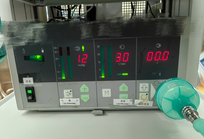
Figure 4: Pneumoperitoneum pressure. Please click here to view a larger version of this figure.

Figure 5: TROCAR position. (A) The examination port is located on the right side of the abdomen, 2 cm away from the navel. (B) The entrance point for the chief surgeon is located 4 cm below the midline of the right clavicle from the rib margin and 4 cm below the right anterior axillary line from the rib margin. (C) The assistant surgical holes were located anteriorly in the midline of the sternum at 2 cm from the inferior border of the xiphoid process and at 8 cm from the inferior border of the xiphoid process. Please click here to view a larger version of this figure.
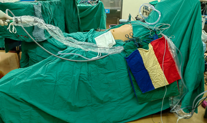
Figure 6: Operating table position with the head elevated at 15-20°. Please click here to view a larger version of this figure.
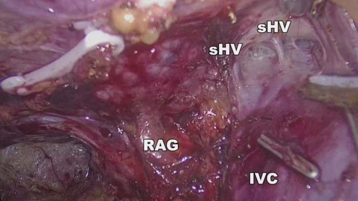
Figure 7: Anatomy of the third hepatic portal of the free retrohepatic tunnel. IVC: inferior vena cava, RAG: right adrenal gland, s-HV: short hepatic vein. Please click here to view a larger version of this figure.

Figure 8: Exposure and severance of the Makuuchi ligament. IVC: Inferior vena cava, s-HV: short hepatic vein. Please click here to view a larger version of this figure.
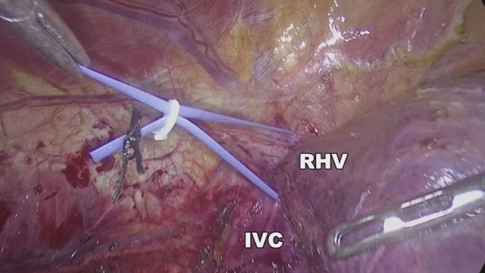
Figure 9: Suspension of the right hepatic vein (RHV). IVC: Inferior vena cava. Please click here to view a larger version of this figure.
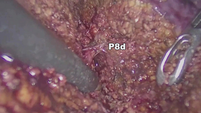
Figure 10: Dorsal Branch of the Hepatic Pedicle of Segment VIII Spanning the Right Hepatic Vein Innervation in Segment VII. The dorsal branch of the hepatic pedicle of segment VIII of the liver spans the area where the right hepatic vein (RHV) innervates segment VII of the liver. P8d: the dorsal branch of the segment VIII portal vein. Please click here to view a larger version of this figure.
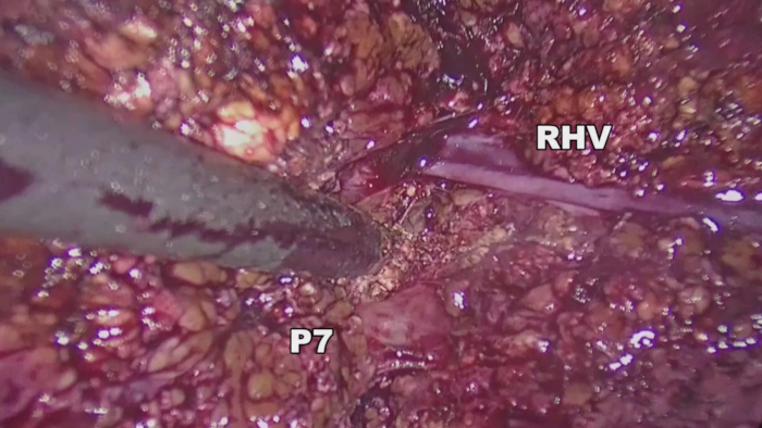
Figure 11: Exposure of the hepatic pedicle of segment VII. The hepatic pedicle of segment VII was exposed behind the main trunk of the right hepatic vein (RHV). P7: segment VII portal vein. Please click here to view a larger version of this figure.

Figure 12: Exposure of the main trunk and various branches of the right hepatic vein (RHV). IVC: inferior vena cava, V7: hepatic vein of segment VII, V8d: dorsal branch of the segment VIII hepatic vein, RHV: right hepatic vein, P8d: the dorsal branch of the segment VIII portal vein. Please click here to view a larger version of this figure.

Figure 13: Resected specimen. Please click here to view a larger version of this figure.

Figure 14: Postoperative computed tomography scans. CT scans showed changes after anatomical liver segment VII resections, with no tumor remnants. Please click here to view a larger version of this figure.
| Age (years) | 54 |
| Tumor diameter (cm) | 2.8 |
| Operative time (min) | 110 |
| Intraoperative blood loss (mL) | 100 |
| Postoperative hospitalization time (days) | 7 |
| Postoperative complications (Yes/No): | |
| bleeding | No |
| biliary leakage | No |
| subphrenic infection | No |
| dropsy of the right chest | No |
| hepatic failure | No |
Table 1: General characteristics and post-operative details of the patient.
| Parameters | Data |
| Age (years, M±SD) | 56.8±13.0 |
| Postoperative pathologic diagnosis: | |
| hepatocellular carcinoma | 25 |
| cholangiocarcinoma | 1 |
| hepatocellular adenoma | 1 |
| Tumor diameter (cm, M±SD) | 3.24±1.47 |
| Operative time (min, M±SD) | 74.5±49.7 |
| Bleeding (mL, M±SD) | 260.9±391.8 |
| Blood transfution rate (%) | 0 |
| Catheter removal (days, M±SD) | 7.7±1.9 |
| Postoperative hospitalization time (days, M±SD) | 8.6±2.1 |
| Postoperative complications: | |
| bleeding | 0 |
| biliary leakage | 0 |
| subphrenic infection | 0 |
| dropsy of the right chest | 2 |
| hepatic Failure | 0 |
Table 2: Baseline characteristics of the study population.
| Date of Operation | Operative time (min) | Bleeding (mL) | Postoperative hospitalization time (days) | Total bilirubin levels on the fifth postoperative day (umol/L) | Pathologic diagnosis |
| 2019.01.16 | 164 | 50 | 7 | 9.1 | Hepatocellular adenoma |
| 2019.04.29 | 270 | 200 | 9 | 22.2 | Hepatocellular carcinoma |
| 2019.06.13 | 240 | 50 | 8 | 16.3 | Hepatocellular carcinoma |
| 2019.07.12 | 90 | 200 | 10 | 20.1 | Hepatocellular carcinoma |
| 2019.09.17 | 172 | 350 | 7 | 24.4 | Hepatocellular carcinoma |
| 2020.08.28 | 157 | 30 | 5 | 21.3 | Hepatocellular carcinoma |
| 2021.03.15 | 82 | 5 | 7 | 11.3 | Hepatocellular carcinoma |
| 2021.04.08 | 255 | 800 | 11 | 14.2 | Hepatocellular carcinoma |
| 2021.05.15 | 180 | 30 | 6 | 15.1 | Hepatocellular carcinoma |
| 2021.07.29 | 158 | 100 | 13 | 20.4 | Hepatocellular carcinoma |
| 2021.08.31 | 165 | 100 | 8 | 26.8 | Hepatocellular carcinoma |
| 2021.12.03 | 161 | 10 | 7 | 14.5 | Hepatocellular carcinoma |
| 2021.12.17 | 180 | 50 | 7 | 15.5 | Hepatocellular carcinoma |
| 2021.12.29 | 195 | 50 | 9 | 11.1 | Hepatocellular carcinoma |
| 2022.02.15 | 104 | 50 | 6 | 7.5 | Hepatocellular carcinoma |
| 2022.02.25 | 200 | 50 | 10 | 23.8 | Hepatocellular carcinoma |
| 2022.02.25 | 205 | 300 | 11 | 9.9 | Hepatocellular carcinoma |
| 2022.03.03 | 180 | 1600 | 8 | 32.3 | Cholangiocarcinoma |
| 2022.05.05 | 193 | 20 | 13 | 13.4 | Hepatocellular carcinoma |
| 2022.06.27 | 127 | 100 | 12 | 13.9 | Hepatocellular carcinoma |
| 2022.08.29 | 215 | 100 | 9 | 17.7 | Hepatocellular carcinoma |
| 2022.08.31 | 168 | 1000 | 8 | 16.2 | Hepatocellular carcinoma |
| 2022.09.01 | 245 | 100 | 9 | 13.6 | Hepatocellular carcinoma |
| 2022.09.30 | 110 | 100 | 10 | 37.7 | Hepatocellular carcinoma |
| 2022.10.13 | 151 | 100 | 8 | 12.4 | Hepatocellular carcinoma |
| 2022.11.29 | 230 | 500 | 8 | 13.4 | Hepatocellular carcinoma |
| 2023.04.13 | 115 | 1000 | 7 | 13.4 | Hepatocellular carcinoma |
Table 3: Detailed data for all patients.
| Innovative surgical approach | Traditional surgical approach | |
| Number of cases (n) | 27 | 24 |
| The average age (years) | 56.8 | 52.2 |
| The average tumor diameter (cm) | 3.24 | 3.4 |
| The average surgical time (min) | 74.5 | 216.5 |
| The average blood loss (mL) | 260.9 | 320 |
| The main postoperative complication (%) | 7.40% | 16.70% |
Table 4: Comparison of data between traditional laparoscopic VII anatomical liver segment resection and the priority hepatic pedicle approach.
Discussion
Determining the surgical section is crucial in anatomical liver resection. Traditional anatomical liver resection techniques rely mainly on blocking the target liver pedicle to display the ischemic line on the liver surface. However, owing to the irregular boundaries between liver segments and variations in intrahepatic blood vessels, the level and extent of liver resection during surgery are often not accurate16. In recent years, many scholars have applied indocyanine green fluorescence technology to anatomical liver resection surgeries15. However, for cases where the dorsal branch of the hepatic pedicle in segment VIII of the liver crosses the area where the right hepatic vein innervates segment VII, laparoscopic ultrasound-guided portal vein puncture is more difficult, and indocyanine green easily diffuses through the intrahepatic communicating branches, affecting the assessment17. Investigators are committed to investigating laparoscopic liver segmentectomy using liver parenchymal transection as a priority technique18. Hepatic vein dissection techniques and strategies have been refined based on the rich experience in early laparoscopic anatomical liver resection surgeries, as demonstrated by the author's examination of the preferred path through the hepatic parenchyma19,20.
Anatomical liver segment VII resections with priority on the liver parenchyma require the use of natural anatomical landmarks of the liver and deep hepatic veins in the liver parenchyma to determine the liver dissection plane, as there is no pre-blocking of the ischemic line of the target liver pedicle. In this procedure, during surgery, the ligaments around the liver were sequentially severed, and the second hepatic hilum was dissected to expose the upper and lower hepatic vena cava and the root of the right hepatic vein. When separating the right liver and right adrenal gland adhesions, if the adrenal gland adhesions were tight, part of the liver capsule was removed with an ultrasound knife because hemostasis is relatively difficult if the adrenal gland is damaged. Liver wound bleeding was stopped by bipolar electrocoagulation, whereas adrenal bleeding was stopped by continuous suturing of blood vessels. Furthermore, we dissected and suspended the root of the right hepatic vein as a guide and located it in the naked area of the liver or marked the course of the right hepatic vein with ultrasound during surgery, cut open the liver parenchyma, searched for the main trunk of the right hepatic vein, prioritized the dissection of the right hepatic vein branch, and followed the direction of the right hepatic vein branch to fully expose the right hepatic vein in the severed liver area. To meet the requirements for anatomical resection, the segment VII hepatic pedicle beneath the main right hepatic vein was exposed and ligated. At the same time, the variant VIII dorsal segment in front of the main right hepatic vein was exposed and ligated, crossing the innervated branch of the right hepatic vein.
Laparoscopic liver segment VII resection with liver parenchyma priority adopts an "easy priority" method. This procedure prioritizes the exposure of the right hepatic vein and its branches, expands the relative gap to expose the cross-section, facilitates the exposure of the segment VII liver pedicle, reduces the risk of injury and bleeding caused by the anatomical separation of the Glisson pedicle under insufficient exposure, and solves the problem of difficulty in determining the level of liver transection owing to liver pedicle variation, fully covering the tumor area, and providing sufficient margins. In contrast, laparoscopic liver segment VII resection with priority to the liver parenchyma follows the right hepatic vein as anatomical guidance, preserving normal and important blood vessels as much as possible and completely removing the tumor, ensuring complete removal of the lesion while avoiding damage to important blood vessels and maximizing the preservation of a functional liver21.
However, this study has some limitations. First, the technique requires a high level of surgical skill from the operator to dissect the right hepatic vein. This requires the operator to have accumulated sufficient experience in laparoscopic hepatectomy. Moreover, intraoperative dissection of the hepatic vein must be performed at a low central venous pressure to minimize surface sieve bleeding caused by venous filling. This requires close collaboration with trained anesthesia teams.
Hepatic segment VII is a challenging area to resect via laparoscopic anatomical liver segmentectomy, with a high risk of substantial hemorrhage, and laparoscopic anatomical liver segmentectomy has a high conversion rate to open surgery. In our study, a new liver parenchyma transection priority method was developed. The liver parenchyma was dissected before it was cut off and disconnected in the event of interrupted whole liver blood flow. Without increasing the scope of the surgery or optimizing liver function, this procedure streamlines the processes involved in preventing blood flow to the liver. Thus, this study provides new evidence for the development and application of anatomical hepatectomy.
Disclosures
There are no financial conflicts of interest to disclose.
Materials
| Name | Company | Catalog Number | Comments |
|---|---|---|---|
| laparoscopic imaging systems | KARL STORZ ENDOSCOPY(SHANGHAI)LTD. | TC201,26003BA,N90X0666 | |
| Laparoscopic curved cutting stapler cartridge | Reach Surgical (Beijing), Inc. | ENDO SRC 4525R | |
| Laparoscopic curved cutting stapler | Reach Surgical (Beijing), Inc. | ENDO SRC | |
| Ultrasonic surgical equipment for soft tissue excision and hemostasia | Reach Surgical (Beijing), Inc. | CH14PD |
References
- Vogel, A., Meyer, T., Sapisochin, G., Salem, R., Saborowski, A. Hepatocellular carcinoma. Lancet. 400 (10360), 1345-1362 (2022).
- Han, B., et al. Cancer incidence and mortality in China, 2022. J Natl Cancer Cent. 4 (1), 47-53 (2022).
- Yoshida, H., et al. Current status of laparoscopic hepatectomy. J Nippon Med Sch. 86 (4), 201-206 (2019).
- Biertho, L., Waage, A., Gagner, M. Hépatectomies sous laparoscopie [Laparoscopic hepatectomy. Ann Chir. 127 (3), 164-170 (2002).
- Cai, X. Laparoscopic liver resection: The current status and the future. Hepatobiliary Surg Nutr. 7 (2), 98-104 (2018).
- Buell, J. F., et al. The international position on laparoscopic liver surgery: the Louisville statement. Ann Surg. 250 (5), 825-830 (2009).
- Wakabayashi, G., et al. Recommendations for laparoscopic liver resection: a report from the second international consensus conference held in MoriokaQ. Ann Surg. 261 (4), 619-629 (2015).
- Anselmo, A., Sensi, B., Bacchiocchi, G., Siragusa, L., Tisone, G. All the routes for laparoscopic liver segment viii resection: a comprehensive review of surgical techniques. Front Oncol. 1 (12), 864867(2022).
- Ishizawa, T., Gumbs, A. A., Kokudo, N., Gayet, B. Laparoscopic segmentectomy of the liver: from segment I to VIII. Ann Surg. 6 (6), 959-964 (2012).
- Morise, Z. Laparoscopic liver resection for posterosuperior tumors using caudal approach and postural changes: A new technical approach. World J Gastroenterol. 22 (47), 10267-10274 (2016).
- Cao, J., et al. Totally laparoscopic anatomic s7 segmentectomy using in situ split along the right intersectoral and intersegmental planes. Surg Endosc. 35 (1), 174-181 (2021).
- Shindoh, J., Mise, Y., Satou, S., Sugawara, Y., Kokudo, N. The intersegmental plane of the liver is not always flat--tricks for anatomical liver resection. Ann Surg. 251, 917-922 (2010).
- Zheng, Z., et al. Laparoscopic central hepatectomy using a parenchymal-first approach: How we do it. Surg Endosc. 36 (11), 8630-8638 (2022).
- Lin, C. X., et al. Optimal central venous pressure during partial hepatectomy for hepatocellular carcinoma. Hepatobiliary Pancreat Dis Int. 12, 520-524 (2013).
- Sun, H., Yang, H., Xu, H. A commentary on "Laparoscopic anatomical versus non-anatomical hepatectomy in the treatment of hepatocellular carcinoma: a randomized controlled trial.". Int J Surg. 104, 106783(2022).
- Otsuka, Y., et al. Gas embolism in laparoscopic hepatectomy: what is the optimal pneumoperitoneal pressure for laparoscopic major hepatectomy. J Hepatobiliary Pancreat Sci. 20, 137-140 (2013).
- Marino, M. V., Builes Ramirez, S., Gomez Ruiz, M. The application of Indocyanine Green (ICG) staining technique during robotic-assisted right hepatectomy: with video. J Gastrointest Surg. 23 (11), 2312-2313 (2019).
- Liu, Q., et al. Liver parenchyma transection-first approach for laparoscopic left hemihepatectomy: A propensity score matching analysis. World J Surg. 45 (2), 615-623 (2021).
- Jiang, Z., et al. A novel method of ultrasound-guided positive staining using indocyanine green fluorescence in laparoscopic anatomical liver resection of segments VII and VIII. Front Oncol. 13, 1138068(2023).
- Kawabata, Y., Hayashi, H., Yano, S., Tajima, Y. Liver parenchyma transection-first approach in hemihepatectomy with en bloc caudate lobectomy for hilar cholangiocarcinoma: A safe technique to secure favorable surgical outcomes. J Surg Oncol. 115 (8), 963-970 (2017).
- You, N., Wu, K., Li, J., Zheng, L. Laparoscopic liver resection of segment 8 via a hepatic parenchymal transection-first approach guided by the middle hepatic vein. BMC Gastroenterol. 22 (1), 224(2022).
Reprints and Permissions
