In this paper, we propose a protocol for hybrid spheroid manufacturing using optical tweezers. This method allows precise control of the early stages of the spheroid formation process, which is unattainable by other methods.
A subscription to JoVE is required to view this content. Sign in or start your free trial.
Method Article
Using Optical Tweezers for the Generation of Hybrid Spheroids
In This Article
Summary
Abstract
Two-dimensional (2D) cell culture is still among the most commonly used models in preclinical cancer research. However, the three-dimensional (3D) in vitro models appear to assess the in vivo environment more accurately, including a more relevant architectural morphology, microenvironment, and responses to drugs.
Optical trapping uses one or more focused laser beams to non-invasively manipulate the position, motion, interaction, and dynamics of structures on nano- and microscale. Optical tweezers (OT) have found numerous applications in cell biology; however, their potential in cancer research is still undervalued. In this paper, we recommend a noninvasive protocol for lymphoma-stromal cell spheroid formation with the use of optical tweezers. This method not only constructs the hybrid spheroids de novo but also enables controlling the number of attached cells and the time of adhesion formation, providing an important source of information for tissue engineering. Furthermore, OT enables the study of the early stages of hybrid spheroid formation, which cannot be achieved with standard bulk techniques. Importantly, the described model can be used to study the minimal changes in adhesion induced by anti-cancer drugs. This protocol can be easily applied to other cell types.
Introduction
Three-dimensional (3D) cellular models are accelerating cancer research because they better resemble tumor tissue in several functional and structural properties. These include, among others, extracellular matrix production, interaction between cells, maintaining nutrient and oxygen gradients, and similar responses to the treatments1,2,3,4. Recently, to better reflect in vivo conditions, advanced heterogeneous 3D models involving various cell types have been developed5,6,7.
B-cell Non-Hodgkin Lymphoma (B-NHL) is a heterogeneous group of malignancies originating from B lymphocytes, integral components of the immune system8. Characterized by a diverse array of clinical presentations and histological features, B-NHL encompasses a spectrum of disorders, ranging from indolent forms with a protracted clinical course to aggressive variants that progress swiftly9,10. The pathogenesis of B-NHL and other hematological malignancies is often linked to the impact of the tumor microenvironment11,12. For instance, lymphoma cells often spread to the bone marrow, potentially resulting in life-threatening complications13. Mesenchymal stromal cells (MSCs), which are the key components of the bone marrow niche, have been recognized to impact tumor growth and progression14, largely through direct cell-cell interactions15,16,17; thus, there is an urgent need to develop models that allow studying such interplays. We previously established that MSCs, when co-cultured on agarose hydrogels with lymphoma cells, self-aggregate into multicellular spheroids, with stromal cells forming the core and being uniformly surrounded by lymphoma cells18. In this paper, we present a protocol for the fabrication of a hybrid lymphoma-stromal cells 3D model using optical tweezers.
Over 35 years ago, Ashkin first reported the phenomenon of non-invasive optical trapping of viruses and bacteria by focused laser beam19. Nowadays, optical tweezers (OT) have a dynamically increasing impact in the biomedical field, including cell biology research20,21,22,23. OT allowed studying not only the physico-mechanical properties of individual cell24,25,26but also the direct cell-cell interactions at a single cell level, and further 2D and 3D cellular arrangement and structuring27,28,29. The optical trapping technique is based on the optical forces of the order of a few piconewtons exerted as electromagnetic radiation (photons), which changes its momentum when it interacts with matter30. In consequence, by using one or more laser beams, operators may selectively manipulate positions, motion, and dynamics of objects in micro- and nanoscale20. In practice, the optical trapping technique allows for the selective and non-destructive trapping of living cells, placing them in contact with the surface of another cell or molecule and triggering the initiation of the adhesion process31. Using the above functionalities, we first attempted to optically place a single lymphoma cell onto a monolayer of stromal cells to investigate its adhesive properties. Next, we proceeded to assess the ability of optical tweezers to physically recreate a multicellular spheroid in real-time mode33. To achieve this, individual lymphoma cells were trapped one by one and attached to a previously prepared 3D stromal spheroid. Another applicability of optical trapping was to measure the minimum cell-cell adhesion time and assess the minute changes in adhesion resulting from the anti-cancer treatment34,35. Although 3D models are widely used in cancer research, studying the early stages of spheroid formation remains challenging. This is mainly due to the limited availability of methods that allow the micro-world to be touched with high precision and minimally invasive impact at the same time. Conversely, the full potential of OT is yet to be achieved within the scope of cellular arrangement and structure in 3D. Thus, in this paper, a combined workflow to generate 3D lymphoma construct using standard 3D techniques and manipulations in optical tweezers is described step by step. Importantly, the protocol may be easily adapted for use with other cell types of importance in hematopathological malignancies.
Access restricted. Please log in or start a trial to view this content.
Protocol
1. Fabrication of agarose gel-based micromolds
NOTE: Proper aseptic techniques are essential when handling all cell cultures. All the reagents used in this protocol are listed in the Table of Materials. Prepare three gels with MSCs for a single experiment as shown in Figure 1A.
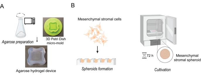
Figure 1: Preparation of mesenchymal stromal cell (MSC) spheroids. (A) Illustration depicting the step for preparing the agarose micromolds. (B) Seeding of mesenchymal stromal cells (MSCs), and cultivation of mesenchymal spheroids. Please click here to view a larger version of this figure.
- Begin by preparing the agarose micromolds. Rinse the micromold 3D Petri Dish (see Table of Materials) with deionized water (3x) and place under the UV lamp for 30 min to perform sterilization.
- First, prepare 2% agarose solution. Measure 1 g of high-quality pure sterile agarose powder (see Table of Materials) and place it in a sterile 50 mL conical tube. Add 50 mL of sterile 0.9% sodium chloride solution (see Table of Materials) to the agarose and mix well. Screw on the cover of the conical tube until it is secure but still loose.
- Use the microwave oven to boil and completely dissolve the agarose powder. Stop microwaving every 10 s and carefully swirl the conical tube to ensure the agarose dissolves. Avoid creating bubbles while mixing or pipetting agarose. Cool agarose to approximately 70 °C.
NOTE: Ensure that there are no remnants of translucent undissolved powder.
CAUTION: Molten agarose is extremely hot and can cause severe skin burns. - Work in the laminar flow cabinet using aseptic techniques. Prepare hydrogels by filling previously sterilized micro-molds with 500 µL of agarose. Aspirate out any bubbles that may be trapped in the small features of the micro-mold using a pipette.
- After 15 min of incubation at room temperature, when the agarose has solidified, carefully remove the gels from micromolds to a sterile 6-well plate. Do not damage the gel structure. Before proceeding to the next step, check under a microscope whether the micromolds have been prepared correctly and ensure that the edges are intact. Check for any gaps through which the medium can leak.
- Add enough 1x Phosphate Buffer Saline (PBS) to completely cover the micromolds before placing the plate under a UV lamp for 30 min for sterilization.
NOTE: The agarose micromolds may be stored before use for up to 2 weeks in a conical tube covered with 1x PBS under refrigerator conditions (4 °C). - Before using the micromolds to produce spheroids, equilibrate the gel by completely covering it in RPMI medium (4.0 mL/well for a 6-well plate) and incubate for a minimum of 15 min.
2. Preparation of mesenchymal stromal cells (MSCs) spheroids
- Culture the HS-5 cell line (see Table of Materials), a human mesenchymal stromal cell line, in RPMI 1640 culture medium supplemented with 10% heat-inactivated fetal bovine serum (FBS) and 1% penicillin-streptomycin (see Table of Materials) in standard condition, ensuring a changing of medium every 48 h.
- When the HS-5 cells are approximately 90% confluent, perform cell passage as follows:
- Remove the culture media using a serological pipette.
- Wash cells by adding 1x PBS and swirling and aspirating to remove any remaining media.
- Detach cells from the bottom of the culture flask by adding 2 mL of trypsin-EDTA solution (see Table of Materials) to the cell culture flask and incubating at 37 °C to facilitate cell detachment.
- Stop trypsinization by adding 2 mL of complete culture medium.
- Using a 5 mL pipette, gently suspend the cells and transfer them into a 15 mL conical tube.
- Centrifuge the cell suspension at 300 x g for 7 min. Remove the supernatant and resuspend cells in 1 mL of fresh culture medium.
- Take 10 µL of the cell pellet and transfer it to a 0.5 mL tube. Then, add 10 µL of 0.4% Trypan blue solution (see Table of Materials) and count the cells in an automatic cell counter (see Table of Materials).
- Based on the cell density obtained above, dilute cells to 2 x 10⁵ cells/mL of medium.
- Remove the cell culture medium from the agarose micromold.
- Carefully seed 190 µL of cell suspension mentioned above (step 2.2.8) into the cell seeding chamber of the agarose micromold. A single well will contain approximately 150 cells (38.000 cells/gel).
- Incubate cells in agarose micromold in an incubator (5% CO2, 37 °C, humid atmosphere) for 30 min until the cells settle at the bottom of the gel. After 30 min, remove the plate containing micromold from the incubator, and slowly add an additional medium to the outside of the gels, covering them entirely (e.g., 4.0 mL/well for the 6-well plate).
NOTE: Do not add the medium directly onto the gel agarose micromold, but into the culture dish, avoiding damaging the freshly formed spheroids. - Place the gel in an incubator at 37 °C in a humid atmosphere of 5% CO2 for 72 h until uniform spheroids are formed. (Figure 1B).
NOTE: It is recommended to observe the spheroid formation for the following 3 days under an inverted microscope. In general, mesenchymal stromal cells begin to form spheroids within 24 h, but this process may be faster or slower with different cell passages and densities. After 72 h of incubation on agarose micromold, mesenchymal stromal cells form relatively uniform spheroids; however, it is worth noting that they may vary in size and shape despite being incubated in identical conditions. The medium needs to be replaced when it changes from a purple-red color to yellow.
3. Culturing of lymphoma cells and preparation for optical trapping
NOTE: Hematological cell lines, including lymphoma cells, grow free-floating in the culture media in flasks designed for suspension culture without any attachment to the culture plate or vessel. To maintain optimal cell properties, it is recommended to start preparing the sample no earlier than 30 min before the procedure of assembling the hybrid spheroids using the optical tweezers. Importantly, the protocol outlined in this section is for the Ri-1 human lymphoma cell line, but a similar protocol applies to other hematological cancer cell lines.
- Culture the Ri-1 lymphoma cell line (see Table of Materials) in RPMI 1640 medium supplemented with 10% FBS and 1% penicillin/streptomycin.
- Keep the lymphoma cells in the incubator under standard culture conditions, including 37 °C and humidified air with 5% CO2. Maintain cultures between 3-5 x 10⁵ cells/mL in the logarithmic phase by passaging every 2-3 days.
- Collect the cells and transfer the suspension to a 15 mL polystyrene tube. Centrifuge the cells at 300 x g for 7 min at room temperature to obtain a cell pellet. Remove the supernatant and add 10 mL of fresh medium, gently pipette until a homogenous suspension is obtained.
- Aspirate 10 µL of cell suspension into a new centrifuge tube and add 10 µL of Trypan blue. Pipette the suspension well before transferring 10 µL into the counting chamber to determine the cell density and viability (typically higher than 95%).
- Dilute the cell suspension to reach a final density of 1 x 10⁴ cells/mL. Keep lymphoma cells at 37 °C until optical manipulations.
4. Setting up the optical tweezers system
NOTE: The protocol below was developed specifically for the custom-built optical tweezers36 (Figure 2A). However, this assay can be used with other commercially available trapping systems. Regardless of what type of equipment is available, the basic steps for system preparation (e.g., starting the laser and computer software, mounting the sample, and testing the possibility of moving the cell in an optical trap), remain the same. All experiments were carried out at room temperature, i.e., 25 °C.
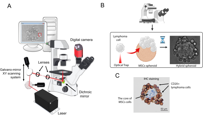
Figure 2: A schematic illustrating the working mechanism of hybrid spheroid generation by optical tweezers. (A) Custom-designed optical tweezers setup. (B) The process of attaching individual lymphoma cells to the surface of MSCs spheroid. A single lymphoma cell was optically trapped and attached to the surface of a stromal spheroid until a stable connection was formed. This process continued until the entire surface of the stromal spheroid was covered by lymphoma cells. (C) A representative image of immunohistochemical staining of the hybrid spheroid section was obtained using a monoclonal mouse anti-CD20 antibody against CD20. The immunostaining was performed using a monoclonal mouse anti-human antibody against CD20 on an autostainer (see Table of Materials), following the manufacturer's instructions. Please click here to view a larger version of this figure.
- Turn on the microscope light source.
- Wear a laser-safe protective glass.
- Turn on the laser and set the power to obtain a trap stiffness of approximately 50 pN/µm in the optical tweezers system. Before proceeding, wait for 10 min, allowing the laser beam to stabilize.
NOTE: In the custom-built system, given trap stiffness is achieved for the laser power of 100 mW at the entrance of the microscope objective measured for 3 µm polystyrene bead. Its minimal invasiveness on lymphoma cells was previously confirmed32,33, while maintaining the ability to manipulate living cells. - In the meantime, start up the computer software. In the software window, an image of the microscope sample will appear. Use the markers visible on the screen to move the optical trap in the desired direction.
- Once the system is ready, add 30 µL of deionized water to the center of the bottom objective.
NOTE: The custom-built system uses a water immersion objective; however, the immersion media may differ depending on the user's setup. - Place a 35 mm cell culture dish (see Table of Materials) with a glass bottom on the microscope stage. Stabilize the dish with the holders.
- Raise the objective using the micrometric microscope screw until the bead of water touches the glass bottom of the dish.
- Using the micrometer screw, gently adjust the microscopic stage until one sees the laser beam sharply focused into a single, bright point on the screen. An image of the microscope sample will be present in the software window. Use the markers visible on the screen to move the optical trap in any desired direction.
- Add 100 µL of previously prepared lymphoma cell suspension (see point 3.5) to the center of the glass-bottom dish. Let them sink to the bottom of the dish for approximately 5 min.
- Using the movements of the microscope stage, find a floating lymphoma cell. Allow the optical trap to hit the sample by clicking on the trap cursor on the screen. Try to move the cell to the desired location. A cell, to be useful for manipulation, needs to be able to move easily in the given direction.
NOTE: As per our observation, between 5 to 10% of lymphoma cells added to the glass dish will permanently attach to the bottom after the incubation period. This percentage will increase with the duration of manipulation and may reach 30% after 60 min. However, despite these losses, the number of cells initially added to the dish will allow for both effective system calibration and coverage of the spheroid with lymphoma cells.
5. Generation of hybrid spheroids using optical tweezers
- Carefully remove a single stromal spheroid from a well of the agarose mold with a pipette tip (100-1000 µL) and place it on a 35 mm microscopy glass bottom dish.
- Add 2 mL of RPMI culture medium and incubate for 10 min at 37 °C under standard conditions. During this time, the spheroid will gently stick to the glass, which prevents movement during manipulation.
- Place the dish containing the spheroid on the microscope stage and stabilize the dish on the microscopic stage with the holders.
- Carefully add 100 µL of lymphoma cell solution at a final density of 1 x 10⁴ cells/mL (step 3.5). Let them sink to the bottom of the dish for approximately 5 min.
- Using the movements of microscope stage, find a floating lymphoma cell. Allow the optical trap to hit the selected cell by clicking on the trap cursor on the screen. Once the cell is trapped and movable, move the microscope stage to deliver the cell into contact with the stromal spheroid surface.
- Start by attaching the lymphoma cell to the surface of the stromal spheroid for 10 s. Next, check if the cell is permanently attached with three repeated attempts of detaching the lymphoma cell using the optical tweezers.
- If the cellular contact is broken, attach the lymphoma cell to the spheroid surface once more using the optical trap; however, this time for a longer period of time, approximately 20 s.
NOTE: It is essential to determine specific time intervals for single-cell trapping for each cell line separately. In preliminary manipulations, it was established that lymphoma cells of Ri-1 cell line exhibit a strong adhesive property to stromal spheroids, predominantly attaching in 10-20 s to its surface. Thus, the specific contact time intervals for single Ri-1 cell trapping were 10, 20, and 30 s. Due to the time-consuming nature of the procedure, it is recommended to use cell lines with a minimum adhesion time of no longer than 30 s for spheroid formation with optical tweezers. - Repeat the procedure with subsequent lymphoma cells until the entire surface of the stromal spheroid is covered by lymphoma cells.
- To uniformly cover the available surface of the stromal spheroid with lymphoma cells, it is necessary to change the position of the optical trap in the volume of the microscopic sample. By changing the imaging plane using the micrometric microscope screw, one can change the position of the optical trap, which enables a more profound movement of the trapped objects in the sample.
- To better represent the spontaneously formed spheroid, cover the core of stromal cells with 2-3 layers of lymphoma cells.
- Transfer the glass-bottom dish containing the spheroid to an incubator for 1 h of incubation under standard conditions.
NOTE: Hybrid spheroid may be cultured long-term in 20 µL of RPMI medium in a hanging drop.
6. Determination of spheroid viability
- Assess the viability of cell spheroid using a Live/dead staining kit (see Table of Materials). Gently remove the culture medium and directly wash the spheroid with 100 µL of 1x PBS.
- Incubate the spheroid in 100 µL of 1x PBS solution containing 1 µM of calcein-AM, and 2 µM of ethidium homodimer-1 for 20 min at 37 °C.
- Observe the spheroids using a fluorescence microscope (see Table of Materials) and capture the images (see Table of Materials).
NOTE: There is no need to wash the spheroid after the staining. Background fluorescence levels are inherently low with this assay because the dyes are virtually non-fluorescent before interacting with cells. - Using the ImageJ program (see Table of Materials), determine spheroid viability by calculating the ratio of the area stained green (live cells) to the total area stained in red (dead cells) and green.
Access restricted. Please log in or start a trial to view this content.
Results
Our previous results showed that mesenchymal stromal cells cultured with lymphoma cells make up the homogenous stromal core to which cancer cells attach uniformly18. In this study, we have successfully engineered the human hybrid spheroid consisting of mesenchymal stromal cells and lymphoma cells using custom-built optical tweezers. Figure 2B shows a representative spheroid in a brightfield microscope, an essential part of the optical tweezers system. In turn,
Access restricted. Please log in or start a trial to view this content.
Discussion
In this study, the hybrid spheroids of mesenchymal stromal core and lymphoma cells were engineered in real-time with the use of optical tweezers. New laser-based technologies, including trapping systems, are now commercially available and are becoming increasingly popular. However, to date, the majority of optical tweezers continue to be custom-built instruments; thus, calibration, operation procedures, and experimental geometries can vary between laboratories38,39
Access restricted. Please log in or start a trial to view this content.
Disclosures
The authors declare no conflicts of interest.
Acknowledgements
Authors sincere gratitude Nicholas Czarnik, whose careful editing significantly improved the clarity of the study.
Access restricted. Please log in or start a trial to view this content.
Materials
| Name | Company | Catalog Number | Comments |
|---|---|---|---|
| µ-Dish 35 mm, high glass bottom | IBIDI | 81158 | |
| 12-256 mico-mold 3D PetriDish | Microtissues Inc., RI, USA | 12-256TR | |
| 3 µm polystyrene microspheres | Polysciences | 17134-15 | |
| anti CD20 antibody (clone L26) | Dako | IS604 | |
| Automated Cell Counter, EVE | NanoEntek | EVE/ EVE-MC | |
| Autostainer Link 48 | Dako | D09634 | |
| Camera | Olympus | U-TV0.63XC | |
| CelCulture CO2 Incubator | Esco Technologies, Inc. | CCL | |
| Cell Counting Slides, EVE | NanoEntek | NE-EVS | |
| Cell line HS-5 | ATCC | CRL-3611 | |
| Cell line Ri-1 | DSMZ | ACC 585 | |
| Fetal Bovine Serum | Gibco | A5256701 | |
| Fluorescence microscope | Olympus | BX43 | |
| ImageJ program | National Institutes of Health, MD, USA | ||
| Inverted microscope | Olympus | IX73 | |
| Live/Dead reagent kit | Thermo Fisher Scientific | L3224 | |
| Penicillin-Streptomycin (10,000 U/mL) | Gibco | 15140122 | |
| Phosphate Buffered Saline, pH 7.4 | Gibco | 10010023 | |
| RPMI 1640 Medium | Gibco | 11875093 | |
| Sodium chloride solution | Sigma-Aldrich | 7647-14-5 | |
| Sodium Chloride solution 0.9 %, sterile | ITW ReITW Reagents, S.R.L.agents, S.R.L. | A1671 | |
| Trypan Blue Solution, 0.4% | Gibco | 15250061 | |
| TrypLE Express Enzyme (1X), no phenol red | Gibco | 12604013 | |
| Trypsin-EDTA (0.05%), phenol red | Gibco | 25300096 | |
| UltraPure Agarose | Invitrogen | 16500500 | |
| Optical tweezers setup | |||
| Dichroic mirrors | Thorlabs | DMSP805R | |
| Digital camera | Mikrotron GmbH | MC1362 | |
| Galvano-mirror XY scanning system | Thorlabs | GVS002 | |
| Inverted microscope | Olympus | IX71 | |
| Lens 1 | Thorlabs | AC254-050-B-ML | |
| Lens 2 | Thorlabs | AC508-200-B-ML | |
| Nd:YAG 1064 nm laser | Laser Quantum | Ventus |
References
- Brancato, V., Oliveira, J. M., Correlo, V. M., Reis, R. L., Kundu, S. C. Could 3D models of cancer enhance drug screening. Biomaterials. 232, 119744(2020).
- Fontoura, J. C., et al. Comparison of 2D and 3D cell culture models for cell growth, gene expression and drug resistance. Mater Sci Eng. C. 107, 110264(2020).
- Kapałczyńska, M., et al. 2D and 3D cell cultures - a comparison of different types of cancer cell cultures. Arch Med Sci. 14 (4), 910-919 (2018).
- Duval, K., et al. Modeling physiological events in 2D vs. 3D cell culture. Physiology (Bethesda). 32 (4), 266-277 (2017).
- Barbosa, M. A. G., Xavier, C. P. R., Pereira, R. F., Petrikaitė, V., Vasconcelos, M. H. 3D cell culture models as recapitulators of the tumor microenvironment for the screening of anti-cancer drugs. Cancers (Basel). 14 (1), 190(2021).
- Jubelin, C., et al. Three-dimensional in vitro culture models in oncology research. Cell Biosci. 12, 155(2022).
- Pape, J., Emberton, M., Cheema, U. 3D cancer models: the need for a complex stroma, compartmentalization and stiffness. Front Bioeng Biotechnol. 9, 660502(2021).
- Singh, R., et al. Non-Hodgkin's lymphoma: A review. J Family Med Prim Care. 9 (4), 1834-1840 (2020).
- Campo, E., et al. The International consensus classification of mature lymphoid neoplasms: a report from the clinical advisory committee. Blood. 140 (11), Erratum in: Blood. 141 (4), 437 (2023). 1229-1253 (2022).
- Swerdlow, S. H., et al. The 2016 revision of the World Health Organization classification of lymphoid neoplasms. Blood. 127 (20), 2375-2390 (2016).
- Ng, W. L., Ansell, S. M., Mondello, P. Insights into the tumor microenvironment of B cell lymphoma. J Exp Clin Cancer Res. 41, 362(2022).
- Mancini, S. J. C., et al. Deciphering tumor niches: lessons from solid and hematological malignancies. Front Immunol. 12, 766275(2021).
- Zhang, Q. Y., Foucar, K. marrow involvement by Hodgkin and non-Hodgkin lymphomas. Hematol. Oncol Clin North Am. 23 (4), 873-902 (2009).
- Gao, Q., et al. marrow mesenchymal stromal cells: Identification, classification, and differentiation. Front Cell Dev Biol. 9, 787118(2022).
- Lwin, T., et al. Lymphoma cell adhesion-induced expression of B cell-activating factor of the TNF family in bone marrow stromal cells protects non-Hodgkin's B lymphoma cells from apoptosis. Leukemia. 23 (1), 170-177 (2009).
- Kumar, A., Bhattacharyya, J., Jaganathan, B. G. Adhesion to stromal cells mediates imatinib resistance in chronic myeloid leukemia through ERK and BMP signaling pathways. Sci Rep. 7 (1), 9535(2017).
- Cuiffo, B. G., Karnoub, A. E. Mesenchymal stem cells in tumor development: emerging roles and concepts. Cell Adh Migr. 6 (3), 220-230 (2012).
- Duś-Szachniewicz, K., Gdesz-Birula, K., Rymkiewicz, G. Development and characterization of 3D hybrid spheroids for the investigation of the crosstalk between B-Cell non-Hodgkin lymphomas and mesenchymal stromal cells. OncoTargets Ther. 15, 683-697 (2022).
- Ashkin, A., Dziedzic, J. M. Optical trapping and manipulation of viruses and bacteria. Science. 235, 1517-1520 (1987).
- Spyratou, E. Advanced biophotonics techniques: The role of optical tweezers for cells and molecules manipulation associated with cancer. Front Phys. 10, 247-285 (2022).
- Choudhary, D., Mossa, A., Jadhav, M., Cecconi, C. Bio-molecular applications of recent developments in optical tweezers. Biomolecules. 9 (1), 23(2019).
- Nussenzveig, H. M. Cell membrane biophysics with optical tweezers. Eur Biophys J. 47 (5), 499-514 (2018).
- Zhang, X., Ma, L., Zhang, Y. High-resolution optical tweezers for single-molecule manipulation. Yale J Biol Med. 86 (3), 367-383 (2013).
- Li, X., et al. Optical tweezers study of membrane fluidity in small cell lung cancer cells. Opt Express. 29 (8), 11976-11986 (2021).
- Wullkopf, L., et al. Cancer cell's ability to mechanically adjust to extracellular matrix stiffness correlates with their invasive potential. Mol Biol Cell. 29, 2378-2385 (2018).
- Yousafzai, M. S., et al. Effect of neighboring cells on cell stiffness measured by optical tweezers indentation. J Biomed Opt. 21 (5), 57004(2016).
- Gupta, S. K., et al. Quantification of cell-matrix interaction in 3D using optical tweezers. Multi-Scale Extracellular Matrix Mechanics and Mechanobiology. Studies in Mechanobiology, Tissue Engineering and Biomaterials. Zhang, Y. , Springer. Berlin/Heidelberg, Germany. 283-310 (2020).
- Ma, Z., et al. Laser patterning for the study of MSC cardiogenic differentiation at the single-cell level. Light Sci Appl. 2, 68(2013).
- Kirkham, G. R., et al. Precision assembly of complex cellular microenvironments using holographic optical tweezers. Sci Rep. 5, 8577(2015).
- Pesce, G., et al. Optical tweezers: theory and practice. Eur Phys J Plus. 135, 949(2020).
- Avsievicha, T., Zhua, R., Popovb, A., Bykova, A., Meglinski, I. The advancement of blood cell research by optical tweezers. Rev Phys. 5, 100043(2020).
- Duś-Szachniewicz, K., et al. Physiological hypoxia (physioxia) impairs the early adhesion of single lymphoma cell to marrow stromal cell and extracellular matrix. Optical tweezers study. Int J Mol Sci. 19, 1880(2018).
- Duś-Szachniewicz, K., Gdesz-Birula, K., Nowosielska, E., Ziółkowski, P., Drobczyński, S. Formation of lymphoma hybrid spheroids and drug testing in real time with the use of fluorescence optical tweezers. Cells. 11 (13), 2113(2022).
- Hu, S., Gou, X., Han, H., Leung, A. Y., Sun, D. Manipulating cell adhesions with optical tweezers for study of cell-to-cell interactions. J Biomed Nanotechnol. 9 (2), 281-285 (2013).
- Johansen, P., Fenaroli, F., Evensen, L., Griffiths, G., Koster, G. Optical micromanipulation of nanoparticles and cells inside living zebrafish. Nat Commun. 7, 10974(2016).
- Drobczyński, S., Duś-Szachniewicz, K. Real-time force measurement in double wavelength optical tweezers. J Opt Soc AmB. 34, 38-43 (2017).
- Pavlasova, G., Mraz, M. The regulation and function of CD20: an "enigma" of B-cell biology and targeted therapy. Haematologica. 105 (6), 1494-1506 (2020).
- Gieseler, J., et al. Optical tweezers- from calibration to applications: a tutorial. Adv Opt Mater. 13, 74-241 (2021).
- Bustamante, C. J., Chemla, Y. R., Liu, S., Wang, M. D. Optical tweezers in single-molecule biophysics. Nat Rev Methods Primers. 1, 25(2021).
- Mitrakas, A. G., et al. Applications and advances of multicellular tumor spheroids: Challenges in their development and analysis. Int J Mol Sci. 24 (8), 6949(2023).
- Malm, M., et al. Evolution from adherent to suspension: systems biology of HEK293 cell line development. Sci Rep. 10, 18996(2020).
- Ling, Y., et al. A cell-laden microfluidic hydrogel. Lab on a Chip. 7 (6), 756-762 (2007).
- Azizipour, N., Avazpour, R., Sawan, M., Ajji, A., Rosenzweig, H. Surface optimization and design adaptation toward spheroid formation on-chip. Sensors. 22 (9), 3191(2022).
- Ryu, N. E., Lee, S. H., Park, H. Spheroid culture system methods and applications for mesenchymal stem cells. Cells. 8 (12), 1620(2019).
- Han, S. J., Kwon, S., Kim, K. S. Challenges of applying multicellular tumor spheroids in preclinical phase. Cancer Cell Int. 21, 152(2021).
- Feng, Q., Li, D., Li, Q., Cao, X., Dong, H. Microgel assembly: Fabrication, characteristics, and application in tissue engineering and regenerative medicine. Bioact Mater. 9, 105-119 (2021).
- Volpe, G., et al. Roadmap for optical tweezers. J Phys Photonics. 5, 022501(2023).
- Pradhan, M., Pathak, S., Mathur, D., Ladiwala, U. Optically trapping tumor cells to assess differentiation and prognosis of cancers. Biomed Opt Express. 7, 943(2016).
- Duś-Szachniewicz, K., et al. Differentiation of single lymphoma primary cells and normal B-cells based on their adhesion to mesenchymal stromal cells in optical tweezers. Sci Rep. 9, 9885(2019).
- Gdesz-Birula, K., Drobczyński, S., Sarat, K., Duś-Szachniewicz, K. Sonidegib Inhibits the adhesion of acute myeloid leukemia to the bone marrow in hypoxia: An optical tweezer study. Biomedicines. 13, 578(2025).
- Matheson, A. B., et al. Optical tweezers with integrated multiplane microscopy (OpTIMuM): A new tool for 3D microrheology. Sci Rep. 11, 5614(2021).
Access restricted. Please log in or start a trial to view this content.
Reprints and Permissions
