The present protocol outlines the application of organotypic tissue models for investigating host-pathogen interactions in vitro. Specifically, the model system described uses a multi-layered oral epithelium exposed to microorganisms associated with biofilms (dental plaque) within the context of oral health versus disease.
Method Article
Organotypic Tissue Model Systems for Investigating Host-Pathogen Interactions In Vitro
In This Article
Summary
Abstract
Recapitulating the host-pathogen interface at the epithelial or mucosal barrier in vitro remains a challenging prospect for infection biologists. While in-house grown 2D epithelial monolayers lack true representation of the in vivo situation, commercially available tissue models are often overlooked due to their cost and practicality. However, with careful planning, such models provide reproducible platforms for a vast array of different applications. Here, we report the use of epithelial models that can be utilized for a wide variety of experimental purposes to investigate host-pathogen interactions in various ecological niches, such as the oral cavity, skin, and vaginal mucosa. From simple planktonic cells to complex biofilm co-culture, epithelial models are used to assess microbial adherence and invasion, and to evaluate the host response at a transcriptional and/or protein level, with scope for more detailed profiling using different omics approaches. Furthermore, these biological systems can be used as more accurate test beds for evaluating conventional and novel antimicrobial activity in a complex host-pathogen microenvironment in vitro. The protocols described herein document how models are handled upon arrival and prepared in the laboratory for co-culture stimulation with biofilm communities. The methods detail how experimental outputs are achieved from the model systems, including the processing of tissue, the co-culture setup, and data generation. These experiments include host gene expression through single- and multiplex qPCR analyses and inflammatory protein detection using ELISAs. In conclusion, epithelial models provide useful in vitro systems for preclinical investigatory studies into simple or complex host-pathogen interactions.
Introduction
In vitro model systems provide excellent testing beds for investigating host-pathogen interactions. These organotypic systems aim to recapitulate the microenvironment of different ecological niches prone to microbial perturbations. A number of research articles have been published utilizing EpiSkin (formerly SkinEthic) tissue models to assess host-pathogen interactions in the oral cavity1,2,3,4,5, skin barrier6,7,8,9,10,11, and the vaginal mucosa5,12,13, amongst others14,15. Unlike "two-dimensional" or "2D" cell culture, which relies on the use of monolayers of epithelial cell lines exposed to microorganisms, these commercially available models consist of multiple layers of differentiated cells grown at an air-liquid interface. Although cheaper and often considered more reproducible, 2D culture systems are prone to cellular damage when cultured with microorganisms, which does not always accurately represent epithelial or mucosal barriers in vivo16,17.
Recent evidence demonstrates that "three-dimensional" or "3D" culturing techniques are becoming more popular in various scientific disciplines, including cancer sciences, stem cell research, and drug discovery16,18. Within the context of infection biology, 3D tissue models can be utilized for investigations into host-bacterial, fungal, or mixed-species interactions at the epithelial or mucosal barrier interface within a controlled microenvironment in vitro. These models hold many advantages to 2D culture systems, allowing for the assessment of tissue colonization and/or invasion by pathogenic organisms or complex biofilm communities17, and evaluating host responses at a multi-component level for different biomarkers where 2D models are sometimes restricted by their transcriptional or proteomics profiles16,17,18. Ultimately, the preclinical applications of such 3D systems are vast, providing important exploratory data that can be taken forward into in vivo models or clinical studies.
Within the context of oral health and disease, understanding the inflammatory pathways involved at the oral mucosal surface is important for clinicians, as this may direct treatment modalities. Previous studies have shown that "health-associated" biofilms can elicit minimal inflammatory responses3,19,20,21. This can arise from a lower microbial bioburden associated with oral health or due to the composition of the biofilm with Streptococcus spp. widely considered immune-modulatory22,23. On the other hand, biofilms comprised of disease-associated microorganisms such as Fusobacterium nucleatum and Porphyromonas gingivalis are pro-inflammatory in nature3,20,24,25,26,27. A review by Mountcastle et al. in 2020 described all relevant co-culture models that existed at the time for the oral microenvironment17. Although such organotypic models are plentiful, with several produced since28,29, there remains a number of challenging obstacles associated with creating such 3D models. To name a few, these models require significant optimization and are highly labor- and resource-intensive to produce; reproducibility can also be highly variable unless carefully controlled16,17. Commercially available models circumvent such issues, providing platforms for investigating host-pathogen interactions within the oral cavity and at other ecological niches in the human body.
To summarize, the current protocol aims to outline the application of organotypic tissue models for investigating host-pathogen interactions in vitro within the context of oral health and disease. Specifically, the model system described uses a multi-layered commercially available oral epithelium exposed to a defined biofilm community representative of the oral inflammatory disease, gingivitis.
Protocol
The following protocol involves the preparation of a multi-species biofilm representative of gingivitis, containing a total of 7 species (spp.)7,30. Three Streptococcus spp., Streptococcus mitis (NCTC 12261), Streptococcus intermedius (DSM 20753), and Streptococcus oralis (NTCC 11427) are included to mimic oral health, acting as initial colonizers of the salivary pellicle. Four anaerobic microorganisms associated with the shift from oral health to disease are next added: Veillonella dispar (NCTC 11831), Actinomyces naeslundii (DSM 17233), and two Fusobacterium spp. Fusobacterium nucleatum (ATCC 10953) and Fusobacterium nucleatum subspecies (subspp.) vincentii (DSM 19507). All steps involved are conducted aseptically either at the flame or in a class II safety cabinet. All media and phosphate-buffered saline (PBS) used for microbiological preparations are autoclaved prior to use, and sterility is assessed at regular intervals during the protocol. The details of the reagents and the equipment used in this study are listed in the Table of Materials.
1. Preparation of microbial communities for co-culture
NOTE: This protocol depicts the generation of a multi-species biofilm representative of consortia associated with inflammation of the gum tissue, also known as gingivitis. Such a model has been used for assessment of the host response in oral health and disease, as previously described3.
- Revive all three Streptococcus species on blood agar plates (Columbia Blood Agar base containing 5% sterile defibrinated horse blood) from frozen stocks of porous beads (commercially obtained) containing the microorganisms stored at -80°C. This is achieved by using an inoculating loop and the streak-plate technique.
- Incubate for 24 h at 37 °C, 5% CO2, then isolate 3-4 colonies for propagation into 10 mL of Tryptone Soya Broth medium. Culture broths for 16-18 h at 37 °C, 5% CO2.
- For the intermediate pathogens, revive V. dispar, A. naeslundii, F. nucleatum and F. nucleatum subspp. vincentii anaerobically on Fastidious Anaerobic Agar base containing 5% sterile defibrinated horse blood for 48 h at 37 °C, prior to culture in Schaedler's broth for an additional 24-48 h under the same conditions.
- After growth, pellet cell suspensions by centrifugation for 5 min, 20 °C at 3000 x g, then wash pellets in 10 mL of sterile PBS (pH 7.2-7.6). Pellet cell suspensions again via centrifugation for 5 min, 20 °C at 3000 x g, then repeat wash steps for a second time. Resuspend washed cells in 10 mL of sterile PBS for standardization.
- Standardize all three Streptococcus spp. individually using a spectrophotometer at 550 nm. Absorbance values of 0.50 (range from 0.45-0.55 acceptable) are indicative of a cell count of ~1 x 10⁸ cells/mL, as previously determined using the Miles and Misra cell count technique31. To achieve this absorbance reading, further dilute 10 mL of washed cell suspensions in sterile PBS.
- Following standardization, dilute all Streptococcus spp. 1:10 to 1 x 10⁷ cells/mL in a 1:1 mix of Todd Hewitt Broth (THB) and Roswell Park Memorial Institute (RPMI) medium. Add 500 μL of cell suspensions by pipetting to a 24-well microtiter tissue culture plate containing a 13 mm diameter hydroxyapatite disc. Leave biofilms to mature for 24 h at 37 °C, 5% CO2.
NOTE: Multi-species biofilms can be grown on different oral-relevant substrates such as enamel, dentin32, and poly(methyl methacrylate) denture surfaces33,34. Alternative media can also be used for these models, such as artificial or synthetic saliva, although careful consideration should be made depending on the consortia of microorganisms used: studies have shown that growth medium selection has important implications for mixed community biofilm growth35,36. - The next day, standardize the four anaerobic microorganisms in a similar manner to the above (steps 1.2-1.4). Pellet cell suspensions, wash twice, then standardize V. dispar to 0.50 absorbance (range from 0.45-0.55) and the three remaining microorganisms to 0.20 absorbance (range from 0.18-0.22). Once standardized, further dilute all suspensions 1:10 to 1 x 10⁷ cells/mL in a 1:1 mix of THB and RPMI.
- Carefully remove non-adhered cells and spent media and discard them from the Streptococcus biofilms by pipetting. Replace microtiter plate wells with 500 μL of standardized 1 x 10⁷ cells/mL suspensions of the four anaerobes. Culture biofilms for 24 h under anaerobic conditions at 37 °C.
- After 24 h, remove non-adhered cells and spent media from the 7-species biofilms and replace with 500 µL of sterile 1:1 mix of THB: RPMI media. Biofilms are left to mature anaerobically at 37 °C for 4 days, with media removed and replenished on a daily basis (four media changes in total).
- On day 7, the multi-species biofilm is fully mature and ready for downstream experiments. Wash biofilms twice with 500 μL of sterile PBS for use in co-culture.
NOTE: Biofilms can be profiled using a range of biological methodologies such as qPCR (for compositional assessment) and microscopic profiling with confocal or electron microscopy as previously described3.
2. Organotypic tissue handling and experimental setup
NOTE: The experimental setup described below involves Human Oral Epithelium (HOE) tissue composed of TR146 cells cultivated on an inert polycarbonate membrane filter. Other models exist, including epidermis models, bladder, oesophageal, corneal, gingival, and vaginal epithelium. All models are handled and prepared in a manner similar to the one described below for investigating host-pathogen interactions.
- Upon arrival, unbox and transfer HOE tissue and media to a class II safety cabinet. Add a total of 1 mL of maintenance media supplied with the tissue to 12-well plates.
- Remove polycarbonate inserts containing the HOE with sterile tweezers from the 24-well plates and nutrient agar used for shipping and transfer them to the 12-well plates containing the media, ensuring no air bubbles remain underneath the tissue. Ensure any excess agar attached to the sides or bottom of the inserts is carefully removed using an additional pair of tweezers or tissue paper.
- Incubate tissue models for 24 h at 37 °C, 5% CO2 prior to experimental setup to acclimatize to laboratory conditions following shipment. It is noteworthy that additional maintenance media or growth media is available for longer maintenance or further maturation of the tissue models.
- Co-culture experiments can now be conducted. For the example provided here, remove the 7-species biofilms created as above (steps 1.1-1.9) from their substrates by sonication. To achieve this, remove HA discs containing biofilms from the bottom of 24-well plates using a 19 G needle and tweezers, then transfer a bijoux containing 1 mL of sterile Dulbecco's PBS. Sonicate at 35 kHz for 10 min in a sonication water bath.
- Carefully remove inserts containing the tissue models using tweezers from the overnight acclimatization, and add 100 µL of biofilm sonicate suspension directly to the tissue by pipetting. Use unstimulated control tissues for comparative purposes. For these control tissue inserts, add 100 µL of sterile Dulbecco's PBS without the biofilm suspension.
NOTE: Planktonic cells, spent biofilm supernatants containing dispersed cells, or whole biofilms can be utilized for the co-culture model in place of biofilm sonicate. Different applications for these host-pathogen models using different microbial stimulants are schematized in Figure 1 as documented elsewhere3,5,6,8,13,37,38. - Following the addition, transfer inserts to another 12-well plate containing 1 mL of fresh maintenance media, again ensuring no air bubbles are present underneath the inserts. Incubate plates containing tissue models for 24 h at 37 °C, 5% CO2 prior to tissue processing for downstream applications.
NOTE: Tissue suppliers can provide additional media to support the continued culture of the tissue following exposure to sonicated aggregates or biofilms. To this end, several previous models investigating prolonged tissue-biofilm inoculation have been published21,28,39. To achieve similar results using the current organotypic model, sonicate the tissue (as above, step 2.6) and leave the microorganisms to attach for 24 h. Discard any remaining microbial suspension and continue culture at the air-liquid interface for the required experimental time course.
3. Tissue processing for experimental outputs
NOTE: Following co-culture, tissue models are processed for experimental outputs. The following steps document how RNA is extracted from the tissue for transcriptional profiling, and how spent tissue media is used for protein detection. Tissue may also be fixed in formalin, paraformaldehyde, or a similar fixative for histological assessment, as previously described3,6.
- Firstly, prepare 350 µL of RLT lysis buffer in 2.0 mL screw-cap O-Ring tubes containing 1% of β-mercaptoethanol and ~100 µL equivalent of 0.5 mm acid-washed glass beads.
- Remove inserts containing the tissue from the media using tweezers, and discard any remaining microbial suspension from the insert. Next, hold, inverted, at eye level for ease. Using a 19 G needle, carefully slice the tissue and the membrane from the bottom of the insert and transfer to the RLT buffer.
- Homogenize tissue at 30 s using a benchtop bead beater homogenizer, then extract RNA from the lysate following the manufacturer's instructions of the RNA extraction kit (see Table of Materials).
NOTE: Extracted RNA is used for cDNA synthesis to profile the expression of cytokine and chemokine genes as markers of inflammation using quantitative PCR (qPCR) or RNA sequencing. qPCR is achieved using multiplex arrays such as the RT2 PCR profiler array containing wells with ready-made primers for specific genes, or SYBR green-based reagent with in-house designed primer sequences and nuclease-free ddH2O3.
- Homogenize tissue at 30 s using a benchtop bead beater homogenizer, then extract RNA from the lysate following the manufacturer's instructions of the RNA extraction kit (see Table of Materials).
- Collect the remaining spent tissue media (~850-900 µL) for proteomic analyses using low- and high-throughput methodologies such as ELISAs, multiplex immunoassays, or multiplex protein biomarker analysis7.
- Using the spent tissue media, assess a range of markers associated with inflammation at the protein level. Spent media can be directly used for the above methodologies (step 3.3) or stored at -20 °C or -80 °C. Avoid multiple freeze-thaw cycles of the media for such analyses.
NOTE: The media may need to be diluted 1:10 depending on tissue stimulant for accurate proteomic profiling using ELISAs.
Results
In this experiment, HOE was exposed to 7-species biofilm sonicate containing organism's representative of the shift from oral health to inflammation of the gums (known as gingivitis). This disease arises from inflammation of the gingival or oral epithelial tissue due to microbial perturbations from dental plaque build-up on the tooth surface. Following stimulation, tissue, and spent media are utilized for transcriptional and proteomic analyses, as discussed above. For this experiment, the gene expression of a panel of inflammatory biomarkers was detected in the tissue post-stimulation with biofilm sonicate (Figure 2). This was achieved by using a custom-made RT2 PCR profiler array containing 16 different genes, and gene expression was shown as fold change relative to unstimulated tissue following normalization to the housekeeping gene GAPDH. Microbial stimulation increased the expression of all genes with the exception of CXCL5, with statistical differences seen for NFKB, CCL2, CXCL1, CXCL3, CSF2, CSF3, TNF, IL1A, IL1B and TLR4 (Figure 2A). The greatest fold change was observed for CCL2, CXCL1, and CSF3, with increases of 16.9, 10.3, and 15.1, respectively (Figure 2B).
Spent HOE media was utilized for proteomic analyses to detect proteins produced and released by the tissue following stimulation. This is useful for determining if gene expression correlates with protein production. To do this, IL8 gene expression and IL-8 protein release were assessed in control tissue and biofilm-sonicate stimulated tissue (Figure 3). Gene expression was assessed using SYBR-green-based qPCR with primers for IL8 and GAPDH as previously described3. IL8 mRNA expression in HOE was increased 8.67-fold following stimulation with biofilm sonicate (Figure 3A). At the protein level, IL-8 levels were quantified using an IL-8 ELISA kit. The concentration of IL-8 in spent media was increased from ~1.005 ng/mL in control tissue to ~4.245 ng/mL in biofilm-sonicate stimulated tissue (Figure 3B).
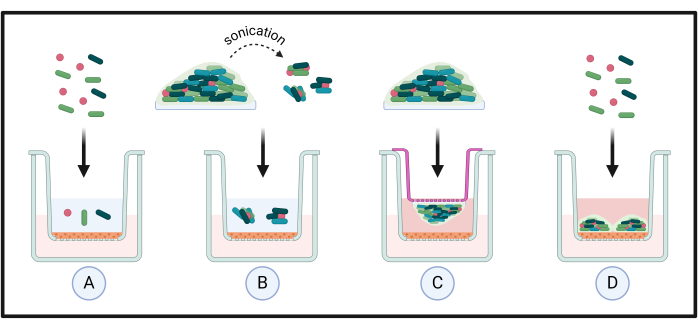
Figure 1: Different applications for organotypic tissue co-culture models. Standardized suspensions of microorganisms are directly applied to the tissue to investigate species-host interactions (A). These are often used for assessing microbial colonization over a short period of time (e.g., <24 h). Complex biofilms are sonicated to investigate the effects of dispersed cells or biofilm aggregates on the tissues (B). This is important as previously it has been shown that such cells have unique phenotypes compared to planktonic or biofilm counterparts; to this end, intact biofilms can be directly added to tissue using additional smaller inserts to create adjacent exposure with the host (C). Finally, planktonic cells are added to tissue in appropriate culture media to assess microbial colonization, biofilm formation, and growth dynamics on the tissue (D). Please click here to view a larger version of this figure.
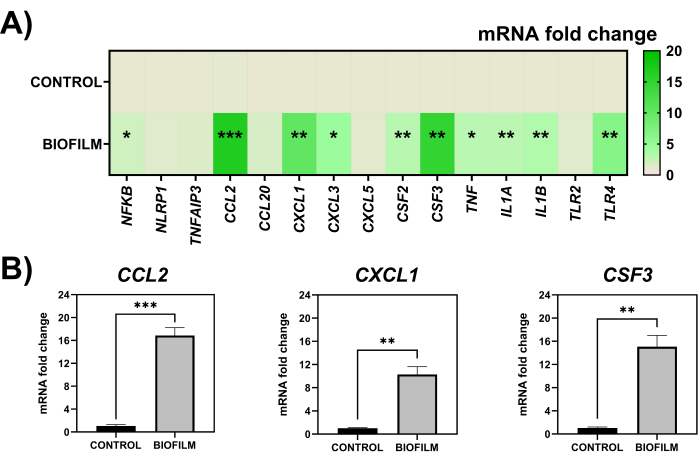
Figure 2: Gene expression of HOE tissue following stimulation with 7-species biofilm sonicate. mRNA expression fold change of a total of 15 inflammatory genes normalized to the housekeeping gene, GAPDH, in HOE tissue in control, unstimulated samples and that exposed to 100 µL of 7-species biofilm sonicate (A). The three genes (CCL2, CXCL1, CSF3) with the highest changes in mRNA expression are shown in (B). Statistical analyses were conducted using a parametric unpaired T-test, with significant changes depicted as *p < 0.05, **p < 0.01 and ***p < 0.001, respectively. Data was plotted and analyzed using statistical and graphing software. Please click here to view a larger version of this figure.
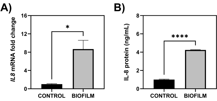
Figure 3: IL8 gene expression and IL-8 protein levels produced by HOE tissue. mRNA expression fold change of IL8 in HOE tissue as determined by SYBR-green-based qPCR (A) and IL-8 protein levels quantified from spent tissue media using the ELISA methodology (B). Statistical analyses were conducted using a parametric unpaired T-test, with significant changes depicted as *p < 0.05 and ****p < 0.0001, respectively. Data was plotted and analyzed using statistical and graphing software. Please click here to view a larger version of this figure.
Discussion
Here, methodologies are described to produce a complex multi-species biofilm model representative of gingivitis for co-culturing with HOE tissue to assess the host response following microbial stimulation. This protocol can be adapted for use in investigating host-pathogen interactions between planktonic cells, single-, dual- or mixed-species biofilms, or biofilm-dispersed cells with epithelial tissue from different ecological niches in the human body. The use of tissue models for investigating host-pathogen interactions provides an important advancement to previous co-culture systems that are restricted to 2D monolayers that don't always truly recapitulate the in vivo situation, whereby epithelial tissue contains multiple cell layers16,17,18. Furthermore, challenges associated with in vitro growth of multi-layered tissue models are plentiful, with the use of multiple cell lines and/or growth of cell layers at an air-liquid interface prone to contamination. The commercially available tissues provide a reproducible platform with consistently high-quality models that are comparable between replicates and batches, as shown in previous publications3,7.
For the "preparation of microbial communities for co-culture" section, the inclusion of these microorganisms for the 7-species gingivitis model were chosen based on commonly identified commensals and pathogens associated with the shift from oral health to disease. As with all complex biofilm model systems in vitro, the inclusion of microorganisms is directed by microbiome studies relating to the particular healthy or diseased ecological niche. To this end, we and others have reported the use of an oral health-associated biofilm model containing commensal microorganisms (e.g., Streptococcus and Rothia spp.)3,40,41. Moreover, additional pathogens can be added to these models to create biofilms associated with other diseases. For example, three microorganisms can be added to the 7-species described here to create a disease model associated with periodontitis3, whilst the fungal pathogen Candida albicans can be added to increase polymicrobiality as well as adding a layer of interkingdom complexity33. Indeed, promoting fungal-bacterial interactions can have huge implications on various experimental outputs using such biofilm models in vitro when compared to bacterial-only biofilms42. Ultimately, it is highly recommended that careful consideration be taken when creating a new multi-species biofilm model for such studies. Whilst the included microorganisms should be easily identifiable from extensive literature searches, for most niches and/or diseases, these may not integrate well into a complex model in vitro for different reasons. The following depicts some other potential considerations that should be made:
Biofilm formation dynamics
Biofilm formation in vivo often involves early, intermediate, and late colonization by particular microorganisms. Supra- and sub-gingival dental plaque formation is heavily characterized by initial attachment of the salivary pellicle found on enamel by pioneering species (e.g., Streptococcus species), with intermediate and later pathogens requiring these as a scaffold for colonization43. A similar phenomenon has been proposed in the vaginal environment during bacterial vaginosis, whereby Gardnerella vaginalis is believed to be the initial colonizer, followed by subsequent anaerobes44. However, it is important to note that this may not be the case for other diseases in other ecological niches.
Seeding density for each microorganism
Microorganisms will have different sizes and/or growth dynamics when cultured in vitro; therefore, it is important to consider this during the standardization process. For example, C. albicans is 100-150 times the size of bacterial cells45,46, therefore, it may warrant addition at a lower concentration, e.g., 1 x 10⁶ cells/mL, to such biofilm models.
Species antagonism
Some microorganisms (including the same species but different isolates, laboratory and/or clinical strains) utilized for these models may compete with others during biofilm formation, leading to inhibition of the growth of some microbial species47. A study by Sadiq et al. highlighted how different combinations of microorganisms can influence biofilm biomass resulting from microbial synergy (or antagonism)48. Although investigating such interactions within a biofilm model may be of interest to research groups (e.g., testing pre- or probiotic treatment), others may not account for such antagonism, which could impact multi-species complexity when creating the model.
Duration of biofilm maturation
It is critical to optimize biofilm maturation timeframes as this can depend on the growth dynamics of the microorganisms included and the model system (including substrata) used for culture. Longer maturation times could result in better colonization for later pathogens but more cell death within the models, particularly of the earlier colonizers. Conversely, shorter incubation times could be important if wanting to investigate the effects of immature biofilm models on the host. A study by Brown et al. described how the same 10-species wound biofilm model had different inflammatory profiles in human THP-1 cells when matured for 24 h, 48 h, and 72 h, suggesting the less mature the biofilm, the more pro-inflammatory it is7. Similar results could be observed in multi-layered tissue models. It is also important to note that regular daily media changes are a necessity to minimize cell death in the biofilm, although this can be amended depending on ongoing treatment regimens, e.g., if assessing the effects of prolonged antimicrobial interventions.
For the "organotypic tissue handling, experimental setup, and tissue processing" section, incubation timeframes can be adjusted according to the researcher's needs. Host-pathogen interactions can be investigated at earlier timepoints, e.g., 1-12 h, to later timepoints of 48 h and 72 h, depending on the research question. Secondly, all maintenance media containing antibiotics is supplied; thus, requests need to be made to the company upon ordering to remove these depending on the experimental design. Although the media underneath the insert does not come in direct contact with the upper periphery of the tissue, unless a wound is inflicted in the model6,8, removal of antibiotics may merit consideration if investigating microbial invasion into the tissue.
The microbial material used for co-culture stimulation can also be changed, as discussed above, with the scope to assess planktonic, spent biofilm supernatants (filtered and un-filtered), whole biofilms, or biofilm sonicate incubations with the tissue. For example, previous evidence has shown that tissue models such as those supplied by EpiSkin provide useful models for investigating planktonic fungal-host interactions: C. albicans, Candida auris, Malassezia furfur, and Trichophyton rubrum cells have been shown to attach and interact with peripheral tissue layers in HOE, Reconstructed Human Epidermis (RHE) or Human Vaginal Epithelium, with some of these studies showing stimulation of a host response following fungal infection1,5,6,11,12,49. Similar publications exist for bacterial-host interactions, e.g., biofilm formation of Cutibacterium acnes, a common pathogen associated with the scalp microbiota in dandruff, has been studied on the surface of RHE, when cultured alone and with the fungal skin organism, Malassezia restricta10. Others have investigated the ability of Staphylococcus spp. to attach to RHE, measuring the physicochemical and microbiological characteristics of this bacterial-host interaction50. N'Diaye and the co-authors explored how the human-derived neuropeptide, Calcitonin Gene-Related Peptide, influenced Staphylococcus aureus virulence in the RHE tissue model51. Others have investigated the protective effects of probiotic interventions on Pseudomonas aeruginosa infection of Human Corneal Epithelium14. Meanwhile, from a polymicrobial perspective, different groups have studied the effects of mixed-species biofilms on different tissue substrates2,3,7,13.
Overall, these protocols and studies referenced above document the vast array of applications for organotypic tissue models to investigate host-pathogen interactions. Although studies have shown that EpiSkin models, particularly the RHE skin model, have good applicability for testing cosmetic products for corrosion/irritation52,53,54,55, some limitations exist between these and real-world ex vivo tissue explants or other suppliers of commercially available tissue56,57. One obvious limitation would be that all commercially available tissue models are generated from cell lines in a sterile, "germ-free" environment, meaning the tissue has never been exposed to microbial perturbations: this may exacerbate any inflammatory response in the host, far beyond what would be seen in vivo or following stimulation of ex vivo tissue explants57. Conversely, these laboratory model systems will not contain underlying connective layers or vasculature that one would associate with in vivo tissue, characteristics that can be preserved during ex vivo tissue explantation, and features that impact inflammatory responses. Indeed, a recent systematic review highlighted that careful consideration should be made to utilize tissue models with appropriate vasculature created using various engineering technologies, including biomaterials58. For example, one recent study used a fibrin-based matrix embedded with gingival fibroblasts and microvascular endothelial cells to create a vascularised gingival tissue equivalent. The authors described a differential inflammatory response in the model following exposure to health or disease-associated microorganisms59. From a commercial standpoint, more complex models now exist, such as the "T-Skin model", which contains a full-thickness tissue consisting of a dermis comprised of fibroblasts overlaid with the epidermis. It would be interesting to see how such complex models compare to the epidermis-only systems.
While no model is perfect, organotypic tissue models represent promising alternatives for preclinical testing, aligning with the framework outlined by the three R's, for Replacement, Reduction, and Refinement in undertaking animal research. To this end, although animal models provide important insights into the complex pathophysiological nature of biofilm-related human diseases, they come with obvious disadvantages. These 3D models are easily manipulatable, allowing for large, subtle changes to investigations without ethical approval. Combining these models with existing complex biofilm systems outlined above can greatly improve our understanding of host-pathogen interactions and better predict the success of novel therapies prior to in vivo investigations.
Disclosures
The authors have nothing to disclose.
Acknowledgements
The authors would like to thank Haleon for funding support for Saeed Alqahtani to run associated projects relating to the tissue models described, as well as the Ministry of Education, Saudi Arabia, for PhD funding of Muhanna Alshehri.
Materials
| Name | Company | Catalog Number | Comments |
|---|---|---|---|
| 13 mm hydroxyapatite (HA) discs | Plasma Biotal Limited | n/a (made to order) | Substrate for oral biofilm formation, mimicking the tooth surface |
| 19 G needle (40 mm) | VWR | 613-2028 | Used to help with removal of HA discs from plates and to cut out tissue from inserts |
| 24 well TC-treated flat bottom microtitre plates with lids | Scientific Laboratory Supplies | 3524 (pack of 100) | Plates for culturing biofilms and tissue co-culture experiments |
| Acid washed glass beads (425-600 μm) | Merck: Sigma-Aldrich | G8772 | For tissue lysis to improve yield of RNA extraction |
| Actinomyces naeslundii DSM 17233 | Leibniz Institute DSMZ | n/a | One of the microorganisms used for the complex biofilm model |
| Bead Mill 24 Homogenizer (or equivalent) | Fisher Scientific | 15-340-163 | For tissue lysis to improve yield of RNA extraction |
| Bijouxes (7 mL) | Greiner Bio-One | 189170 (700 per bag) | Used for biofilm sonication |
| cDNA synthesis kit (high-capacity) | Thermo Fisher Scientific | 4368814 (for 200 reactions) | cDNA synthesis from tissue RNA |
| Columbia Blood Agar (CBA) base | Thermo Fisher Scientific | CM0331B (for 500 grams) | Used to prepare CBA plates for culturing aerobic microorganisms |
| Defibrinated horse blood | E&O Laboratories | DHB | Used to supplement CBA and FAA plates for culturing aerobic and anaerobic microorganisms |
| Dulbecco's Phosphate Buffered Saline (dPBS: without CaCl2 and MgCl2) | Merck: Sigma-Aldrich | DB537 | dPBS for cell co-culture with biofilm sonicate |
| Fusobacterium nucleatum ATCC 10953 | American Type Culture Collection | n/a | One of the microorganisms used for the complex biofilm model |
| Fusobacterium nucleatum subspecies (subspp.) vincentii DSM 19507 | Leibniz Institute DSMZ | n/a | One of the microorganisms used for the complex biofilm model |
| GraphPad Prism (Version 10) | GraphPad Software, Inc | n/a | For graph creation and data analyses |
| Human IL-8 ELISA kit (uncoated ELISA kit with plates) | Thermo Fisher Scientific (Invitrogen) | 88-8086-86 (for 10 x 96 tests) | ELISA kit and plates for protein assessment of spent tissue media |
| Human Oral Epithelium (HOE) | Episkin | n/a (made to order) | The epithelial tissue utilised for the co-culture model system |
| Inoculating loops (disposable, 10 µL, or equivalent) | Fisher Scientific | 12870155 (pack of 1000) | For microbiological culture in solid and liquid media |
| MicroAmp Fast Reaction 96-well qPCR plates (0.1 mL) | Thermo Fisher Scientific | 4346907 (for 10 plates) | For qPCR gene expression profiling of tissue |
| Microbank beads | Pro-Lab Diagnostics | PL.170 | Storage and cyropreservation of microorganisms |
| Nuclease-free water | Thermo Fisher Scientific | AM9935 (10 x 1.5 mL) | RT-PCR grade water for qPCR experiments |
| Petri Dishes (90 mm) | Fisher Scientific (Sterilin) | 11309283 (pack of 500) | Used for preparation of agar plates for microbial culture |
| Phosphate-buffered saline (PBS) | Thermo Fisher Scientific | 18912014 (pack of 100 tablets) | PBS for microorganism standardization and biofilm wash steps |
| Rneasy mini kit | Qiagen | 74106 (for 250 reactions) | Column-based RNA extraction kit for tissue |
| Roswell Park Memorial Institute 1640 medium | Merck: Sigma-Aldrich | R7755-10L (for 10 L) | Media for biofilm culture |
| RT2 profiler array | Qiagen | 330171 (custom-made) | qPCR array containing ready-made primers for gene expression profiling |
| Schaedler anaerobe agar base | Merck: Sigma-Aldrich (Millipore) | 91019 (for 500 g) | Used to prepare Fastdious Anaerobic Agar plates |
| Schaedler anaerobe broth | Thermo Fisher Scientific | CM0497B (for 500 g) | Liquid media for growth of anaerobic microorganisms |
| Screw-cap Beadbug O-Ring tube | Scientific Laboratory Supplies | Z763837-1000EA (pack of 1000) | For use in Bead Mill 24 homogenizer, to lyse tissue |
| Sonication bath | Fisher Scientific (Fisherbrand) | n/a | For sonication of biofilms from HA discs |
| Streptococcus intermedius DSM 20753 | Leibniz Institute DSMZ | n/a | One of the microorganisms used for the complex biofilm model |
| Streptococcus mitis NCTC 12261 | National Collection of Type Cultures | n/a | One of the microorganisms used for the complex biofilm model |
| Streptococcus oralis NTCC 11427 | National Collection of Type Cultures | n/a | One of the microorganisms used for the complex biofilm model |
| SYBR-green (qPCRBIO SyGreen Mix Hi-ROX) | PCR Biosystems | PB20.12 | SYBR-green mix for qPCR |
| Todd Hewitt broth | Merck: Sigma-Aldrich | T1438 | Media for biofilm culture |
| Tryptone soya broth (TSB) medium | Merck: Sigma-Aldrich | 22092 (for 500 g) | Liquid media for growth of aerobic microorganisms |
| Tweezers (150 mm, Stainless Steel, Serrated) | RS-Pro | 545-187 | Used to move HA discs and tissue inserts/tissue extracts |
| Universal tubes (30 mL) | Greiner Bio-One | 201150 (400 per bag) | Vessels for culturing microorganisms in liquid culture |
| Veillonella dispar NCTC 11831 | National Collection of Type Cultures | n/a` | One of the microorganisms used for the complex biofilm model |
| β-metacapthoethanol | Merck: Sigma-Aldrich | M3148 | RNase deactivation in tissue samples |
References
- Moyes, D. L., et al. A biphasic innate immune MAPK response discriminates between the yeast and hyphal forms of candida albicans in epithelial cells. Cell Host Microbe. 8 (3), 225-235 (2010).
- Morse, D. J., et al. Denture-associated biofilm infection in three-dimensional oral mucosal tissue models. J Med Microbiol. 67 (3), 364-375 (2018).
- Brown, J. L., et al. Biofilm-stimulated epithelium modulates the inflammatory responses in co-cultured immune cells. Sci Rep. 9 (1), 15779(2019).
- Yadev, N. P., Murdoch, C., Saville, S. P., Thornhill, M. H. Evaluation of tissue engineered models of the oral mucosa to investigate oral candidiasis. Microb Pathog. 50 (6), 278-285 (2011).
- Schaller, M., Zakikhany, K., Naglik, J. R., Weindl, G., Hube, B. Models of oral and vaginal candidiasis based on in vitro reconstituted human epithelia. Nat Protoc. 1 (6), 2767-2773 (2006).
- Brown, J. L., et al. Candida auris phenotypic heterogeneity determines pathogenicity in vitro. mSphere. 5 (3), e00371-e00420 (2020).
- Brown, J. L., et al. Assessing the inflammatory response to in vitro polymicrobial wound biofilms in a skin epidermis model. NPJ Biofilms Microbiomes. 8 (1), 19(2022).
- Butcher, M. C., et al. Antifungal-loaded calcium sulfate beads as a potential therapeutic in combating candida auris. Antimicrob Agents Chemother. 66 (1), e0171321(2022).
- Duckney, P., et al. The role of the skin barrier in modulating the effects of common skin microbial species on the inflammation, differentiation and proliferation status of epidermal keratinocytes. BMC Res Notes. 6, 474(2013).
- Meloni, M., Balzaretti, S., Collard, N., Desaint, S., Laperdrix, C. Reproducing the scalp microbiota community: Co-colonization of a 3D reconstructed human epidermis with c. Acnes and m. Restricta. Int J Cosmet Sci. 43 (2), 235-245 (2021).
- Pedrosa, A. F., et al. Malassezia colonisation on a reconstructed human epidermis: Imaging studies. Mycoses. 62 (12), 1194-1201 (2019).
- Schaller, M., Weindl, G. Models of oral and vaginal candidiasis based on in vitro reconstituted human epithelia for the study of host-pathogen interactions. Methods Mol Biol. 470, 327-345 (2009).
- Rosca, A. S., et al. Six bacterial vaginosis-associated species can form an in vitro and ex vivo polymicrobial biofilm that is susceptible to thymbra capitata essential oil. Front Cell Infect Microbiol. 12, 824860(2022).
- Paterniti, I., et al. Protective effect of probiotics against pseudomonas aeruginosa infection of human corneal epithelial cells. Int J Mol Sci. 25 (3), 1770(2024).
- Pellegatta, G., et al. Evaluation of human esophageal epithelium permeability in presence of different formulations containing hyaluronic acid and chondroitin sulphate. Med Devices (Auckl). 13, 57-66 (2020).
- Jensen, C., Teng, Is it time to start transitioning from 2D to 3D cell culture. Front Mol Biosci. 7, (2020).
- Mountcastle, S. E., et al. A review of co-culture models to study the oral microenvironment and disease. J Oral Microbiol. 12 (1), 1773122(2020).
- Cacciamali, A., Villa, R., Dotti, S. 3D cell cultures: Evolution of an ancient tool for new applications. Front Physiol. 13, 836480(2022).
- Langfeldt, D., et al. Health- and disease-associated species clusters in complex natural biofilms determine the innate immune response in oral epithelial cells during biofilm maturation. FEMS Microbiol Lett. 360 (2), 137-143 (2014).
- Ramage, G., et al. The epithelial cell response to health and disease associated oral biofilm models. J Periodontal Res. 52 (3), 325-333 (2017).
- Shang, L., et al. Multi-species oral biofilm promotes reconstructed human gingiva epithelial barrier function. Sci Rep. 8 (1), 16061(2018).
- Myers, S., et al. Immunomodulatory streptococci that inhibit cxcl8 secretion and NF-kappaB activation are common members of the oral microbiota. J Med Microbiol. 70 (3), 001329(2021).
- Tang, Y. L., Sim, T. S., Tan, K. S. Oral streptococci subvert the host innate immune response through hydrogen peroxide. Sci Rep. 12 (1), 656(2022).
- Peyyala, R., Kirakodu, S. S., Novak, K. F., Ebersole, J. L. Oral microbial biofilm stimulation of epithelial cell responses. Cytokine. 58 (1), 65-72 (2012).
- Yee, M., Kim, S., Sethi, P., Duzgunes, N., Konopka, K. Porphyromonas gingivalis stimulates il-6 and il-8 secretion in gsm-k, hsc-3 and h413 oral epithelial cells. Anaerobe. 28, 62-67 (2014).
- Groeger, S., Jarzina, F., Domann, E., Meyle, J. Porphyromonas gingivalis activates NF-kappaB and MAPK pathways in human oral epithelial cells. BMC Immunol. 18 (1), 1(2017).
- Groeger, S., Zhou, Y., Ruf, S., Meyle, J. Pathogenic mechanisms of fusobacterium nucleatum on oral epithelial cells. Front Oral Health. 3, 831607(2022).
- Li, X., et al. Saliva-derived microcosm biofilms grown on different oral surfaces in vitro. NPJ Biofilms Microbiomes. 7 (1), 74(2021).
- Krishnamoorthy, A. L., Lemus, A. A., Solomon, A. P., Valm, A. M., Neelakantan, P. Interactions between Candida albicans and Enterococcus faecalis in an organotypic oral epithelial model. Microorganisms. 8 (11), 1771(2020).
- Brown, J. L., et al. Generation of multi-species oral bacteria biofilm models. Methods Mol Biol. 2588, 187-199 (2023).
- Miles, A. A., Misra, S. S., Irwin, J. O. The estimation of the bactericidal power of the blood. J Hyg (Lond). 38 (6), 732-749 (1938).
- Lukic, D., Karygianni, L., Flury, M., Attin, T., Thurnheer, T. Endodontic-like oral biofilms as models for multi-species interactions in endodontic diseases. Microorganisms. 8 (5), 674(2020).
- Sherry, L., et al. Viable compositional analysis of an eleven-species oral polymicrobial biofilm. Front Microbiol. 7, 912(2016).
- Brown, J. L., et al. An in vitro evaluation of denture cleansing regimens against a polymicrobial denture biofilm model. Antibiotics (Basel). 11 (1), 113(2022).
- Ammann, T. W., Gmur, R., Thurnheer, T. Advancement of the 10-species subgingival zurich biofilm model by examining different nutritional conditions and defining the structure of the in vitro biofilms. BMC Microbiol. 12, 227(2012).
- Montelongo-Jauregui, D., Srinivasan, A., Ramasubramanian, A. K., Lopez-Ribot, J. L. An in vitro model for oral mixed biofilms of candida albicans and streptococcus gordonii in synthetic saliva. Front Microbiol. 7, 686(2016).
- Rumbaugh, K. P., Sauer, K. Biofilm dispersion. Nat Rev Microbiol. 18 (10), 571-586 (2020).
- Chua, S. L., et al. Dispersed cells represent a distinct stage in the transition from bacterial biofilm to planktonic lifestyles. Nat Commun. 5, 4462(2014).
- Buskermolen, J. K., Janus, M. M., Roffel, S., Krom, B. P., Gibbs, S. Saliva-derived commensal and pathogenic biofilms in a human gingiva model. J Dent Res. 97 (2), 201-208 (2018).
- Rosier, B. T., Buetas, E., Moya-Gonzalvez, E. M., Artacho, A., Mira, A. Nitrate as a potential prebiotic for the oral microbiome. Sci Rep. 10 (1), 12895(2020).
- Rosier, B. T., Moya-Gonzalvez, E. M., Corell-Escuin, P., Mira, A. Isolation and characterization of nitrate-reducing bacteria as potential probiotics for oral and systemic health. Front Microbiol. 11, 555465(2020).
- Young, T., et al. Candida albicans as an essential "keystone" component within polymicrobial oral biofilm models. Microorganisms. 9 (1), 59(2020).
- Marsh, P. D., Lewis, M. A., Rogers, H., Williams, D., Wilson, M. Marsh and Martin's oral microbiology-e-book: Marsh and Martin's oral microbiology-e-book. , Elsevier Health Sciences. (2016).
- Johnston, W., et al. In vitro bacterial vaginosis biofilm community manipulation using endolysin therapy. Biofilm. 5, 100101(2023).
- Janus, M. M., Willems, H. M., Krom, B. P. Candida albicans in multi-species oral communities; a keystone commensal. Adv Exp Med Biol. 931, 13-20 (2016).
- Delaney, C., et al. Fungi at the scene of the crime: Innocent bystanders or accomplices in oral infections. Curr Clin Microbiol Rep. 5, 190-200 (2018).
- Luo, A., Wang, F., Sun, D., Liu, X., Xin, B. Formation, development, and cross-species interactions in biofilms. Front Microbiol. 12, 757327(2021).
- Sadiq, F. A., De Reu, K., Burmolle, M., Maes, S., Heyndrickx, M. Synergistic interactions in multi-species biofilm combinations of bacterial isolates recovered from diverse food processing industries. Front Microbiol. 14, 1159434(2023).
- Liang, P. P., et al. A trichophyton rubrum infection model based on the reconstructed human epidermis - EpiSkin(r). Chin Med J (Engl). 129 (1), 54-58 (2016).
- Lerebour, G., Cupferman, S., Bellon-Fontaine, M. N. Adhesion of staphylococcus aureus and staphylococcus epidermidis to the EpiSkin reconstructed epidermis model and to an inert 304 stainless steel substrate. J Appl Microbiol. 97 (1), 7-16 (2004).
- N'diaye, A. R., et al. Skin-bacteria communication: Involvement of the neurohormone calcitonin gene-related peptide (cgrp) in the regulation of staphylococcus epidermidis virulence. Sci Rep. 6, 35379(2016).
- Géniès, C., et al. Comparison of the metabolism of 10 cosmetics-relevant chemicals in EpiSkin s9 subcellular fractions and in vitro human skin explants. J Appl Toxicol. 40 (2), 313-326 (2020).
- Alépée, N., Grandidier, M. -H., Cotovio, J. Usefulness of the EpiSkin reconstructed human epidermis model within integrated approaches on testing and assessment (IATA) for skin corrosion and irritation. Toxicol Vitro. 54, 147-167 (2019).
- Spielmann, H., et al. The ECVAM international validation study on in vitro tests for acute skin irritation: Report on the validity of the EpiSkin and epiderm assays and on the skin integrity function test. Alternatives to Laboratory Animals. 35 (6), 559-601 (2007).
- Cotovio, J., Grandidier, M. -H., Portes, P., Roguet, R., Rubinstenn, G. The in vitro acute skin irritation of chemicals: Optimisation of the EpiSkin prediction model within the framework of the ECVAM validation process. Alt Lab Anim. 33 (4), 329-349 (2005).
- Netzlaff, F., Lehr, C. M., Wertz, P. W., Schaefer, U. F. The human epidermis models EpiSkin, SkinEthic and epiderm: An evaluation of morphology and their suitability for testing phototoxicity, irritancy, corrosivity, and substance transport. Eur J Pharm Biopharm. 60 (2), 167-178 (2005).
- Larson, P. J., Chong, D., Fleming, E., Oh, J. Challenges in developing a human model system for skin microbiome research. J Invest Dermatol. 141 (1), 228-231.e4 (2021).
- Dechelette, C., Smirani, R., Medina, C., Naveau, A. Cellularized biomaterials used as gingival connective tissue substitutes in vivo: A systematic review. Tissue Eng Part B Rev. , (2024).
- Makkar, H., Lim, C. T., Tan, K. S., Sriram, G. Modeling periodontal host-microbe interactions using vascularized gingival connective tissue equivalents. Biofabrication. 15 (4), (2023).
Reprints and Permissions
