Here we provide an overview of the anteromesial temporal lobectomy procedure, used in the treatment of patients with medically refractory temporal lobe epilepsy. In particular, we describe herein, the details of the operative method and related surgical technique, in addition to summarizing the indications and outcomes of the procedure.
Method Article
Anteromesial Temporal Lobectomy for Medically Intractable Temporal Lobe Epilepsy: An Operative Study
In This Article
Summary
Abstract
Anteromesial temporal lobectomy, including surgical resection of the mesial temporal structures, is an important surgical procedure for the treatment of medically refractory temporal lobe epilepsy. Given the widespread use of this technique in appropriately screened patients with intractable focal epilepsy (not to mention other non-epileptic neurosurgical conditions, including brain tumors and vascular lesions), it is important for the treating neurosurgeon to have a comprehensive understanding of the complex anatomy and surgical technique for performing a successful resection.
Here, we describe the key steps and important technical pearls for a standard anteromesial temporal lobectomy procedure. This begins with appropriate patient selection and presurgical optimization. In the operating room, patient positioning, careful opening of the scalp layers, and a well-planned craniotomy are essential to setting up the subsequent critical steps of surgery. Subsequent removal of the temporal lobe is achieved firstly with resection of the lateral temporal neocortex, followed by the mesial temporal structures (including removal of the amygdala and hippocampus). Wound closure requires proper attention to hemostasis and approximation of tissue layers. These steps may be further modified in some cases based on the patient's anatomy and/or the type of pathology encountered. Meticulous execution of the surgical techniques presented here is imperative to achieving successful seizure-free outcomes in this patient population while mitigating against the risk of surgical complications.
Introduction
Epilepsy remains a significant global healthcare burden, impacting 1% of the population in the United States1. Approximately 40% of all patients with epilepsy (equivalent to nearly 1 million patients) remain resistant to medication, referred to as medically refractory epilepsy (MRE). MRE is defined by the failure of two or more appropriately utilized anti-seizure medication regimes (ASM) to provide sustained seizure freedom2. Patients with MRE experience a significant decline in the quality of life, neurocognitive function, as well as psychological and physical well-being. Surgical intervention, therefore, becomes critical in the long-term management of MRE for afflicted patients and offers a chance at seizure freedom, in addition to the possibility of weaning off anti-seizure medications (which themselves carry cumulative side effects and medical risks).
Anteromesial temporal lobectomy (ATL) is the most widely performed surgery for the treatment of MRE, yielding excellent seizure-free outcomes and significant improvements in quality of life3,4,5,6,7. There is some variability in surgical approaches and techniques for performing the procedure, ranging from a more extensive resection of the lateral and mesial temporal structures (corticoamygdalohippocampectomy) to a more limited resection focusing on the mesial structures via a smaller surgical window (selective amygdalohippocampectomy). This may be further influenced by the underlying pathology, baseline neurocognitive function, hemispheric dominance for speech/language function, and surgeon preference. Common temporal lobe pathologies that warrant consideration of ATL include mesial temporal sclerosis, focal cortical dysplasia, neoplastic or vascular lesions, and non-lesional cases where electrocorticography studies may also be a helpful strategy. In the latter case, a pre-surgical invasive evaluation may be first completed using stereoelectroencephalography or subdural grid mapping, with/without intra-operative electrocorticography as a surgical adjunct at the time of resection.
Importantly, the decision to perform epilepsy surgery (including ATL, if indicated) is conventionally made by a multidisciplinary team at a tertiary or quaternary care epilepsy center, involving multiple specialists specialized in the management of patients with complex MRE8. This may include epileptologists (neurologists specializing in epilepsy), neurosurgeons, neuroradiologists, nuclear medicine specialists, neuropsychologists, ethicists, social workers, pharmacists, EEG technicians, and nurses, amongst other participants. A careful understanding of the clinical work-up and surgical decision-making process for a patient with focal temporal lobe epilepsy, including the implications, indications, and risks of ATL surgery, must be carefully discussed. A formal consent discussion is also required before surgery can be offered to the patient. Of relevance to this project, the neurosurgeon offering the procedure must be well-versed in the anatomy and surgical technique of ATL, in addition to the growing literature and outcomes-based research on the subject. These details will be highlighted in this article on the standard ATL procedure for intractable epilepsy with a step-by-step approach.
Protocol
Surgical consent was given by the patient for the procedure, as part of standard medical care. Written permission was obtained from the patient to allow for the video recording during their surgery, for the purpose of publishing for educational use.
1. Positioning in the operating room and wound opening
- Anesthesia and positioning
- In the operating room (OR), place the patient under full general anesthesia with endotracheal intubation. Administer pre-operative antibiotic medication and perform hyperventilation.
NOTE: Steroids and anti-seizure medication (ASM) may be given at the discretion of the anesthesia and neurosurgical team. Hyperventilation per the anesthesia team should be done to promote brain relaxation, typically using an end-tidal PaCO2 level of 26-30 mm Hg. - Position the patient supine on the OR table, with the head rotated toward the contralateral shoulder and extended slightly to facilitate visualization along the antero-posterior axis of the intracranial mesial temporal structures.
NOTE: Depending on the surgeon's preference, the head may or may not be rigidly fixed in a headframe. - Perform disinfection of the surgical field with a preferred agent of choice (e.g., betadine, alcohol-based scrub) and drape the field in sterile fashion.
- In the operating room (OR), place the patient under full general anesthesia with endotracheal intubation. Administer pre-operative antibiotic medication and perform hyperventilation.
- Incision and exposure
- Make a reverse question-mark (curvilinear) incision using a #10 scalpel over the temporal region, starting from the zygoma inferiorly, extending back toward the posterior limit of the ear, and up to a few centimeters above the superior temporal line, dorsally. Elevate the scalp flap with preservation of the underlying temporalis muscle.
- Open the muscle in a T-shaped or curvilinear fashion. With a T-shaped opening, make a bisection to the level of the zygoma and leave a small cuff superiorly to allow for the later closure of the muscle.
NOTE: This also reduces muscle bulk anteriorly in the working field. - Minimize the use of electrocautery and preserve the deeper fascia to maintain blood supply and reduce muscle atrophy.
- Use hooks for gentle retraction of the musculocutaneous flap as the exposure is further developed. The temporal and inferior frontal bone surface should now be visible.
2. Craniotomy and dural opening
- Craniotomy
- Make a frontotemporal craniotomy to provide access down to the middle cranial fossa and anteriorly to the keyhole, with minimal exposure to the supra-Sylvian frontal operculum.
- Make burr holes using a perforator drill or a cutting burr at the zygoma, keyhole, and posteriorly.
NOTE: This allows multiple access points to strip the dura from the inner cortical table. This may be especially important in older patients and those patients who have undergone pre-operative stereoelectroencephalography (SEEG), which can produce multiple dural adhesion points that require additional dissection and release. - Use a craniotome to make cuts between burr holes. The dura is stripped off the inner cortex of the bone flap, which is then carefully removed. Gently cauterize major dural vessels with bipolar cautery.
- If required, perform additional bony removal using a rongeur along the sphenoid wing and inferior squamous temporal bone to optimize the opening further.
- Wax off exposed air cells to avoid CSF leak complications. Place tack-up sutures at this time to minimize the chance of post-operative epidural hematoma formation.
- Dural opening
- Open the dura in a curvilinear fashion using a #11 or #15 scalpel. Reflect the dura anteriorly, avoiding injury to any underlying vessels or cortex. Retraction sutures are placed to keep the dural edges open.
- Visualize the dural opening of the anterolateral surface of the temporal lobe and Sylvian fissure.
3. Temporal cortical resection
- Lateral temporal neocortical resection
- Make a posterior corticectomy using bipolar cautery and micro-scissors to open the pial surface.
NOTE: In a typical non-dominant (conventionally right-sided) resection, the posterior disconnection is made ~5.5 to 6 cm from the temporal pole, as measured along the middle temporal gyrus. On the language-dominant (conventionally left) side, a more conservative measurement of ~4 to 4.5 cm is used. - Identify and preserve the large draining Vein of Labbé. The posterior cut is carried down to the middle cranial fossa floor to visualize the tentorium.
- Make the superior corticectomy along the superior temporal gyrus and carry it anteriorly towards the temporal pole.
- Perform subpial dissection and microsurgical aspiration to visualize the anatomy and pial planes throughout the case. This can be achieved using bipolar cautery and micro-suction devices. Aspirate the superior temporal gyrus to expose the underlying insula and MCA branches, which are carefully preserved.
- Follow the superior temporal gyrus anteriorly into the temporal pole and remove this tissue in a subpial fashion, as well.
- Identify the inferior circular sulcus, the lower border of the insula. Safely follow the white matter at this level into the temporal stem, often taking a 30-45° angle depending on the anatomy.
NOTE: The temporal horn of the lateral ventricle is often encountered, typically further posteriorly, and a cottonoid patty is inserted to prevent blood from entering the ventricular system. - Visually identify the hippocampus, with a small intervening lateral ventricular sulcus and the more laterally situated collateral eminence (i.e., the bulge lateral to the hippocampus), which is an identifiable prominence caused by the collateral sulcus underneath.
NOTE: This is a safe landmark that keeps the surgeon above the tentorium. - As one completes the posterior disconnection laterally around and down to the middle cranial fossa floor, make the resection line traverse the lateral occipital temporal sulcus and fusiform gyrus to meet up at the collateral sulcus.
- At this point, follow the collateral eminence/sulcus anteriorly to connect with the anterior disconnection line. Sharply disconnect the pia and complete the lateral temporal neocortical resection en bloc.
- Carefully inspect the cortical draining veins along the temporal pole and coagulate as appropriate, with the disconnection to release the tissue. The specimen is tagged for orientation and sent to pathology.
- Make a posterior corticectomy using bipolar cautery and micro-scissors to open the pial surface.
- Resection of the mesial temporal structures (i.e., Amygdalohippocampectomy)
- Further open the temporal horn to identify the hippocampus and choroidal point. Inside the ventricle, identify the hippocampal head and body along the inferior aspect, within the floor of the ventricle. The choroid plexus and inferior choroidal point are located behind the head of the hippocampus.
- Place a cottonoid in this hippocampal region to protect the anterior choroidal artery entering the choroidal fissure.
- Identify the amygdala as anterior and superior to the hippocampal head, separated by a small sulcus, i.e., the uncal recess.
NOTE: The amygdala extends superiorly to the striatum with no clear demarcation, and, therefore, its superior extent of resection is limited based on a line drawn from the inferior choroidal point to the middle cerebral artery (MCA) bifurcation, effectively within the same plane as the choroidal fissure itself. - Then resect the amygdala. Divide the uncal recess and perform the amygdalar resection mesially through the anterior uncus to reach the mesial pia, which should be preserved.
- Resect the parahippocampal gyrus situated beneath the hippocampus in a subpial fashion to undermine the hippocampus and allow it to mobilize (and rotate) outwardly, following disconnection of its posterior limit further back along the tail.
- To complete the hippocampal resection, divide the alveus and fimbria of the fornix (located mesially on the curvature of the hippocampus, as seen from above) down to the pia.
- Coagulate the perforating vessels of Uchimura, arising mesially from the posterior cerebral artery as small arterial branches supplying the hippocampus. Then, sharply divide these as well when the hippocampal body is further dissected and rotated laterally.
NOTE: In many cases, the hippocampus may be peeled off the hippocampal sulcus using a Penfield instrument; when firmly adherent and stuck, it may be resected in a piece-meal fashion using cautery and suction. It should be noted that samples of the amygdala and hippocampus are routinely sent to pathology, as well. - Finally, further resect the residual tail of the hippocampus posteriorly, up to the point of curvature behind the midbrain. Resect the remaining posterior uncus in subpial fashion.
NOTE: The tentorial edge and oculomotor nerve will now be in view, often with the posterior cerebral artery visualized further posteriorly. Meticulous hemostasis is maintained throughout using a combination of bipolar cautery and hemostatic materials.
4. Cranioplasty and wound closure
- Close the dura and reinforce with a dural substitute (onlay) agent. Replace the bone flap and secure it with titanium cranial plate fixation.
- Re-approximate the temporalis muscle with sutures and place a subgaleal drain and tunnel out of the scalp.
- Close the scalp in layers, principally closing the galea followed by the subcutaneous tissues. Close the wound primarily with a running suture.
- Following skin closure, apply a sterile dressing and a clean headwrap to further minimize subgaleal fluid collection.
- Postoperatively, monitor the patient in a neuro-stepdown unit or an intensive care unit. Administer antibiotics postoperatively, along with a short course of steroids (up to 48 h) and the patient's anti-seizure medication.
NOTE: An early post-operative CT scan (Figure 1) may be obtained within the next few hours to assess for hematoma formation, and to rule out any complications.
Results
At approximately six months following surgery, an EEG and MRI are often obtained to clinically reassess the patient. The MRI (Figure 2) is performed to radiographically document the complete removal of the lateral temporal neocortex and mesial temporal structure.
The success of ATL in the treatment of refractory temporal lobe epilepsy has been documented extensively in the literature, with the most important goal being seizure-freedom9. The extent of the disconnection and complete resection of the lateral and mesial temporal structures are essential to optimizing these outcomes. In a randomized-controlled trial by Wiebe et al, 80 patients with MRE arising from the temporal lobe were randomly assigned to ATL (40 patients) or continued treatment with ASM alone (40 patients), with a minimum follow-up of one year7. The primary outcome was freedom from seizures impairing awareness, which was noted to be 58% for the surgical group versus 8% for the medical group. The surgical group also experienced improved quality of life. In another trial by Engel et al, patients with temporal lobe MRE were similarly assigned to either ATL (15 patients) or medical treatment alone (23 patients)10. At two years, 73% of patients in the surgical arm were seizure-free, while none of the patients treated with ASM alone achieved seizure-freedom.
Suboptimal outcomes and complications primarily relate to the surgical risks in this brain region. Post-operative visual field deficit, typically a contralateral superior quadrantanopsia, may occur in 28% to 52% of patients as a result of manipulation of the visual fibers of Meyer's loop coursing along the superior/lateral wall of the temporal horn11. This may be minimized with reduced retraction, use of intraoperative neuro-navigation, and pre-operative tractography imaging. Naming deficit is seen in 25% to 60% of patients following a dominant (typically left) side temporal lobectomy, especially notable in patients without pre-existing language deficits9. It remains difficult to precisely predict which patients may develop this symptom, and therefore, extensive pre-operative neuropsychological testing and patient counseling are important. Other surgical complications include the risk for infection, hemorrhage (requiring transfusion), neurological deficits (weakness, cranial nerve dysfunction, etc.), hydrocephalus, seizure recurrence, medical complications (pneumonia, blood clots, myocardial infarction), and/or death. Again, careful pre-operative counselling and discussion of the indications, steps, and risks of surgery during the consent process is imperative for the patient (and their family) to be well-informed about the expectations and risks of surgery.
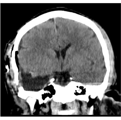
Figure 1: Representative post-operative computed tomography (CT) scan. This is a typical CT scan obtained in the immediate post-operative period. There is no evidence of hematoma or other post-operative complication. Please click here to view a larger version of this figure.

Figure 2: Representative magnetic resonance imaging (MRI) scan. Here, we see a pre- (left image) and post-operative MRI scan (right image) highlighting successful removal of the right temporal lobe, including the mesial structures. Please click here to view a larger version of this figure.
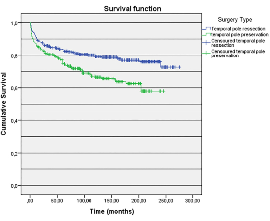
Figure 3. Kaplan-Meier curve of long-term seizure outcomes. Kaplan-Meier curve of long-term seizure outcomes (up to 23 years) in 621 refractory temporal lobe epilepsy patients, stratified by type of surgery (anteromesial temporal lobectomy, i.e., ATL, versus selective amygdalohippocampectomy), as reported in a study of 621 patients with hippocampal sclerosis. The findings support that standard ATL led to better outcomes (78.6%, Engel Class I), compared to the more selective procedure, which spared the temporal pole (67.2%, Engel Class 1; p = 0.002). This figure is reproduced from Dalio et al. (2022), with permissions granted by the JNS Publishing Group12. Please click here to view a larger version of this figure.
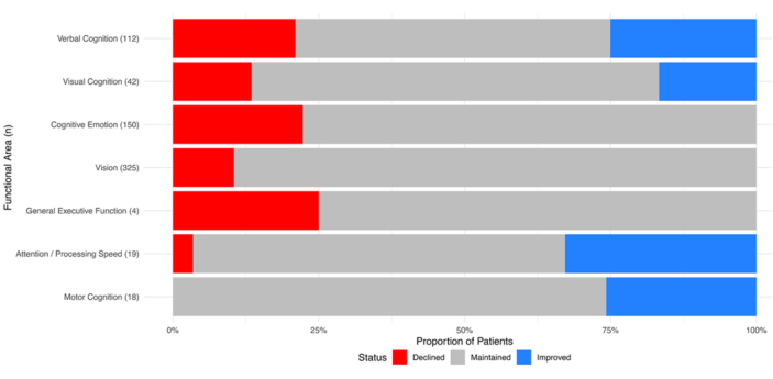
Figure 4. Distribution of patient outcomes across multiple cognitive domains. Distribution of patient outcomes across multiple cognitive domains following stereotactic laser ablation in 408 refractory temporal lobe epilepsy patients, as reported in a systematic review and meta-analysis of 14 studies. Proportional percentages are shown on the horizontal axis for patients who have declined, maintained or improved in the various functional test areas shown on the vertical axis. This figure is reproduced from Brenner et al. (2024), an open-access article distributed under the terms of the Creative Commons Attribution License (CC BY)13. Please click here to view a larger version of this figure.
Discussion
The structural and functional anatomy of the temporal lobe is complex, and this requires careful study to be well understood by the aspiring neurosurgeon. In brief, from rostral to caudal, the lateral temporal neocortex is comprised of five gyri (superior temporal gyrus, middle temporal gyrus, inferior temporal gyrus, fusiform gyrus, and parahippocampal gyrus) and four intervening sulci (superior temporal sulcus, inferior temporal sulcus, lateral occipitotemporal sulcus, and collateral sulcus)14. The superior temporal gyrus is most dorsal, lying against the Sylvian fissure, and in its anteroposterior orientation is comprised of the planum polare, anterior transverse temporal gyrus (Heschl's gyrus), and the planum temporale (middle and posterior transverse temporal gyri)15. The superior temporal gyrus is also continuous anteriorly with the temporal pole, which encapsulates the amygdala (the latter forming the anterior wall of the temporal horn of the lateral ventricle). In addition to the amygdala, other important mesial temporal structures include the hippocampus, the choroidal fissure (containing the choroid plexus and anterior choroidal artery), and the underlying parahippocampal gyrus (including the uncus)16.
The technique of the standard anterior temporal lobectomy (ATL) procedure has evolved over time. Penfield and Baldwin (1952) reported on their early experience with subtotal temporal lobectomy for patients with temporal lobe seizures, identifying a number of individuals with 'incisural sclerosis' of the superior temporal gyrus, temporal pole, and mesial structures17. The modern 'standard' ATL procedure entails the removal of the overlying neocortex, along with resection of the mesial structures, most notably the amygdala and hippocampus. In general, ATL carries a high seizure-freedom rate for adult and pediatric patients who are carefully screened and selected for the procedure and may be used in the treatment of different pathologies including mesial temporal sclerosis, neocortical temporal lobe epilepsy (including focal cortical dysplasia); temporal encephaloceles, cavernomas, brain tumors, and other neurosurgical or pathological conditions. Concomitantly, the procedure may follow the results of a pre-surgical invasive evaluation, whether by stereoelectroencephalography or subdural grid monitoring, or may be performed in conjunction with intra-operative electrocorticography for in vivo guidance during resective surgery18,19.
Additionally, for select cases of hippocampal sclerosis (confirmed radiographically via MRI), and where there is concordance of EEG and other clinical data supporting resection, selective amygdalohippocampectomy is another alternative resective option that spares the lateral temporal neocortex20. As per Figure 1, seizure-free outcomes are correlated to the extent of tissue resected, with improved outcomes reported with standard temporal lobectomy versus more selective procedures that spare the temporal pole12. In the modern era, this latter selective procedure is gradually being supplanted by stereotactic laser ablation therapy, using a fiberoptic laser to perform a minimally-invasive, MR-activated cauterization of tissue that may be tracked in near real-time. A number of studies comparing these methods have begun to define the emerging role of less invasive options in the treatment of temporal lobe epilepsy, with a number of studies identifying that seizure-freedom rates are generally lower with laser ablation as compared to the standard ATL procedure, while offering a better preservation of speech/language and neuropsychological function (owing to less disruption of surrounding networks), as shown in Figure 213,21,22. Nevertheless, the standard ATL procedure as described herein remains the workhorse procedure of the consummate epilepsy neurosurgeon, who must become increasingly familiar with an ever-expanding armamentarium of open and minimally resective techniques, in addition to stereotactic laser ablation and emerging neuromodulation-based treatments for refractory epilepsy.
Acknowledgements
No outside funding was utilized for this study.
Materials
| Name | Company | Catalog Number | Comments |
|---|---|---|---|
| Bipolar cautery forceps | Aesculap | US801SU | Non-stick, insulated forceps |
| Dressing materials | Kendall | 6725 | Kerlix gauze rolls |
| Dural substitute onlay | Stryker | DMOP33 | DuraMatrix-Onlay Plus |
| Mayfield Head Holder | Integra | A1059 | Mayfield Skull Clamp |
| Monopolar cautery forceps | Valleylab | E2516H | Valleylab pencil electrode |
| Needleholder | Symmetry | 36-2001 | Crile-Wood needleholder |
| Penfield dissectors | V Mueller | NL1090 | Penfield #1 dissector |
| Pneumatic surgical drill | Stryker | 5400-201-000 | Maestro drill |
| Rhoton microscissors | Boss Instruments | 71-5300T | Rhoton microscissors - 7" |
| Scalpel - #10, 11, and 15 blades | Symmetry | 11-5530, 32-5000 | Surgical steel scalpel and blades |
| Suctions, including microsuctions | Ruggles | R8988, R8983 | Fukushima tapered/teardrop suction |
| Surgical drain | Zimmer Biomet | 00-2500-700-10 | Hemovac 10 French drain |
| Surgical Forceps | Symmetry | 30-1186 | Adson tissue forceps |
References
- Engel, J. What can we do for people with drug-resistant epilepsy? The 2016 Wartenberg Lecture. Neurology. 87 (23), 2483-2489 (2016).
- Kwan, P., et al. Definition of drug resistant epilepsy: consensus proposal by the ad hoc Task Force of the ILAE Commission on Therapeutic Strategies. Epilepsia. 51 (6), 1069-1077 (2010).
- Al-Otaibi, F., Baeesa, S. S., Parrent, A. G., Girvin, J. P., Steven, D. Surgical techniques for the treatment of temporal lobe epilepsy. Epilepsy Res Treat. 2012, 374848(2012).
- Alonso Vanegas, M. A., Lew, S. M., Morino, M., Sarmento, S. A. Microsurgical techniques in temporal lobe epilepsy. Epilepsia. 58 Suppl 1, 10-18 (2017).
- Ghizoni, E., et al. Modified anterior temporal lobectomy: anatomical landmarks and operative technique. J Neurol Surg A Cent Eur Neurosurg. 76 (5), 407-414 (2015).
- Schaller, K., Cabrilo, I. Anterior temporal lobectomy. Acta Neurochir (Wien). 158 (1), 161-166 (2016).
- Wiebe, S., Blume, W. T., Girvin, J. P., Eliasziw, M. Effectiveness and Efficiency of Surgery for Temporal Lobe Epilepsy Study Group. A randomized, controlled trial of surgery for temporal-lobe epilepsy. N Engl J Med. 345 (5), 311-318 (2001).
- Bulacio, J. C., et al. Determinants of seizure outcome after resective surgery following stereoelectroencephalography. J Neurosurg. 136 (6), 1638-1646 (2022).
- Muzumdar, D., et al. Mesial temporal lobe epilepsy - An overview of surgical techniques. Int J Surg. 36 (Pt B), 411-419 (2016).
- Engel, J., et al. Early surgical therapy for drug-resistant temporal lobe epilepsy: a randomized trial. JAMA. 307 (9), 922-930 (2012).
- Grewal, S. S., Tatum, W. O., Brazis, P. W., Shih, J. J., Wharen, R. E. Preoperative visual field deficits in temporal lobe epilepsy. Epilepsy Behav Case Rep. 7, 37-39 (2017).
- Pereira Dalio, M. T. R., et al. Long-term outcome of temporal lobe epilepsy surgery in 621 patients with hippocampal sclerosis: clinical and surgical prognostic factors. Front Neurol. 13, 833293(2022).
- Brenner, D. A., et al. Functional outcomes in MRI-guided laser interstitial therapy for temporal lobe epilepsy: a systematic review and meta-analysis. J Neurosurg. 141 (2), 362-371 (2024).
- Ono, M., Kubik, S., Abernathey, C. D. Atlas of the Cerebral. Journal of the Neurological Sciences. 100 (1990), Georg Thieme Verlag. Stuttgart, New York. (1990).
- Kucukyuruk, B., Richardson, R. M., Wen, H. T., Fernandez-Miranda, J. C., Rhoton, A. L. Microsurgical anatomy of the temporal lobe and its implications on temporal lobe epilepsy surgery. Epilepsy Res Treat. 2012, 769825(2012).
- Duvernoy, H. M., et al. The Human Hippocampus: Functional Anatomy, Vascularization and Serial Sections with MRI. , Springer-Verlag. (2005).
- Penfield, W., Baldwin, M. Temporal lobe seizures and the technic of subtotal temporal lobectomy. Ann Surg. 136 (4), 625-634 (1952).
- Almojuela, A., Xu, Q., O'Carroll, A., Ritchie, L., Serletis, D. Paediatric epilepsy surgery: Techniques and outcomes. J Paediatr Child Health. 58 (11), 1952-1957 (2022).
- Albert, G., Serletis, D. Surgical Management of Epilepsy in Adolescent Patients. J. Pediatr. Epilepsy. 04 (03), 102-108 (2015).
- Wieser, H. G., Yaşargil, M. G. Selective amygdalohippocampectomy as a surgical treatment of mesiobasal limbic epilepsy. Surg Neurol. 17 (6), 445-457 (1982).
- Alomar, S. A., Moshref, R. H., Moshref, L. H., Sabbagh, A. J. Outcomes after laser interstitial thermal ablation for temporal lobe epilepsy: a systematic review and meta-analysis. Neurosurg Rev. 46 (1), 261(2023).
- Youngerman, B. E., et al. Long-term outcomes of mesial temporal laser interstitial thermal therapy for drug-resistant epilepsy and subsequent surgery for seizure recurrence: a multi-centre cohort study. J Neurol Neurosurg Psychiatry. 94 (11), 879-886 (2023).
Reprints and Permissions
