Here, we demonstrate the characterization method for extracellular vesicles (EVs) collected from biological fluids, such as tears and saliva, of human subjects. The scanner used in this method is capable of detecting the phenotype, size, and total particle count of EVs from 1 µL of the sample.
Method Article
Characterizing Extracellular Vesicles from Biological Fluids
In This Article
Summary
Abstract
Extracellular vesicles (EVs) are structures that are produced from cells and participate in intercellular communication by transporting biomolecules from one cell to another. EVs have been shown to travel short and far distances in the body and are tissue-specific. EVs are not only found in tissues, but they can also be found in practically all bodily fluids, such as tears, saliva, cerebral spinal fluid, blood, etc. Even though EVs can be collected non-invasively from tears and saliva, only small volumes can be collected at a time, which can cause issues in obtaining enough EVs to analyze proteins. The scanner discussed in this paper is a nanoparticle analyzer that provides a solution to this problem, allowing us to characterize and study the phenotype, size, and total particle count of EVs from as little as 1 µL of biological fluid. This protocol will expand the knowledge of EVs from small volumes of samples that are difficult to extract from patients. This could enhance patient comfort and potentially identify new therapeutic targets for a range of diseases and disorders.
Introduction
Cells communicate with neighboring cells through various signaling mechanisms, including the release of extracellular vesicles (EVs), which play a key role in intercellular communication. EVs participate in intercellular communication by passing genetic cargo, such as DNA, RNA, and proteins, from one cell to another1,2,3,4,5. Currently, there are three categories of EVs: exosomes, microvesicles, and apoptotic bodies, which are characterized by their size. Exosomes are the smallest, with a diameter of 30-150 nm6,7,8, and are formed from the endosomal membrane system9,10,11 (Figure 1). Microvesicles are larger than exosomes in that they range from 100-1000 nm12,13,14 and buds off the plasma membrane11,12,13 (Figure 1). Apoptotic bodies are the largest of the EVs and range from 1000-5000 nm12,14,15, and they also bud off the plasma membrane12,16,17 (Figure 1). Apart from the size, EVs can be classified based on other biophysical characteristics, which include density, molecular markers such as CD63, CD81, CD9, and also biogenesis mechanism18. EVs can travel short distances between adjacent cells and long distances throughout the body19,20,21,22,23. EVs can be found in biological fluids such as blood24,25,26, cerebral spinal fluid27,28,29, tears30,31,32, and saliva33,34,35, to name a few.
To date, ultracentrifugation is one of the best-known methods for isolating EVs from samples36,37,38. This method requires multiple rounds of centrifugations and ultracentrifugation which can be carried out by increasing the speed to isolate the EVs from cells and cell debris. This method can be started with the low speed with pellet cells that will be followed by medium speed to eliminate larger vesicles and, finally an ultracentrifugation step to pellet EVs18. While ultracentrifugation is considered the best method for isolation, some limitations still exist in that it changes the morphology of the EVs39,40,41. Another technique used to isolate EVs is flow cytometry, which highlights the advantages of multiple time point and endpoint evaluation and high throughput single EV analysis. However, the limitations of flow cytometry include but are not limited to, clogging of the pores and weak signals. Another approach used is gradient centrifugation, which uses materials with different densities to be centrifuged with the EVs and allows better separation of the EVs compared to ultracentrifugation. Although this technique improves separation, it is labor intensive, time-consuming, and can lead to significant loss of sample. Additionally, precipitation and filtration can also be used to isolate EVs. Both these techniques are simple and fast, but both can lead to sample contamination. While there are several techniques to isolate EVs, each technique has its advantages and limitations, which are listed below (Table 1) 42,43,44,45,46,47,48,49,50,51,52,53,54,55, 56,57,58,59,60,61,62,63,64,65,66,67.
Once the EVs have been isolated, molecular assays can be performed to characterize the EVs. Western blots are a common assay to look for surface and cargo protein expression68,69,70 and polymerase chain reaction (PCR) is used for miRNA expression71,72,73 for EVs74,75,76. These assays are established and can generate intriguing results. A limitation of these methods is that they require a large quantity of proteins or RNA from EVs to get a reading77, which is a problem for samples that have a small volume or EV concentration, to begin with.
The nanoparticle analyzer discussed in this paper allows the user to overcome many of the limitations stated in Table 1 and Table 2 78,79,80,81,82,83,84,85,86,87,88,89,90. This method does not require the utilization of isolation techniques, which will help overcome the reduced yield of EVs. This method also allows the user to analyze the surface and cargo proteins, total EV count, and EV size from a sample volume of as little as 1 µL. This is done by using tetraspanin chips provided by the company that uses an antibody microarray with tetraspanin antibodies, CD63, CD81, and CD9, to identify EVs in a solution, as shown in Figure 2. Fluorescent antibodies confirm the presence of EVs as well as preventing contaminating particles from skewing the results.
The overall goal of this technique is to provide a less time-consuming method to analyze EVs as well as analyze EVs from a small volume of sample. Using this nanoparticle analyzer allows the users to analyze the size, total particle count, and surface proteins from as little as 1 µL of a sample, which is ideal for biological fluids such as tears and saliva.
Protocol
All studies described adhered to the Declaration of Helsinki. Written consent was obtained from each subject prior to being included in the study. Institutional Review Board (IRB) approval from Aarhus University Hospital (1-10-72-77-14) and Dean McGee Institute (1576837-2) was received according to the federal and institutional guidelines. Before processing, all tear and saliva samples were de–identified. All studies were reviewed and approved by the North Texas Regional Institutional Review Board (#2020-030). The following protocol adheres to all guidelines and has been approved, as mentioned above.
1. Day 1: Sample preparation and incubation
- Collect biological fluid sample(s), i.e., tears and saliva from a subject(s) or thaw sample(s) from the freezer.
- Tears: Collect tears passively from the lateral meniscus using a glass capillary tube.
- Place the glass capillary tube on the lower eyelid, being careful not to touch the cornea.
- Collect a volume of 10 μL of tears within 10 min.
- Use the tears immediately or store them at -20 °C.
- Saliva: Collect saliva from passive drool in a 1.5 mL microcentrifuge tube
- Allow the donor to pool saliva in their mouth.
- Ask the donor to drool into the tube with a funnel.
- Use saliva immediately or store at -20 °C.
- Tears: Collect tears passively from the lateral meniscus using a glass capillary tube.
- Place the tetraspanin chip(s) in the chip washer plate for 15 min before adding samples to allow them to reach room temperature. While placing the chips in the well, ensure the numbers are on the bottom side facing up and that the chip(s) do not touch the side walls of the well(s).
- Record the ID number and chip number in the notebook to keep track of the chip(s).
- Plug in the universal serial bus (USB) that comes with the kit, and extract and save the folder with the associated kit number to a designated area of your choosing.
- Pipette 99 μL of Solution B in a 1.5 mL tube.
- Add 1 μL of biological fluid into the 1.5 mL tube prepared in the previous step.
- Spin down the tubes.
- Take 70 μL of the solution from step 1.6 and pipette on the tetraspanin chip.
- Place the film on the chip washing plate to prevent the sample from evaporating.
- Incubate for 16 h at room temperature (RT) on a vibration–free bench.
2. Day 2: Chip washing
- Turn on the chip washing machine.
- Remove the film from step 1.9 from the plate.
- Place the plate in the chip washer, making sure it locks into place, and close the lid.
NOTE: The plate will not move easily if correctly locked into place. - Press CW-TETRA v0 option on the chip washer.
- Select the starting row and the number of rows to wash.
- Press Continue on the chip washing machine.
- While the chip(s) are being washed, prepare the blocking solution containing the antibodies in a tube as follows:
- Prepare 300 µL of Blocking solution per chip.
- Add 0.6 µL or each antibody per chip (vortex and spin down antibodies before adding to the blocking solution)
- Vortex the blocking solution + antibody cocktail.
NOTE: Cover the blocking solution + antibody cocktail from light to ensure antibody stability. This must be made on the day of use.
- When the chip washer chirps, add 250 µL of the blocking solution + antibody cocktail on top of the chip(s).
- Close the lid and press Continue.
NOTE: The chip washer will now do a 1–h incubation along with several additional rounds of washing and rinsing. - When the chip washer chirps, remove the chip washing plate and move the chip(s) up the ramp.
- Place the plate back into the chip washer, making sure it locks into place. Close the lid and press Continue.
- When the chip washer completes the program, remove the plate and place the chip(s) onto a paper towel. Cover the chip(s) from the light.
3. Day 2: Scanning the chip(s)
- Turn on the scanner and open the scanning software.
NOTE: The scanner will undergo self–checks to indicate if there are any errors or if scanning can be continued. - Select Save Folder and choose the location to save the files.
- Select ChipFile Folder and choose the folder saved in step 1.4.
- Select the drop-down option to choose each chip and locate it in Chip Position on the chuck. Repeat this step for all the chips that need to be scanned.
- Turn on the fluorescence by clicking the three squares below Chip Position. Squares will turn yellow, blue, and red, respectively.
- Remove the chuck lid by pressing the lid down and pull it.
- Place the chip(s) on the chuck, ensuring they are firmly secured.
- Place the chuck lid back on by placing the pegs in the holes, press down, and slide the lid to lock it into place.
- Place the chuck onto the stage, making sure it locks into place.
- Select Scan Chips.
- Select OK.
- Once the stage has fully moved into the machine, close the door.
NOTE: It takes approximately 12 min to scan a single chip. - Once the program is done scanning the chip(s), open the door to eject the stage check to see if the scans were successful.
- If the scans were successful, discard the chip(s) and place the next set of chips on the chuck, or return the empty chuck to the stage. Then, exit the software and turn off the machine when done.
- If the scans were not successful, look at the error code and correct the error.
4. Data processing
- Open the analyzing software.
- Select Prescan Data and choose the folder that has the scan from Day 2 step 3.2.
- Select Next.
- Label the chip(s) in the Sample Name column with the name of the sample by clicking the cell and typing in the sample ID.
- Next, label the tetraspanins in the Channel Name column. Red = CD63, Green = CD81, and Blue = CD9.
- Select Next.
- Select High CV from the Select Spot group to inspect the drop–down box and turn off the high CVs for each chip by disabling one or two spots.
- Next, select High Count and try to turn off the high–count warnings.
NOTE: Some samples may have too high of a count, but they are still able to be analyzed. - Select All Spots from the drop-down menu, select Spot Montage, and visually check each chip for any deformities or scratches that may be present.
- Select Next.
- In the CD63 channel, set the minimum to 300 to get rid of red cells in the Avg % Included row; yellow cells are fine, and proceed to the CD81 channel. If setting the minimum value to 300 does not change the red cell to yellow or white, proceed to step 4.14 for troubleshooting.
- In the CD81 channel, set the minimum to 300 to get rid of red cells in the Avg % Included row; yellow cells are fine, and proceed to the CD9 channel. If setting the minimum value to 300 does not change the red cell to yellow or white, proceed to step 4.14 for troubleshooting.
- In the CD9 channel, set the minimum to 400 to get rid of red cells in the Avg % Included row; yellow cells are fine, and proceed to step 4.15. If setting the minimum value to 400 does not change the red cell to yellow or white, proceed to step 4.14 for troubleshooting.
- Troubleshooting
- Increase the minimum value by 100 units.
- If step 4.14.1 did not work, then select the chip in the Pick Chips to work on section and choose the chip. Select Particle Count and disable the spot(s) that has high particle counts. Select Intensity/Size, select All Chips, and see if this resolves the issue.
- If step 4.14.2 did not work, select the chip in the Pick Chips to work on section and choose the chip. Select Single-Chip Cutoffs and increase the minimum by 100 units. Then select All Chips to see if the red cell is now yellow. If this does not work, repeat this step until the Avg % Included cell is now yellow or white.
- Select Next.
- Deselect the IM channel.
- Set the heatmap value in the bottom right-hand corner to include all particles and select Add Plot to Report.
- Adjust the maximum number in the drop-down box and manually enter a value.
- Next, select the drop-down menu in the top middle under Capture Probes and select CD63.
- Select Add Plot to Report to add the total particle count to the report.
- Select the drop–down menu under Analysis Mode, choose Colocalization, and Add Plot to Report.
- Next, select Size, select Change view to Plot, and select Add Plot to Report.
- Finally, select Images and select Add Plot to Report.
- Repeat steps 4.18–4.22 for CD81 and CD9, making sure to deselect the IM channel each time.
- Once all items have been added to the report, select Export Report, make sure it saves to the correct file, and select Folder.
NOTE: Excel files and images have been generated and can now be used for further analysis.
Results
When analyzing the spots on the chips, aim for an optimal concentration of EVs by observing fluorescently labeled EVs in the CD63, CD81, and CD9 channels, and the MIgG channel30,91 should remain black, as this is the control channel as seen in Figure 3A. CD81 will be reduced for biological fluids. If there is no fluorescence in the CD63, CD81, and CD9 channels, there is an absence of EVs (Figure 3B). Be careful not to oversaturate the chip (Figure 3C). This will make it difficult for the analyzing software to calculate the accuracy of the size, total particle count, and phenotypes of the EVs. From the spots of the optimal concentration, the analyzing software will be able to measure the size (50-200 nm), total particle count, and tetraspanin expression of the EVs (Figure 4) and will be exported into individual spreadsheets for further analysis. The tetraspanin analysis will include the colocalization of the surface proteins on the EVs. The colocalization will be represented with a "/", for example, "CD63/CD81". This does not mean that CD63 and CD81 are combined; it means that both CD63 and CD81 are located on the surface of the EV.
These results will provide valuable insight between healthy and diseased samples. We will be able to determine if healthy samples or diseased samples produce more EVs, larger or smaller EVs, and the phenotype of the EVs. Any or all of these characteristics could play a role in the disease biogenesis and/or progression. With these results, we will be able to see if there is an absence or increase of the tetraspanin levels, which can provide insight into cell processes and the formation of EVs.

Figure 1: The types and sizes of the different EVs. Please click here to view a larger version of this figure.
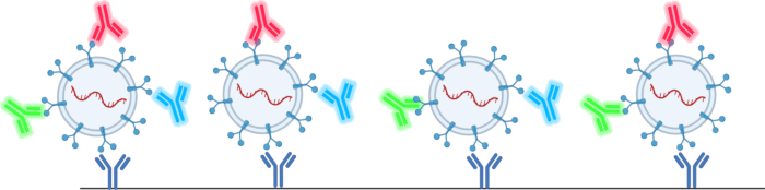
Figure 2: Schematic of how the tetraspanin chips capture and detect EVs. Please click here to view a larger version of this figure.
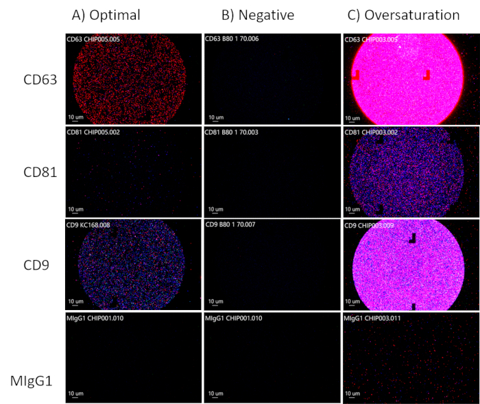
Figure 3: Optimizing EV concentrations. (A) Optimal concentration of EVs. (B) Negative result: no EVs present. (C) Oversaturation of EVs. Please click here to view a larger version of this figure.
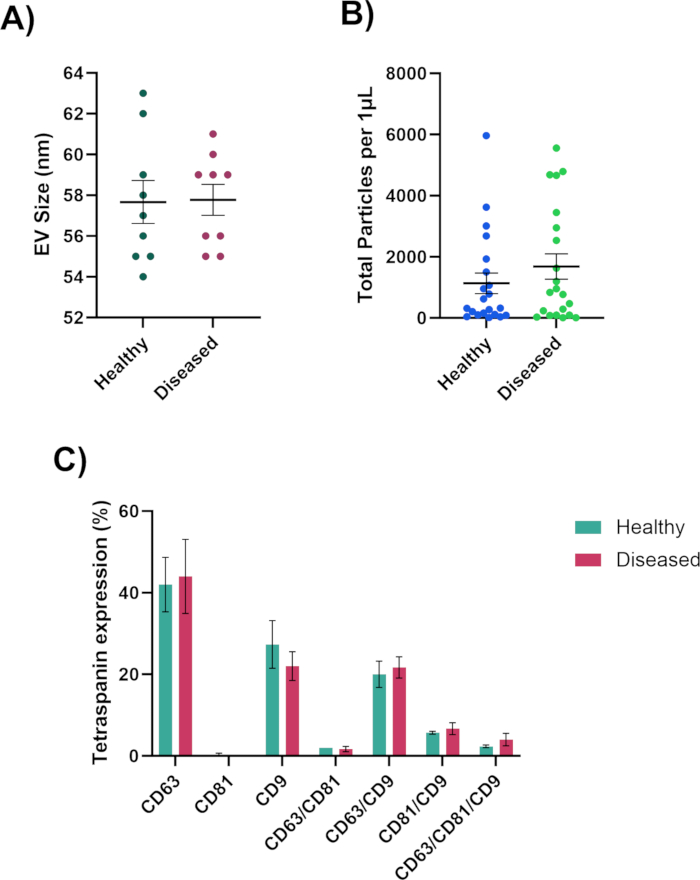
Figure 4: Data generated by the nanoparticle analyzer. (A) EV diameter size measured in nanometers. (B) Total particle count of EVs. (C) EV colocalized phenotype. Please click here to view a larger version of this figure.
| Technique | Advantages | Limitations |
| Ultracentrifuge | High Purity42,43 | Changes EV morphology42,44 |
| Homogeneity42,45 | Requires large volume42,44,46 | |
| Functionality42,43 | Expensive42,43 | |
| Flow Cytometry | Evaluate samples at multiple time points47 | Weak signal48,49 |
| Multiple endpoints47,50 | Clogged pores51 | |
| High throughput single EV analysis52,53 | ||
| Density Gradient Centrifugation | Produce highly pure samples54 | Labor intensive55,56 |
| Variety of samples55,57 | Time-consuming55,58 | |
| Significant yield loss54 | ||
| Precipitation | Simple59,60 | Contamination36,44,61 |
| Fast61,62 | Non-Specific63 | |
| High Yield44,59,62 | ||
| Neutral pH36 | ||
| Filtration | Simple64 | Trap exosomes65 |
| Fast64 | Damages large vesicles36 | |
| Inexpensive64 | Filter cake66,67 | |
| Exosome clogging65 |
Table 1: Advantages and Limitations of EV isolation techniques. A list of the various ways to isolate EVs, including their advantages and limitations.
| Technique | Advantages | Limitations |
| Dynamic light scattering | Measure particles ranging in size 1 nm to 6 µM78 | Not suitable for measuring complex exosome samples with large size range79 |
| The lower limit is 10 nm suitable for monodisperse systems79 | Not able to distinguish contaminated proteins from exosomes79 | |
| Transmission electron microscopy (TEM) | Observe morphology of exosomes79,80 | Complicated sample preparations79 |
| Observe internal structure81 | Not able to distinguish exosomes based on size and shape because of exaggerated fluorescence signals82 | |
| Nanoparticle Tracking analysis | Measure concentration, size and size distribution of exosomes in the 10 nm to 2 µM range78 | Cannot differentiate the EVs from protein aggregates and other contaminants83 |
| Quick sample preparation and measurement78,84 | Expensive NTA instrument85 | |
| Samples can be recovered in native form84 | Sensitive to vibrations85 | |
| Western Blot (WBs) | can qualitatively and quantitatively analyze marker proteins79,86 | Complicated and time consuming79 |
| Analyzing exosomes from cell culture media79 | EV isolates may contain lipoproteins and other contaminants86 | |
| Flow cytometry | Higher sensitivity and high-resolution imaging to | Time-consuming and laborious with detection limit of 400 nm79,88 |
| distinguish stained exosomes from containments87 | ||
| Required low sample concentration79 | Optical signals hinder the accuracy and resolution88 | |
| Exoview | Measure Tetraspanins (CD9, CD63, and CD81) on exosomes89 | Does not measure larger EV sizes75 |
| Measures EV cargo proteins90 |
Table 2: Advantages and limitations of EV characterization techniques.
Discussion
The most critical step in this protocol is to make sure that an optimal concentration of EVs is achieved. There needs to be enough EVs present to obtain a reading, but not too many EVs that will oversaturate the chip. The best way to determine the optimal EV concentration is to do an optimization run with 1 µL of sample and see if the concentration needs to be adjusted. Another critical step is to see if there is an abundance of cell debris in the sample, which can be determined by viewing large chunks on the chips in the analyzing software. If the sample has cell debris, a simple centrifugation or filtering of the sample should resolve this issue.
An additional, crucial step is ensuring that the chip(s) do not touch the walls of the well and avoiding contact with the center area of the chip(s) when placing them up the ramp. The scanner will read from the square at the center of the chip, so it is essential to avoid touching that area with the forceps to prevent disrupting the EVs.
This method does have some limitations such as having insufficient concentration of EVs for detection by the instrument. The concentration could be improved by either drying the sample or using a concentrator tube. Another limitation is that the analyzing software only measures EVs in the range of 50-200 nm, excluding some microvesicles and all apoptotic bodies from the size measurements75.
There are many ways to isolate (Table 1 42,43,44,45,46,47,48,49,50,51,52, 53,54,55,56,57,58,59,60, 61,62,63,64,65,66,67) and analyze EVs (Table 2 78,79,80,81,82,83,84,85,86,87,88,89,90), and the current gold standard for EV isolation is ultracentrifugation36,37,38to pellet the cells for assays such as Western Blots68,69,70and PCR71,72,73,74,75,76. While this protocol works well for large samples42,44,46, obtaining biological fluids can be challenging, and often, only a small volume of samples can be gathered at a time, which is not ideal for ultracentrifugation. In contrast, using this nanoparticle analyzer allows the user to generate valuable data, such as size, total particle count, and phenotype of the EVs, in as little as 1 µL of sample, making it ideal for biological fluids. We will be able to expand the knowledge of EVs from small volumes of samples that are difficult to extract, which can increase patient comfort and possibly find potential therapeutic targets for various diseases and disorders.
Disclosures
The authors have no competing financial interests or other conflicts of interest to disclose.
Acknowledgements
We would like to thank the NIH for the funding (EY031316 and EY034714). We would also like to thank UNTHSC and NTERI for the lab space.
Materials
| Name | Company | Catalog Number | Comments |
|---|---|---|---|
| ChipWasher 100 | NanoView | EV-CW100 | Incubates, washes, rinses and dries the tetraspanin chips. This current model is no longer available. Price at time of purchase: $9,995.00 |
| ExoView Analyzer software | NanoView | N/A | Analyzes the chip informations and produces excel files for further analysis. No longer available. |
| ExoView R100 | NanoView | EV-R100 | Used to scan the tetraspanin chips at 3 wavelengths. This current model is no longer available. Price at time of purchase: $110,000.00 |
| ExoView Scanner software | NanoView | N/A | Scans the chips at 3 different wavelengths. No longer available. |
| Human Tetraspanin Kits | Unchained Labs | EV-TETRA-C | Includes 8 tetraspanin chips, Incubation Solution, Blocking Solution, CD63 antibody, CD81 antibody, CD9 antibody, Solution A, Solution B, USB, and plate cover. |
References
- Abels, E. R., Breakefield, X. O. Introduction to extracellular vesicles: Biogenesis, RNA cargo selection, content, release, and uptake. Cell Mol Neurobiol. 36 (3), 301-312 (2016).
- Radler, J., Gupta, D., Zickler, A., Andaloussi, S. E. Exploiting the biogenesis of extracellular vesicles for bioengineering and therapeutic cargo loading. Mol Ther. 31 (5), 1231-1250 (2023).
- Gupta, D., Zickler, A. M., El Andaloussi, S. Dosing extracellular vesicles. Adv Drug Deliv Rev. 178, 113961(2021).
- Dixson, A. C., Dawson, T. R., Di Vizio, D., Weaver, A. M. Context-specific regulation of extracellular vesicle biogenesis and cargo selection. Nat Rev Mol Cell Biol. 24 (7), 454-476 (2023).
- Keshtkar, S., Azarpira, N., Ghahremani, M. H. Mesenchymal stem cell-derived extracellular vesicles: novel frontiers in regenerative medicine. Stem Cell Res Ther. 9 (1), 63(2018).
- Zhang, H., et al. Exosome-induced regulation in inflammatory bowel disease. Front Immunol. 10, 1464(2019).
- Barzin, M., et al. Application of plant-derived exosome-like nanoparticles in drug delivery. Pharm Dev Technol. 28 (5), 383-402 (2023).
- Zhou, C., et al. Stem cell-derived exosomes: emerging therapeutic opportunities for wound healing. Stem Cell Res Ther. 14 (1), 107(2023).
- Kalluri, R., LeBleu, V. S. The biology, function, and biomedical applications of exosomes. Science. 367 (6478), aau6977(2020).
- Mondal, J., et al. Hybrid exosomes, exosome-like nanovesicles and engineered exosomes for therapeutic applications. J Control Release. 353, 1127-1149 (2023).
- van Niel, G., D'Angelo, G., Raposo, G. Shedding light on the cell biology of extracellular vesicles. Nat Rev Mol Cell Biol. 19 (4), 213-228 (2018).
- Paul, N., Sultana, Z., Fisher, J. J., Maiti, K., Smith, R. Extracellular vesicles- crucial players in human pregnancy. Placenta. 140, 30-38 (2023).
- Zaldivia, M. T. K., McFadyen, J. D., Lim, B., Wang, X., Peter, K. Platelet-derived microvesicles in cardiovascular diseases. Front Cardiovasc Med. 4, 74(2017).
- Tamasi, V., Nemeth, K., Csala, M. Role of extracellular vesicles in liver diseases. Life (Basel). 13 (5), 1117(2023).
- Fu, Y., et al. Emerging understanding of apoptosis in mediating mesenchymal stem cell therapy. Cell Death Dis. 12 (6), 596(2021).
- Santavanond, J. P., Rutter, S. F., Atkin-Smith, G. K., Poon, I. K. H. Apoptotic bodies: Mechanism of formation, isolation and functional relevance. Subcell Biochem. 97, 61-88 (2021).
- Yu, L., et al. Apoptotic bodies: bioactive treasure left behind by the dying cells with robust diagnostic and therapeutic application potentials. J Nanobiotechnology. 21 (1), 218(2023).
- Momen-Heravi, F., Getting, S. J., Moschos, S. A. Extracellular vesicles and their nucleic acids for biomarker discovery. Pharmacol Ther. 192, 170-187 (2018).
- Liu, Y. J., Wang, C. A review of the regulatory mechanisms of extracellular vesicles-mediated intercellular communication. Cell Commun Signal. 21 (1), 77(2023).
- Grange, C., Bussolati, B. Extracellular vesicles in kidney disease. Nat Rev Nephrol. 18 (8), 499-513 (2022).
- Cocucci, E., Meldolesi, J. Ectosomes and exosomes: shedding the confusion between extracellular vesicles. Trends Cell Biol. 25 (6), 364-372 (2015).
- Iannotta, D. A. A., Kijas, A. W., Rowan, A. E., Wolfram, J. Entry and exit of extracellular vesicles to and from the blood circulation. Nat Nanotechnol. 19 (1), 13-20 (2024).
- Saint-Pol, J., Gosselet, F., Duban-Deweer, S., Pottiez, G., Karamanos, Y. Targeting and crossing the blood-brain barrier with extracellular vesicles. Cells. 9 (4), 851(2020).
- Alberro, A., Iparraguirre, L., Fernandes, A., Otaegui, D. Extracellular vesicles in blood: Sources, effects, and applications. Int J Mol Sci. 22 (15), 8163(2021).
- Nieuwland, R., Siljander, P. R. A beginner's guide to study extracellular vesicles in human blood plasma and serum. J Extracell Vesicles. 13 (1), e12400(2024).
- Thangaraju, K., Neerukonda, S. N., Katneni, U., Buehler, P. W. Extracellular vesicles from red blood cells and their evolving roles in health, coagulopathy and therapy. Int J Mol Sci. 22 (1), 153(2020).
- Li, C., et al. Cerebrospinal fluid-derived extracellular vesicles after spinal cord injury promote vascular regeneration via PI3K/AKT signaling pathway. J Orthop Translat. 39, 124-134 (2023).
- Deng, Y., et al. Phosphoproteome analysis of cerebrospinal fluid extracellular vesicles in primary central nervous system lymphoma. Analyst. 148 (15), 3594-3602 (2023).
- Hirschberg, Y., et al. Proteomic comparison between non-purified cerebrospinal fluid and cerebrospinal fluid-derived extracellular vesicles from patients with Alzheimer's, Parkinson's and Lewy body dementia. J Extracell Vesicles. 12 (12), e12383(2023).
- Hefley, B. S., et al. Revealing the presence of tear extracellular vesicles in Keratoconus. Exp Eye Res. 224, 109242(2022).
- Cross, T., et al. RNA profiles of tear fluid extracellular vesicles in patients with dry eye-related symptoms. Int J Mol Sci. 24 (20), 15390(2023).
- Ma, H., et al. Metabolic signatures of tear extracellular vesicles caused by herpes simplex keratitis. Ocul Surf. 31, 21-30 (2024).
- Han, P., Bartold, P. M., Ivanovski, S. The emerging role of small extracellular vesicles in saliva and gingival crevicular fluid as diagnostics for periodontitis. J Periodontal Res. 57 (1), 219-231 (2022).
- Han, P., Li, X., Wei, W., Ivanovski, S. Saliva diagnosis using small extracellular vesicles and salivaomics. Methods Mol Biol. 2588, 25-39 (2023).
- Reseco, L., et al. Characterization of extracellular vesicles from human saliva: Effects of age and isolation techniques. Cells. 13 (1), 95(2024).
- Tiwari, S., Kumar, V., Randhawa, S., Verma, S. K. Preparation and characterization of extracellular vesicles. Am J Reprod Immunol. 85 (2), e13367(2021).
- Tomiyama, E., Fujita, K., Nonomura, N. Urinary extracellular vesicles: Ultracentrifugation method. Methods Mol Biol. 2292, 173-181 (2021).
- Xu, K., Jin, Y., Li, Y., Huang, Y., Zhao, R. Recent progress of exosome isolation and peptide recognition-guided strategies for exosome research. Front Chem. 10, 844124(2022).
- Brennan, K., et al. A comparison of methods for the isolation and separation of extracellular vesicles from protein and lipid particles in human serum. Sci Rep. 10 (1), 1039(2020).
- Gao, J., et al. Recent developments in isolating methods for exosomes. Front Bioeng Biotechnol. 10, 1100892(2022).
- Mol, E. A., Goumans, M. J., Doevendans, P. A., Sluijter, J. P. G., Vader, P. Higher functionality of extracellular vesicles isolated using size-exclusion chromatography compared to ultracentrifugation. Nanomedicine. 13 (6), 2061-2065 (2017).
- Ansari, F. J., et al. Comparison of the efficiency of ultrafiltration, precipitation, and ultracentrifugation methods for exosome isolation. Biochem Biophys Rep. 38, 101668(2024).
- Patel, G. K., et al. Comparative analysis of exosome isolation methods using culture supernatant for optimum yield, purity and downstream applications. Sci Rep. 9 (1), 5335(2019).
- Stam, J., Bartel, S., Bischoff, R., Wolters, J. C. Isolation of extracellular vesicles with combined enrichment methods. J Chromatogr B Analyt Technol Biomed Life Sci. 1169, 122604(2021).
- Le Roy, A., et al. AUC and small-angle scattering for membrane proteins. Methods Enzymol. 562, 257-286 (2015).
- Chhoy, P., Brown, C. W., Amante, J. J., Mercurio, A. M. Protocol for the separation of extracellular vesicles by ultracentrifugation from in vitro cell culture models. STAR Protoc. 2 (1), 100303(2021).
- McDorman, K. S., Chan, C., Rojko, J., Satterwhite, C. M., Morrison, J. P. Chapter 7 - Special Techniques in Toxicologic Pathology. Haschek and Rousseaux's Handbook of Toxicologic Pathology (Third Edition). , Academic Press. (2013).
- Woud, W. W., et al. An imaging flow cytometry-based methodology for the analysis of single extracellular vesicles in unprocessed human plasma. Commun Biol. 5 (1), 633(2022).
- Gul, B., Syed, F., Khan, S., Iqbal, A., Ahmad, I. Characterization of extracellular vesicles by flow cytometry: Challenges and promises. Micron. 161, 103341(2022).
- Black, C. B., Duensing, T. D., Trinkle, L. S., Dunlay, R. T. Cell-based screening using high-throughput flow cytometry. Assay Drug Dev Technol. 9 (1), 13-20 (2011).
- Safford, H. R., Bischel, H. N. Flow cytometry applications in water treatment, distribution, and reuse: A review. Water Res. 151, 110-133 (2019).
- Kobayashi, H., et al. Precise analysis of single small extracellular vesicles using flow cytometry. Sci Rep. 14 (1), 7465(2024).
- Welsh, J. A., et al. A compendium of single extracellular vesicle flow cytometry. J Extracell Vesicles. 12 (2), e12299(2023).
- Carnino, J. M., Lee, H. Extracellular vesicles in respiratory disease. Adv Clin Chem. 108, 105-127 (2022).
- Sun, Y., Sethu, P. Low-stress microfluidic density-gradient centrifugation for blood cell sorting. Biomed Microdevices. 20 (3), 77(2018).
- Strachan, B. C., Xia, H., Voros, E., Gifford, S. C., Shevkoplyas, S. S. Improved expansion of T cells in culture when isolated with an equipment-free, high-throughput, flow-through microfluidic module versus traditional density gradient centrifugation. Cytotherapy. 21 (2), 234-245 (2019).
- Malvezzi, H., Sharma, R., Agarwal, A., Abuzenadah, A. M., Abu-Elmagd, M. Sperm quality after density gradient centrifugation with three commercially available media: a controlled trial. Reprod Biol Endocrinol. 12, 121(2014).
- Sun, Y., Sethu, P. Microfluidic adaptation of density-gradient centrifugation for isolation of particles and cells. Bioengineering (Basel). 4 (3), 67(2017).
- Karttunen, J., et al. Precipitation-based extracellular vesicle isolation from rat plasma co-precipitate vesicle-free microRNAs. J Extracell Vesicles. 8 (1), 1555410(2019).
- De Sousa, K. P., et al. Isolation and characterization of extracellular vesicles and future directions in diagnosis and therapy. Wiley Interdiscip Rev Nanomed Nanobiotechnol. 15 (1), e1835(2023).
- Sidhom, K., Obi, P. O., Saleem, A. A review of exosomal isolation methods: Is size exclusion chromatography the best option. Int J Mol Sci. 21 (18), 6466(2020).
- Coughlan, C., et al. Exosome isolation by ultracentrifugation and precipitation and techniques for downstream analyses. Curr Protoc Cell Biol. 88 (1), e110(2020).
- Konoshenko, M. Y., et al. Isolation of extracellular vesicles from biological fluids via the aggregation-precipitation approach for downstream miRNAs detection. Diagnostics (Basel). 11 (3), 384(2021).
- Drozdz, A., et al. Low-vacuum filtration as an alternative extracellular vesicle concentration method: A comparison with ultracentrifugation and differential centrifugation. Pharmaceutics. 12 (9), 872(2020).
- Akbar, A., Malekian, F., Baghban, N., Kodam, S. P., Ullah, M. Methodologies to isolate and purify clinical grade extracellular vesicles for medical applications. Cells. 11 (2), 186(2022).
- Chen, J., et al. Review on strategies and technologies for exosome isolation and purification. Front Bioeng Biotechnol. 9, 811971(2021).
- Helling, A., et al. Investigation of microbial cell deformability by filter cake compressibility using ultrafiltration membranes. Colloids Surf B Biointerfaces. 185, 110626(2020).
- Bagci, C., et al. Overview of extracellular vesicle characterization techniques and introduction to combined reflectance and fluorescence confocal microscopy to distinguish extracellular vesicle subpopulations. Neurophotonics. 9 (2), 021903(2022).
- Thery, C., et al. Minimal information for studies of extracellular vesicles 2018 (MISEV2018): a position statement of the International Society for Extracellular Vesicles and update of the MISEV2014 guidelines. J Extracell Vesicles. 7 (1), 1535750(2018).
- van Maanen, J. C., et al. A combined western and bead-based multiplex platform to characterize extracellular vesicles. Tissue Eng Part C Methods. 29 (11), 493-504 (2023).
- Xu, D., et al. MicroRNAs in extracellular vesicles: Sorting mechanisms, diagnostic value, isolation, and detection technology. Front Bioeng Biotechnol. 10, 948959(2022).
- Lee, H., He, X., Le, T., Carnino, J. M., Jin, Y. Single-step RT-qPCR for detection of extracellular vesicle microRNAs in vivo: a time- and cost-effective method. Am J Physiol Lung Cell Mol Physiol. 318 (4), L742-L749 (2020).
- Kim, J. A., et al. Small RNA sequencing of circulating small extracellular vesicles microRNAs in patients with amyotrophic lateral sclerosis. Sci Rep. 13 (1), 5528(2023).
- Fan, Y., et al. Differential proteomics argues against a general role for CD9, CD81 or CD63 in the sorting of proteins into extracellular vesicles. J Extracell Vesicles. 12 (8), e12352(2023).
- Silva, A. M., et al. Quantification of protein cargo loading into engineered extracellular vesicles at single-vesicle and single-molecule resolution. J Extracell Vesicles. 10 (10), e12130(2021).
- Salunkhe, S., Basak Dheeraj, M., Chitkara, D., Mittal, A. Surface functionalization of exosomes for target-specific delivery and in vivo imaging, tracking: Strategies and significance. J Control Release. 326, 599-614 (2020).
- Silva, A. K. A., et al. Development of extracellular vesicle-based medicinal products: A position paper of the group "Extracellular Vesicle translatiOn to clinicaL perspectiVEs - EVOLVE France". Adv Drug Deliv Rev. 179, 114001(2021).
- Gurunathan, S., Kang, M. H., Jeyaraj, M., Qasim, M., Kim, J. H. Review of the isolation, characterization, biological function, and multifarious therapeutic approaches of exosomes. Cells. 8 (4), 307(2019).
- Zhang, Y., et al. Exosome: A review of its classification, isolation techniques, storage, diagnostic and targeted therapy applications. Int J Nanomedicine. 15, 6917-6934 (2020).
- Colombo, M., Raposo, G., Thery, C. Biogenesis, secretion, and intercellular interactions of exosomes and other extracellular vesicles. Annu Rev Cell Dev Biol. 30, 255-289 (2014).
- Pisitkun, T., Shen, R. F., Knepper, M. A. Identification and proteomic profiling of exosomes in human urine. Proc Natl Acad Sci U S A. 101 (36), 13368-13373 (2004).
- Zaborowski, M. P., Balaj, L., Breakefield, X. O., Lai, C. P. Extracellular vesicles: Composition, biological relevance, and methods of study. Bioscience. 65 (8), 783-797 (2015).
- Kowkabany, G., Bao, Y. Nanoparticle tracking analysis: An effective tool to characterize extracellular vesicles. Molecules. 29 (19), 4672(2024).
- Li, H. Metastatic characteristics of SY86B human gastric carcinoma in athymic nude mice. Zhonghua Zhong Liu Za Zhi. 10 (6), 421-423 (1988).
- Zhao, Z., Wijerathne, H., Godwin, A. K., Soper, S. A. Isolation and analysis methods of extracellular vesicles (EVs). Extracell Vesicles Circ Nucl Acids. 2, 80-103 (2021).
- Gandham, S., et al. Technologies and standardization in research on extracellular vesicles. Trends Biotechnol. 38 (10), 1066-1098 (2020).
- van der Vlist, E. J., Nolte-'t Hoen, E. N., Stoorvogel, W., Arkesteijn, G. J., Wauben, M. H. Fluorescent labeling of nano-sized vesicles released by cells and subsequent quantitative and qualitative analysis by high-resolution flow cytometry. Nat Protoc. 7 (7), 1311-1326 (2012).
- Pospichalova, V., et al. Simplified protocol for flow cytometry analysis of fluorescently labeled exosomes and microvesicles using dedicated flow cytometer. J Extracell Vesicles. 4, 25530(2015).
- Breitwieser, K., et al. Detailed characterization of small extracellular vesicles from different cell types based on tetraspanin composition by ExoView R100 platform. Int J Mol Sci. 23 (15), 8544(2022).
- An, H. J., Cho, H. K., Song, D. H., Kee, C. Quantitative analysis of exosomes in the aqueous humor of Korean patients with pseudoexfoliation glaucoma. Sci Rep. 12 (1), 12875(2022).
- Hefley, B. S., McKay, T. B., Hutcheon, A. E. K., Ciolino, J. B., Karamichos, D. Corneal epithelial-stromal constructs to study differences associated with diabetes mellitus. Exp Eye Res. 248, 110100(2024).
Reprints and Permissions
