This protocol describes a one-day method for the isolation of human fallopian tube epithelial cells. Isolated epithelial cells can be plated in 2-dimensional (2D) culture or dissociated into single-cell suspensions and utilized in downstream experiments, including flow cytometry and single-cell RNA sequencing.
Method Article
An Efficient Method for Extracting Human Fallopian Tube Epithelia for Single-cell Analyses
In This Article
Summary
Abstract
Human fallopian tubes are intrinsic to reproduction. The purpose of the fallopian tubes is to allow for the transit of sperm, the ovum, and if fertilization is successful, the embryo. The epithelial cells that line the inner surface of fallopian tubes are integral to both normal and abnormal fallopian tube processes, including disease initiation. After menopause, fallopian tubes cease to have a significant role in the body and the intraepithelial cell makeup changes. We describe a method in which these epithelial cells can be isolated from fresh fallopian tubes with minimal stromal cell contamination into a single-cell suspension. These cells can be grown in culture or used for further analysis such as flow cytometry and single-cell RNA sequencing. This isolation protocol can be achieved in 4-6 h and yields viable cells that can be used for immediate downstream analysis. This efficient protocol facilitates the isolation of fallopian tube epithelial cells with an enriched epithelial population.
Introduction
The fallopian tubes are constructed of multiple parts. From the ovary to the uterus, the fallopian tube is composed of the fimbriae, ampulla, isthmus, and the intramural portion. The fimbriae extend from the end of the fallopian tube where they seize the ovum released by the ovary. The ovum then travels through the ampulla, where it is most likely to be fertilized, to the isthmus and finally is transferred to the uterus through the intramural portion1. The innermost mucosa of the fallopian tube that facilitates ovum transportation is made up of a layer of luminal epithelium, including ciliated and secretory cells. Ciliated cells tend to be more concentrated at the fimbriae2. They play an integral role in physically moving the ovum through the fallopian tube from the ovary to the uterus. Their appendages allow the ciliated cells to not only move the ovum along but also to clear out genotoxic stress following ovulation3.
Secretory fallopian tube epithelial cells secrete a fluid that aids in nutrition and gamete assembly4. The proportion of ciliated and secretory cells along the fallopian tube epithelium differs in the post-menopausal state with a decrease in ciliated cells as the fallopian tube no longer serves a critical function in transportation5. Furthermore, in the absence of estrogen, the fallopian tubes are thought to become vestigial1,6. This loss of ciliated cells in the fallopian tube is posited to heighten the risk of developing serous carcinomas7. Additionally, fallopian tube secretory epithelial cells are thought to give rise to serous tubal intraepithelial carcinoma (STIC) lesions, a well-known precursor to the most aggressive subtype of tubo ovarian cancers, high grade serous carcinoma7,8.
The purpose of this protocol is to isolate epithelial cells from human fallopian tubes and dissociate them into single-cell suspensions. This protocol yields an enriched single-cell epithelial population that can be used for many analyses. As shown in this manuscript, we have performed flow cytometry analysis and plated cells in 2D after isolation. Flow cytometry analysis demonstrates the presence of single cells, which are mostly viable and epithelial in nature. In these analyses, we included four markers, e506 for viability, EpCAM for epithelial cells, CD45 for immune cells, and CD10 for stromal cells. Dead cells were excluded using the e506 viability marker, and immune cells were gated out using CD45. It is possible for the suspension to have an immune cell population; however, to achieve a relatively pure population of epithelial cells, CD45-positive cells can be depleted using a CD45 depletion kit. Further, when plated in culture, the CD45-positive cells often do not proliferate. Cells isolated via this method and grown in 2D show adherent cobblestone-like epithelial populations. This method can be used to generate cellular preparations, which can be developed into single-cell RNA libraries.
Research to define the cellular lineage of fallopian tube epithelia, alterations in these lineages during different phases of reproductive life, and inciting events that lead to malignant transformation and genesis has become more prominent within the last four years6,9,10,11,12.This protocol will significantly benefit research in this arena by providing an efficient way to isolate fallopian tube epithelia and process them into single cells.
Protocol
This protocol was adapted from a uterine epithelial cell isolation method described previously13,14. Fresh specimens of deidentified fallopian tubes were collected through our University of California, Los Angeles (UCLA) IRB-approved protocol #10-0727 and digested into a single-cell suspension within approximately 4–6 h.
1. Fallopian tube collection and preparation
- Collect fresh human fallopian tubes (Figure 1A) in dissociation media (DMEM [4.5 g/L D-glucose] containing 10% fetal bovine serum (FBS), 5 mL L-glutamine, and 5 mL of penicillin/streptomycin) in 15 mL or 50 mL tubes and transport on ice to the laboratory.
- Transfer the fallopian tubes into a sterile Petri dish containing fresh dissociation media.
- Under a dissecting microscope, dissect away fat, connective tissue, and any vessels surrounding the tubes using fine point forceps and Vannas Tubingen scissors (Figure 1B).
- Under the dissecting microscope, cut through the coronal plane of fallopian tubes with scissors to form small cylinders, ~3–5 mm each in diameter (Figure 1C), using the fine point forceps to stabilize the tissue.
2. Epithelial cell isolation and dissociation
- Carefully aspirate the dissociation media from the dish.
- Wash the tissue pieces 2x with cold 1x phosphate-buffered saline (PBS) by gently pipetting ~2 mL to cover the pieces and lightly rotating the plate.
- Aspirate the final 1x PBS wash, incubate the tissue pieces for 5 min in cold 5 mM ethylenediaminetetraacetic acid (EDTA) at room temperature, and repeat for a total of two incubations. Aspirate the 5mM EDTA after each incubation.
- Suspend the tissue fragments in cold 1% Trypsin/Hanks' Balanced Salt Solution (HBSS) and incubate at 4 ˚C for 40 min (Figure 1D).
- Aspirate the 1% trypsin/HBSS and wash the tissue pieces 2x in cold dissociation media to inactivate the trypsin.
- Incubate the tissue fragments in 2 mL of dissociation media with 0.4 mg of DNase for at least 30 min at room temperature.
NOTE: A longer incubation period may be necessary, but do not exceed 45 min before starting step 2.7. - Under a dissecting microscope, while still in the media with DNase, hold a fragment of the fallopian tube, the lumen parallel to the bottom of the dish, in place with forceps. With the other hand, repeatedly press on the fallopian tube piece with forceps to expel epithelial cells from the lumen of the tube. Confirm that a halo of cells is seen around the tissue as cells are released from the lumen of the tube. Repeat for each fragment of fallopian tube in the dish (Figure 1E).
- Transfer the media containing cells into a sterile 15 mL tube, wash the Petri dish with dissociation media at least 2x, and combine the washes into the 15 mL tube. Discard the stromal tube fragments.
- Harvest cells by centrifugation at 500 × g for 5 min. Aspirate the supernatant and resuspend the pellet in 1 mL of dissociation media.
- Count cells by aliquoting 10 µL of the cell suspension and mixing 1:1 with trypan blue. Pipette 10 µL of the mixture into a chamber counting slide and insert it into a cell counter (Figure 1F).
3. Digestion to single-cell suspension
- Resuspend the cells in 9 mL of dissociation media, 1 mL of 8 mg/mL collagenase (type 1), and 0.4 mg of DNase. Incubate at 37 ˚C for 30–45 min in an orbital shaker at 200 rpm (Figure 1G).
- Harvest the cells by centrifugation at 500 × g for 5 min. Aspirate the supernatant and resuspend the pellet in 5 mL of dissociation media to wash off collagenase.
- Pass the cell suspension through a 100 µm cell strainer into a 50 mL tube (Figure 1H).
- Harvest the cells by centrifugation at 500 × g for 5 min.
- If the cell pellet appears red, carry out red blood cell (RBC) lysis as described in steps 3.6–3.8. If the cell pellet is not red, skip to step 3.9.
- Dilute 500 µL of RBC lysis buffer in 4.5 mL of ultrapure water.
- Aspirate the media, add 5 mL of the diluted RBC lysis buffer, and incubate on ice for 3 min.
- To stop RBC lysis, add 45 mL of 1x PBS to the 50 mL tube.
- Harvest the cells by centrifugation at 500 × g for 5 min.
- Count the cells by aliquoting 10 µL of the cell suspension and mixing it 1:1 with trypan blue. Pipette 10 µL of the mixture into a chamber counting slide and insert it into the cell counter (Figure 1I).
4. Flow cytometry staining
- Aliquot 250,000 cells into two separate 5 mL polystyrene round bottom tubes for primary conjugated antibody and isotype control to use for surface marker flow cytometry.
NOTE: Fewer cells can be used to accommodate for the cell count and number of cells needed for downstream analysis. To achieve optimal flow results, a minimum of 50,000 cells should be included in the analysis. - Dilute the antibody (e.g., anti-human EpCAM and CD10) and isotype according to the manufacturer’s recommended dilution in 1x PBS. Include fixable viability dye (e506) and a conjugated anti-human CD45 antibody to gate out live human immune cells. Make enough antibody mixture for at least 50 µL per tube. Leave in the dark on ice until step 4.4.
- Wash the cells with 1 mL of 1x PBS. Harvest the cells by centrifugation at 500 × g for 5 min at 4 ˚C.
- Aspirate the 1x PBS. Resuspend the pellet in the antibody or isotype panel and incubate at 4 ˚C for 15 min in the dark.
- After incubation, dilute in 1 mL of 1x PBS and harvest the cells by centrifugation at 500 × g for 5 min at 4 ˚C.
- Aspirate the 1x PBS and resuspend the cell pellet in 200 µL of 1x PBS.
5. Flow cytometry data collection
- Log into the FACS machine.
NOTE: We present the protocol utilizing the referenced flow cytometer (see the Table of Materials). For other flow cytometers, adhere to the manufacturer's instructions. The voltage setting of the flow cytometer has been preconfigured for optimal performance with human PBMCs, eliminating the need for voltage adjustments before each FACS run. - If the machine is in acquisition mode, skip to step 5.3. If the machine is in data analysis mode, change to acquisition mode by pressing the power button in the top right. When a popup displays, press the button that reads acquisition mode.
- In the bottom left, if the machine reads Calibration Required, then proceed to step 5.3.1. If the machine reads Calibration ok, skip to step 5.4.
- To calibrate the machine, take the calibration beads out of the 4 °C fridge and vortex.
- Place 1 drop of calibration beads in a 5 mL polystyrene round bottom tube and place the tube in the single tube holder.
- Press the barcode button in the top right and scan the calibration bead bottle. A pop-up will appear. If everything is set up, click Yes | OK and let the calibration run.
- If the calibration is successful, log out and log back into acquisition mode. Proceed to step 5.4.
- Click open | new workspace in top left.
- Select chill 5 rack; select three of the wells. Select the first well and give it a description (e.g., FTX Ab). Select the next well and give it a unique description (e.g., FTX Iso). Select the last well and give it the description bleach.
NOTE: Always include a bleach sample after every 4–5 experimental samples to ensure timely cleaning of the cell samples. - Select all the wells and set up the experimental conditions as follows: instrument settings: Human PBMCs; Flow rate: high; Mix sample: gentle mixing; Mode: standard; Sample total volume 200 µL; Sample uptake 150 µL; Annotation: input antibodies and their fluorophore labeling.
- Select edit | options and give the file a name under description| apply | ok.
- Take the rack out of the fridge and place it on the tray with the barcode facing the machine. Place the sample tubes in the rack to reflect the order of samples on the screen and press the play button to start the experiment.
- Once the experiment has finished, plug in the USB, select File | copy, expand the private folder, select the experiment, and click copy and eject.
- After data collection, use software to analyze the data sheet.
- Start with the isotype control. Use the polygon tool to select cells on the forward scatter (FSC) versus side scatter (SSC) plot.
- Gate out dead cells by changing the Y-axis to e506 and the X-axis to FSC. Use the polygon tool to select the e506-negative live cell population.
- Double-click on the selected population. Change the Y-axis to human CD45. Use the polygon tool to select the cell population. This is the human CD45-negative cell population.
- Double-click on the CD45-negative cell population. Change the Y axis to histogram and X axis to human EpCAM. Use the range selection tool to click on the end of the histogram and select the empty space until the end of the graph. This represents the EpCAM-positive population. Repeat for human CD10 stain.
- Drag isotype gating under the antibody workspace. CD45-positive cells will be gated out and EpCAM-positive and CD10-positive populations will be selected for.
- To create representative histograms, select layout editor. Drag the desired histograms to the layout page. To compare isotype and antibody staining, overlay the isotype histogram with the antibody histogram. Gate the positive cells based on the isotype control.
6. Immunocytochemistry
- Culture cells after the final isolation step in the desired chamber glass slide until confluent.
- To fix the cells, add 500 µL of cold 4% Paraformaldehyde (PFA) Incubate at room temperature for 10 min.
- Aspirate the 4% PFA and wash 3x with 1x PBS.
- To permeabilize the cells, add at least 200 µL of 0.25% Triton-X in PBS and incubate at room temperature for 10 min.
- During permeabilization, prepare the blocking buffer (4% normal goat serum [NGS] for antibodies not produced in goat).
- Aspirate the 0.25% Triton-X and wash 3x with 1x PBS.
- Add at least 200 µL of blocking buffer (4% NGS in PBS). Block for 1 h at room temperature.
- Prepare primary antibody dilutions in 1% NGS in 1x PBS according to the manufacturer’s recommendations.
- After blocking, add at least 200 µL of the primary antibody solution. Incubate at room temperature for 2 h.
NOTE: Some primary antibodies may prefer a 4 °C overnight incubation; check antibody data sheet. - Prepare secondary antibody solutions in 1% NGS in 1x PBS. Wrap the tubes in tin foil and prepare solutions in the dark as the secondary antibodies are light-sensitive. Store in the dark until ready.
- After primary antibody incubation, wash 3x with 1x PBS.
- Add at least 200 µL of the secondary antibody solution. Incubate in the dark at room temperature for 1 h.
NOTE: During the following steps, try to keep the slide in the dark as much as possible. - After secondary antibody incubation, wash 3x with 1x PBS.
- Remove the chambers, either by simply removing them by hand or using the tool that comes with the slides. Follow the directions printed on the chamber slide packaging for removal.
- Add a drop of 4’,6-diamidino-2-phenylindole (DAPI) mounting serum and place a coverslip on the top. Limit the number of bubbles and make sure all the cells are covered. Optional step: use nail polish around the edges to seal coverslip.
- Lay the slide flat in a slide box to dry and place at 4 °C until imaging.
- Image on a microscope equipped with correct fluorescence based on secondary antibody conjugation to ensure clean image.
Results
We have included seven fallopian tube collections where we have isolated an enriched epithelial cell population (Figure 2A and Supplemental Figure S1A). To assess the viability and epithelial cell enrichment of this single-cell suspension method, flow cytometry was performed. To measure the cell viability, cells were stained with the viability marker, e506. This also allowed for gating out all debris and dead cells when analyzed.
To determine the composition of cells isolated via this method, samples were stained with an epithelial cell marker (EpCAM), a stromal cell marker (CD10), and an immune cell marker (CD45). As seen in Figure 2B, we isolated a viable and enriched epithelial cell population from fallopian tube tissue. Sample viability was at an average of 82%. For all samples, after gating out CD45-positive cells, we observed an enriched epithelial cell population with EpCAM-positive cells averaging 80% of the sample. There was some stromal cell contamination but only 7.8% on average. Figure 2C shows isolated cells in 2D 4-6 days after plating. It is apparent that epithelial cells were isolated as they formed consistent adherent cobblestone-looking clusters. In Supplemental Figure S1B, cells were plated after isolation and cultured for 2-6 days. Immunocytochemistry was used to characterize the cells growing in culture. Most cells in the culture stained positive for EpCAM and the secretory cell marker, PAX8. Only a few cells were identified as vimentin-positive. Ciliated cells were seen in culture as captured by video, shown in Supplemental Video S1.
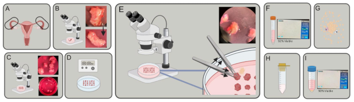
Figure 1: Experimental schema. (A) Fallopian tubes are acquired. (B) Excess fat and connective tissue are removed. (C) Tubes are cut into ~3-5 mm pieces. (D) The pieces are washed in PBS, then incubated in EDTA 2x for 5 min, and incubated in 1% trypsin for 40 min at 4 ˚C. (E) Using two pairs of forceps, one to hold and one to push, expel epithelial cells from the fallopian tube pieces. (F) Transfer the cells to a 15 mL tube, spin, and count. (G) Resuspend in DMEM, collagenase, and DNase. Digest for 30-45 min. (H) Strain with a 100 µm strainer. (I) Harvest by centrifugation and count. Please click here to view a larger version of this figure.
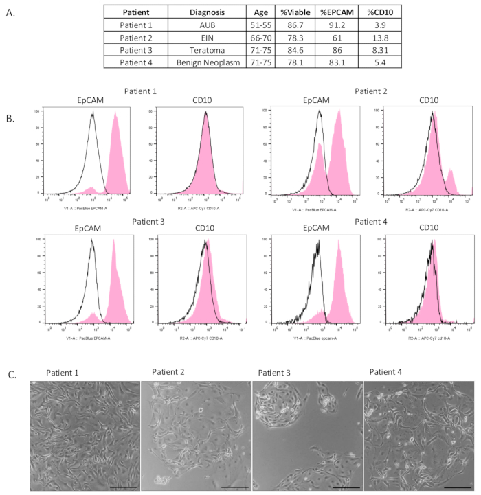
Figure 2: Characterization of isolated epithelial cells. (A) Table of patient clinical information and flow cytometry results. (B) Flow cytometry was performed after isolation to show that an enriched population of epithelial cells (EpCAM-positive) had been isolated. Isotype negative control is indicated in black, and the experimental result is indicated in pink. (C) Cells were plated after isolation. Photos were taken 4-6 days after plating. Scale bars = 100 µm. Abbreviations: AUB = Abnormal uterine bleeding; EIN = Endometrial intraepithelial neoplasia. Please click here to view a larger version of this figure.
Supplemental Figure S1: Additional characterization of isolated epithelial cells in culture. (A) Table of patient clinical information. (B) Isolated cells were cultured and stained for EpCAM, a secretory cell marker (PAX8), and a stromal cell marker (Vimentin) 2-6 days after plating to characterize cell type in culture. Scale bars = 50 µm. Abbreviations: EIN: Endometrial intraepithelial neoplasia; DAPI = 4',6-diamidino-2-phenylindole. Please click here to download this Figure.
Supplemental Video S1: Video of beating ciliated cells were taken at 30x magnification to demonstrate ciliated cells in culture. Please click here to download this Video.
Discussion
There is a considerable amount of interest in studying fallopian tube epithelium as fallopian tubes play a significant role in reproduction and are the site of origin for most HGSOC. To that end, many investigators have described protocols to isolate fallopian tube cells in both human and mouse models10,11,12,15,16,17,18,19,20,21. The method we describe to extract and enrich for fallopian tube epithelial cells adds to existing fallopian tube cell isolation protocols. Although overlaps exist within these protocols, there are two general types of methods reported in mice and humans. The first involves mincing and enzymatically digesting the whole fallopian tube that gives a total cell suspension10,12,15,16,17. The second involves sloughing with agitation or scraping, which results in sheets of tissue11,18,19,20,21. Both methods allow epithelial cells to be analyzed via downstream experiments such as flow cytometry, sequencing, and in-vitro culture. A major advantage of our method is that the protocol yields a population of fallopian tube cells that are already enriched for epithelial cells through enzymatic digestion and mechanical pushing steps. This population enriched for epithelia can be further digested resulting in a single-cell suspension that can be used for many applications such as flow cytometry, 2D culture, immunocytochemistry, and single-cell RNA sequencing.
Key steps we found to impact cell yield include the duration for which the fallopian tube fragments incubate in 1% trypsin/HBSS and DMEM/DNase. Overincubation in either solution will degrade the cells and significantly decrease the viability of the cells. However, insufficient incubation time will inhibit the researcher's ability to push out many epithelial cells during protocol step 2.6 as they will continue to adhere to each other. It is also important to use a 1% trypsin solution, as both lower and higher concentrations of trypsin solutions reduced the yield of viable fallopian tube epithelial cells. By combining chemical and mechanical isolation and a digestion step (protocol step 3.1), we can exponentially shorten the period it takes to go from tissue to single-cell suspension. This ensures good viability and time to perform downstream analysis on the same day.
In protocol step 1.4, it is critical to ensure that the pieces of fallopian tube cut are 3-5 mm thick. If the pieces are too large, it will be difficult to perform protocol step 2.7 and ultimately decrease the cell yield as a longer incubation time in DMEM/DNase will be necessary.
Although the cell suspension is enriched for epithelial cells, stromal cell contamination is inevitable. If the preparation needs to be purely epithelial cells, sorting using flow cytometry and the markers we described can be performed to isolate epithelial cells and deplete stromal cells.
Postmenopausal fallopian tubes were used in this study. However, this method has been successfully utilized in our lab on premenopausal fallopian tubes. The main difference between pre- and postmenopausal fallopian tubes is the composition of ciliated and secretory epithelial cells1. This protocol works for all reproductive stages.
This efficient protocol will facilitate investigating cell types of the fallopian tube epithelium, including delineating cellular lineages, their dynamic changes during reproductive cycles as well as after menopause, and their role in initiating high-grade serous ovarian cancer.
Disclosures
The authors have no conflicts of interest to declare.
Acknowledgements
Authors Ruegg L, James-Allan LB and DiBernardo G are partially supported by the Greater Los Angeles Veterans Association projects 1I01BX006019-01A2 and I01BX006411-01 to Memarzadeh S. Author Ochoa C is supported by UCLA Eli and Edythe Broad Center of Regenerative Medicine and Stem Cell Research Rose Hills Foundation Graduate Scholarship Training Program. We want to thank the Translational Pathology Core Laboratory at UCLA and specifically Ko Kiehle and Chloe Yin for assistance in tissue procurement. We also want to thank Ken Yamauchi and the BSCRC Microscopy core for their assistance in imaging. Figure 1 was created with BioRender.com (agreement number JL27QWDYNT). Lastly, we would like to thank Felicia Cordea and the BSCRC flow cytometry core for assistance in flow cytometry.
Materials
| Name | Company | Catalog Number | Comments |
|---|---|---|---|
| 1% Trypsin | Thermo Scientific | J63993.09 | |
| 100 µm Cell Strainer | Corning | 431752 | |
| 4% Paraformaldehyde | Electron Microscopy Sciences | 15710-S | |
| 5 mL round bottom tubes | Corning | 352008 | |
| 60 mm cell culture plate | Corning | CLS430589-500EA | |
| 6-well Cell Culture Plate | Corning | 353046 | |
| Anti-CD10 | BioLegend | 312212 | |
| Anti-CD326 (Ep-CAM) | BioLegend | 324218 | |
| Anti-CD45 | BioLegend | 304026 | |
| Anti-EpCAM | Abcam | ab223582 | |
| Anti-IgG2a PerCP | BioLegend | 400250 | |
| Anti-PAX8 | Sigma-Aldrich | 363M-15 | |
| Anti-Vimentin | Agilent Technologies | M072501-2 | |
| Chamber Slide System | Thermo Scientific | 154917PK | |
| Collagenase | Thermo Scientific | 17100017 | |
| DMEM | Thermo Scientific | 10569-010 | |
| DNase I | Sigma | 10104159001 | |
| EDTA | Sigma | 3690 | |
| eFluor 506 | Invitrogen | 65-0866-14 | |
| FBS | Sigma | F2442 | |
| Fine point forceps | VWR | 102091-526 | Any finepoint forceps of your choice will work |
| Fixable Viability Dye eFluor 506 | Invitrogen | 65-0866-14 | |
| FlowJo software version 9 | BD Biosciences | Data analysis software | |
| GlutaMAX | Gibco | 35050-061 | |
| HBSS | Thermo Scientific | 14175-095 | |
| MACSQuant Analyzer 10 flow cytometer | Miltenyi Biotec | ||
| MACSQuant Calibration Beads | Miltenyi Biotec | 130-093-607 | |
| Mammocult | Stemcell Technologies | 5620 | |
| Normal Goat Serum | Fisher Scientific | PI31873 | |
| PBS | Thermo Scientific | 14190-144 | |
| Penicillin-Streptomyocin | Gibco | 15140-122 | |
| PerCP Conjugated CD45 | BioLegend | 304026 | |
| Red Blood Cell lysis buffer | Tonbo Biosciences | TNB-4300-L100 | |
| Triton X-100 | Thermo Scientific | BP151-100 | |
| Vannas-Tubingen Spring Scissors | Fine Science Tools | 15003-08 | |
| VECTASHIELD with DAPI | Fisher Scientific | NC9524612 |
References
- Crow, J., Amso, N. N., Lewin, J., Shaw, R. W. Morphology and ultrastructure of fallopian tube epithelium at different stages of the menstrual cycle and menopause. Hum Reprod. 9 (12), 2224-2233 (1994).
- Patek, E., Nilsson, L., Johannisson, E. Scanning electron microscopic study of the human fallopian tube. Report II. Fetal life, reproductive life, and postmenopause. Fert Steril. 23 (10), 719-733 (1972).
- Coan, M., et al. Exploring the role of fallopian ciliated cells in the pathogenesis of high-grade serous ovarian cancer. Int J Mol Sci. 19 (9), 2512(2018).
- Leese, H., Tay, J., Reischl, J., Downing, S. Formation of fallopian tubal fluid: role of a neglected epithelium. Reproduction. 121 (3), 339-346 (2001).
- Donnez, J., Casanas-Roux, F., Ferin, J., Thomas, K. Changes in ciliation and cell height in human tubal epithelium in the fertile and post-fertile years. Maturitas. 5 (1), 39-45 (1983).
- Lengyel, E., et al. A molecular atlas of the human postmenopausal fallopian tube and ovary from single-cell RNA and ATAC sequencing. Cell Rep. 41 (12), 111838-111838 (2022).
- Tao, T., et al. Loss of tubal ciliated cells as a risk for "ovarian" or pelvic serous carcinoma. Am J Cancer Res. 10 (11), 3815(2020).
- Li, J., Fadare, O., Xiang, L., Kong, B., Zheng, W. Ovarian serous carcinoma: recent concepts on its origin and carcinogenesis. J Hematol Oncol. 5 (1), 8(2012).
- Hu, Z., et al. The repertoire of serous ovarian cancer non-genetic heterogeneity revealed by single-cell sequencing of normal fallopian tube epithelial cells. Cancer Cell. 37 (2), 226-242.e7 (2020).
- Dinh, H. Q., et al. Single-cell transcriptomics identifies gene expression networks driving differentiation and tumorigenesis in the human fallopian tube. Cell Rep. 35 (2), 108978(2021).
- Ulrich, N., et al. Cellular heterogeneity of human fallopian tubes in normal and hydrosalpinx disease states identified using scRNA-seq. Dev Cell. 57 (7), 914-929.e7 (2022).
- Brand, J., et al. Fallopian tube single cell analysis reveals myeloid cell alterations in high-grade serous ovarian cancer. iScience. 27 (3), 108990-108990 (2024).
- Memarzadeh, S., et al. Cell-autonomous activation of the PI3-kinase pathway initiates endometrial cancer from adult uterine epithelium. Proc Natl Acad Sci USA. 107 (40), 17298-17303 (2010).
- Cunha, G. R. Stromal induction and specification of morphogenesis and cytodifferentiation of the epithelia of the mullerian ducts and urogenital sinus during development of the uterus and vagina in mice. J Exp Zool. 196 (3), 361-369 (1976).
- Xie, Y., Park, E. -S., Xiang, D., Li, Z. Long-term organoid culture reveals enrichment of organoid-forming epithelial cells in the fimbrial portion of mouse fallopian tube. Stem Cell Res. 32 (1873-5061), 51-60 (2018).
- Karst, A. M., Drapkin, R. Primary culture and immortalization of human fallopian tube secretory epithelial cells. Nat Protoc. 7 (9), 1755-1764 (2012).
- Ford, M. J., Harwalkar, K., Yamanaka, Y. Protocol to generate mouse oviduct epithelial organoids for viral transduction and whole-mount 3D imaging. STAR Protocols. 3 (1), 101164(2022).
- Radecki, K. C., Lorenson, M. Y., Carter, D. G., Walker, A. M. Microdissection and dissociation of the murine oviduct: Individual segment identification and single cell isolation. J Vis Exp. (177), e63168(2021).
- Feng, L., et al. Protocol for the detection of organoid-initiating cell activity in patient-derived single fallopian tube epithelial cells. Methods Mol Bio. 2429, 445-454 (2022).
- Kessler, M., et al. The Notch and Wnt pathways regulate stemness and differentiation in human fallopian tube organoids. Nat Commun. 6 (1), 8989(2015).
- Fotheringham, S., Levanon, K., Drapkin, R. Ex vivo culture of primary human fallopian tube epithelial cells. J Vis Exp. (51), e2728(2011).
Reprints and Permissions
