This is a method for sampling and isolating culturable bacteria from the cutaneous microbiota of European plethodontid salamanders (Speleomantes genus). Here, we present a protocol for sampling salamander skin with swabs and processing them using culturable approaches. We describe sampling, isolation, and establishment of axenic culture with bacterial strain characterization.
A subscription to JoVE is required to view this content. Sign in or start your free trial.
Method Article
Isolation and Identification of Bacterial Strains from Skin of Terrestrial Amphibians
In This Article
Summary
Abstract
The microbiota is a vital element of higher organisms, with the cutaneous microbiota significantly contributing to the organism's defense against external agents. There is no standard method to sample Speleomantes (Amphibia: Plethodontidae) skin microbial communities for the microbial culturing approach. To address this, we have developed a practical protocol for isolating bacteria from the skin of individuals and a best practice for handling and storing samples. The steps are straightforward: swabs are stored in a physiological saline solution supplemented with glycerol upon sampling. Once transferred in the laboratory, swabs can be stored at +4 °C for a maximum of 10 days to ensure the integrity of the microbial community. This study offers interesting insights into the cutaneous microbiota investigation of terrestrial amphibian species, such as those belonging to the genus Speleomantes. This approach contributes to implementing, monitoring and conserving measures for endangered species and might improve the knowledge of microbiota's function in the health and ecology of amphibians. This protocol may also have broader significance for microbiome research in many wildlife conservation scenarios.
Introduction
All organisms harbor a variety of microbial symbionts on their epithelial surfaces, tissues, and organs1. The skin is the primary interface between the host and its environment, providing a habitat for diverse microorganisms2. The skin is the largest organ of vertebrates and acts as a barrier to mechanical, chemical, and microbial aggressions3. However, this wide exposure of skin to environmental conditions likely makes it the most critical ecological frontier that mediates the interaction of the organism with external biotic and abiotic factors4.
The skin microbiota of amphibians has been intensively studied in the last decade, and the relationship between this microbial community and its host has been deeply explored5,6,7,8,9,10. The biodiversity of skin microbiota is largely determined by the type of host (i.e., species-specific community), but further environmental factors can promote higher interspecific diversity11. The skin microbiota of amphibians has a distinctive composition from other animals. It is known that amphibians have been shown to have a mutualistic relationship with their skin microbes, being able to biosynthesize antimicrobial compounds or protect their host against emerging infectious diseases12.
The isolation of bacteria from the skin is usually reached after accomplishing different steps. The first step includes obtaining a representative sample collection by swabbing and avoiding external contamination. The microbial samples are suspended in sterile saline or buffer. Supplementing suspension liquids with protectants (e.g., glycerol) might improve bacterial community integrity during laboratory transfer13,14. Once transferred to the laboratory, samples can be processed immediately or after a short time to maintain microbial community integrity. The classic microbial approach involves sample processing by serial dilution to reduce microbial density and ensure better colony separation on the Petri plates. Sample and serial dilutions are then streaked onto non-specific, non-selective media such as agar Luria-Bertani (LBA)15, Tryptic Soy (TSA)16, Tryptone Yeast extract Agar (TYEA)17,18,19, or Nutrient (NA)20 that support the growth of a wide range of bacteria. Non-selective enrichments during sampling and processing are used since it is necessary to culture a broad range of microbes. The streak or spread plate methods distribute the microbial suspension evenly over the agar surface21. The even distribution allows different microbial species present in the sample to form distinct colonies. After incubation at 20-37 °C for 2-7 days, the microbial colonies are counted and distinguished based on morphology. Morphological characterization of the bacterial colony was performed on the basis of pigmentation, form, elevation, margin, opacity, and surface. Well-isolated colonies on the highest dilutions are sub-cultured to fresh agar plates to purify microbial cultures. The purity of the colonies is assured based on cell uniformity and affinity for Gram-staining. Several biochemical or molecular tests can also be performed to check the purity of colonies22. The pure cultures of isolated strains can be stored either on agar slants at 4 °C for short-term use or preserved long-term at -80°C in cryoprotectants (e.g., a glycerol-to-medium ratio of 20:80 or 50:50)23.
In this protocol, we report the best practice approach to isolating bacteria from the skin microbiota of Speleomantes cave salamanders, strictly terrestrial amphibians that can live both in surface and subterranean environments24. Given the lack of universally applied protocols in the literature, this study aimed to develop a best practice for sampling and preserving cutaneous microbiota samples from these terrestrial amphibians. To test this protocol, we used the Italian cave salamander (Speleomantes italicus) as a model, sampling 12 individuals from a cave population located in Abruzzo (Italy). Individuals were collected by hand and repeatedly swabbed (at least 5 times) on their dorsal, ventral sides and flanks. The used swabs were soaked in sterile distilled water. Swabs were then immersed in physiological 0.9% saline solution with and without glycerol supplementation. Upon transfer to the laboratory, sample preservation was tested under various conditions: uncontrolled room temperature, 4 °C, and -20 °C for 10 days. Culturable microbial communities were compared using culture-based methods on generic and semi-selective media (LBA, TSYEA, TSA, NA).
Access restricted. Please log in or start a trial to view this content.
Protocol
This protocol presents the best practices for obtaining and handling Speleomantes skin samples for a microbiological culturable approach to isolate bacterial strains. The material useful in the following steps is listed in the Table of Materials. The protocol follows the animal care guidelines of article 11 of DPR 357/97 upon the authorization of the Italian Ministry of Environment (PNM 74554 of 22/04/2024).
1. Media preparation
- Calculate and weigh the amount of ingredients powder (see the manufacturer's instructions for the amount of powder used for LBA, TSYEA, TSA, NA, or other media).
- Follow the manufacturer's instructions to dissolve the ingredients in distilled water. Depending on the medium and purchased product, boiling steps might be required.
- Bring the medium to the final volume when the ingredients fully dissolve and control the pH. If the value is higher or lower than expected, adjust it using the solutions suggested in the manufacturer's instructions. HCl and NaOH solutions are commonly used to decrease and increase pH, respectively.
- Add agar (2% w/v), heat until 95 °C, and boil for about 1 min to dissolve the agar before autoclavation.
- Sterilize the medium by autoclaving, following the manufacturer's instructions. Common autoclaving processes include 121-134 °C for 15-30 min treatment.
- After autoclaving, cool down the agar medium to around 45-50°C and pour it into sterile Petri dishes under a previously sterilized laminar flow hood
- After solidification, label the plates according to the medium type and preparation date.
2. Sample collection
- Rinse the salamander's skin (genus Speleomantes) in sterile distilled water to remove the transient microbial communities.
- Collect skin swabs from amphibians using sterile cotton tip swabs in a test tube. Gently rub the swabs over the skin surface to collect microbial samples without harming the individual.
- Pass a swab soaked in sterile distilled water on each side of the whole body, including all extremities, five times (five back-and-forth strokes) to sample the Individuals.
NOTE: Supplementation with additives is not recommended since it might harm individuals. - Place the swabs in tubes containing physiological saline solution (0.9% NaCl) supplemented with glycerol (20%).
- Pass a swab soaked in sterile distilled water on each side of the whole body, including all extremities, five times (five back-and-forth strokes) to sample the Individuals.
- Mix the sample with the solution, pushing the swab towards tube walls while squeezing the tube and rotating the swab thoroughly (10 times clockwise and 10 times anti-clockwise).
- After mixing, close the swab inside the tube and transfer the microbiological samples to the laboratory, maintaining the sample at 4 °C until the microbial analysis.
NOTE: All steps are performed using new sterile material and gloves for each individual to avoid contamination and the operator's exposure to biological and chemical contaminants.
3. Inoculation and Incubation
- Mix the microbiological sample thoroughly before processing.
- Prepare serial dilutions of the cutaneous sample with saline solution (0.9% NaCl), ranging from 10-1 to 10-6.
- Inoculate 100 µL of as-is sample and serial dilutions onto the prepared agar plates. Using a sterile L-shaped loop, streak the sample across the surface of the agar medium, distributing it evenly without applying excessive pressure to avoid damaging the agar.
- Invert the inoculated plates in an incubator at 20-25 °C for 3-5 days. This temperature range is favorable for growing many environmental mesophilic and psychotropic bacteria that inhabit amphibian skin.
NOTE: All steps are performed using sterile material under a previously sterilized laminar hood. Five independent replicates are prepared for each experimental unit. Since the starting density is unknown and unpredictable, including a broad range of dilutions is always helpful.
4. Strain purification and identification
- Select bacterial colonies with distinct morphologies for further analysis after incubation.
- First, choose well-isolated colonies to avoid contamination or overlap and avoid crowded areas where colonies could merge. Moreover, choose colonies with different appearances (e.g., color, size, shape, edge, surface, or texture) to ensure the sampling of potentially different species or strains.
NOTE: This step is crucial to selecting the different cultivable bacterial strains of the skin microbiota.
- First, choose well-isolated colonies to avoid contamination or overlap and avoid crowded areas where colonies could merge. Moreover, choose colonies with different appearances (e.g., color, size, shape, edge, surface, or texture) to ensure the sampling of potentially different species or strains.
- Subculture the selected colonies.
- Transfer a well-isolated colony of the original agar plate aseptically to a fresh agar plate using an inoculating loop. Streak the colony to isolate it and then incubate it appropriately.
- Repeat this step several times on fresh agar plates to obtain pure cultures from each bacterial isolate until the colonies isolated appear similar. Repeat subculturing using selective and generic media.
NOTE: Combining aseptic handling, visual and microscopic examination, and molecular identification ensures contamination-free strain.
- Check the purity of the microbial cultures by microscopic observations (from 5x to 100x objectives), Gram staining affinity (colonies fixing, coloring, and observation), and biochemical tests (e.g., catalase, oxidase, urease, and sugar fermentation test)21,25.
NOTE: Pure cultures should show uniform colony morphology, consistent microscopic characteristics, and biochemical traits. - Perform molecular identification.
- Extract DNA from purified bacterial colonies and perform 16S rRNA gene sequencing to identify the bacterial species. Perform PCR amplification of the 16S rRNA gene using bacterial universal primers, followed by sequencing and comparison with known sequences in databases such as GenBank26.
- Establish biobank cultures.
- Test axenic (or pure) cultures for sterility by inoculating them into a non-selective medium and checking for the absence of growth of contaminants. Store axenic cultures for medium-term and long-term use.
NOTE: Standard methods include streaking colonies on agar slants of the selective medium and room-temperature storage, suspending the culture in stock solutions prepared with a mixture of the selective medium and glycerol (20:80 or 50:50), or broth culture lyophilization (freeze-drying) and freezing at -80 °C.
- Test axenic (or pure) cultures for sterility by inoculating them into a non-selective medium and checking for the absence of growth of contaminants. Store axenic cultures for medium-term and long-term use.
Access restricted. Please log in or start a trial to view this content.
Results
The workflow of the experimental approach is illustrated in Figure 1.
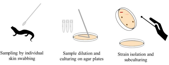
Figure 1: Workflow of the sampling, handling, and processing of the amphibian skin samples.
Access restricted. Please log in or start a trial to view this content.
Discussion
We developed a best practice protocol to obtain cultivable bacteria community from amphibian skin microbiota and demonstrated this on Speleomantes italicus. As demonstrated in the application above, several steps must be taken to ensure successful microbial isolation from amphibian skin. Collection, using a gentle swab technique and sterile materials, is very important to collect sufficient microbial material and avoid contamination. Transport and storage at 4 °C, with the swabs covered in a s...
Access restricted. Please log in or start a trial to view this content.
Disclosures
The authors have nothing to disclose.
Acknowledgements
We acknowledge financial support from the University of L'Aquila within projects "Progetti di Ateneo per la Ricerca di base 2024 UNIVAQ: Ecotoxicology of the endangered European cave salamanders" and "Analisi delle divergenze inter- e intraspecifiche del microbiota dei pletodontidi europei - FFORIC24.35" by Biodiversa+, the European Biodiversity Partnership, in the context of the Sub-BioMon - Developing and testing approaches to monitor subterranean biodiversity in karst project under the 2022–2023 BiodivMon joint call, and the National Recovery and Resilience Plan (NRRP), Mission 4, Component 2, Investment 1.1, Call for tender No. 1409 published on 14.9.2022 by the Italian Ministry of University and Research (MUR), funded by the European Union - NextGenerationEU- Project Code: P2022CYF9L, Project Title Are facultative cave species drivers of metal contaminants into subterranean environments? (METALCAVE) - CUP E53D23015380001.
Access restricted. Please log in or start a trial to view this content.
Materials
| Name | Company | Catalog Number | Comments |
|---|---|---|---|
| Lauria-Bertani agar | Merk Millipore | L3027 | |
| Tryptic Soy Agar | Merk Millipore | 22091 | |
| Tryptone Soy Yeast extract Agar | Merk Millipore | 17221 | |
| Nutrient Agar | Merk Millipore | 70148 | |
| Glycerol solution | Merk Millipore | 49781 |
References
- McFall-Ngai, M., et al. Animals in a bacterial world, a new imperative for the life sciences. Proc Natl Acad Sci U S A. 110 (9), 3229-3236 (2013).
- Belkaid, Y., Tamoutounour, S. The influence of skin microorganisms on cutaneous immunity. Nat Rev Immunol. 16 (6), 353-366 (2016).
- Kligman, A. M. What is the 'true' function of skin. Exp Dermatol. 11 (2), 159-187 (2002).
- Ross, A. A., Rodrigues Hoffmann, A., Neufeld, J. D. The skin microbiome of vertebrates. Microbiome. 7 (1), 79(2019).
- Kueneman, J. G., et al. Community richness of amphibian skin bacteria correlates with bioclimate at the global scale. Nat Ecol Evol. 3 (3), 381-389 (2019).
- Rebollar, E. A., MartínezUgalde, E., Orta, A. H. The amphibian skin microbiome and its protective role against chytridiomycosis. Herpetologica. 76 (2), 167(2020).
- Wuerthner, V. P., HernándezGómez, O., Hua, J. Amphibian skin microbiota response to variable housing conditions and experimental treatment across space and time. J Herpetol. 53 (4), 324(2019).
- HernándezGómez, O., Wuerthner, V., Hua, J. Amphibian host and skin microbiota response to a common agricultural antimicrobial and internal parasite. Microb Ecol. 79 (1), 175-191 (2020).
- SotoCortés, E., et al. Host species and environment shape the skin microbiota of Mexican axolotls. Microb Ecol. 87 (1), 98(2024).
- Cramp, R. L., McPhee, R. K., Meyer, E. A., Ohmer, M. E., Franklin, C. E. First line of defence: the role of sloughing in the regulation of cutaneous microbes in frogs. Conserv Physiol. 2 (1), cou012-cou012 (2014).
- Colston, T. J., Jackson, C. R. Microbiome evolution along divergent branches of the vertebrate tree of life: what is known and unknown. Mol Ecol. 25 (16), 3776-3800 (2016).
- Rebollar, E. A., et al. The skin microbiome of the neotropical frog Craugastor fitzingeri: inferring potential bacterialhostpathogen interactions from metagenomic data. Front Microbiol. 9, 466(2018).
- Li, X., Ricke, S. C. Comparison of cryoprotectants for Escherichia coli lysine bioavailability assay culture. J Food Process Preserv. 28 (1), 39-50 (2004).
- Patra, J. K., Das, G., Das, S. K., Thatoi, H. Isolation, Culture, and Biochemical Characterization of Microbes. In A Practical Guide to Environmental Biotechnology. , Springer. Singapore. (2020).
- Bertani, G. Studies on lysogenesis i. J Bacteriol. 62 (3), 293-300 (1951).
- Forbes, B. A., Sahm, D. F., Weissfeld, A. S. Diagnostic Microbiology. , Mosby. St Louis. (2007).
- O'Reilly, T., Niven, D. F. Tryptoneyeast extract broth as a culture medium for Haemophilus pleuropneumoniae and Haemophilus parasuis to be used as challenge inocula. Can J Vet Res. 50 (3), 441(1986).
- O'Reilly, T., Rosendal, S., Niven, D. F. Porcine haemophili and actinobacilli: characterization by means of API test strips and possible taxonomic implications. Can J Microbiol. 30 (10), 1229-1238 (1984).
- Niven, D. F. The cytochrome complement of Haemophilus parasuis. Can J Microbiol. 30 (6), 763-773 (1984).
- Lapage, S., Shelton, J. E., Mitchell, T. Methods in Microbiology. , Academic Press. London. (1970).
- Smibert, R. M. General Characterization. Manual of Methods for General Bacteriology. , American Society for Microbiology. Washington, DC. (1981).
- Sharma, N., Sharma, N., Sharma, S., Sharma, P., Devi, B. Identification, Morphological, Biochemical, and Genetic Characterization of Microorganisms. Basic Biotechniques for Bioprocess and Bioentrepreneurship. , Elsevier, Academic Press. (2023).
- Hubálek, Z. Protectants used in the cryopreservation of microorganisms. Cryobiology. 46 (3), 205-229 (2003).
- Lanza, B., Pastorelli, C., Laghi, P., Cimmaruta, R. A review of systematics, taxonomy, genetics, biogeography and natural history of the genus Speleomantes Dubois, 1984 (amphibia caudata plethodontidae). Atti Museo Civ St Natl, Trieste. 52, 5-135 (1984).
- Cappuccino, J. G., Welsh, C. T. Microbiology: A Laboratory Manual, Global Edition. , Pearson Education Limited: Pearson. Essex, England. (2017).
- Farda, B., et al. The "Infernaccio" gorges: microbial diversity of black deposits and isolation of manganesesolubilizing bacteria. Biology (Basel). 11 (8), 1204(2022).
- Lunghi, E., et al. Thermal equilibrium and temperature differences among body regions in European plethodontid salamanders. J Therm Biol. 60, 79-85 (2016).
- Woodhams, D. C., et al. Hostassociated microbiomes are predicted by immune system complexity and climate. Genome Biol. 21 (1), 23(2020).
- Stolp, H., Starr, M. P. Principles of Isolation, Cultivation, and Conservation of Bacteria. The Prokaryotes. , Springer. Berlin, Heidelberg. (1981).
- El Baz, S., et al. Resistance to and accumulation of heavy metals by actinobacteria isolated from abandoned mining areas. Sci World J. 2015 (1), 761834(2015).
- Le Pioufle, O., Declerck, S. Reducing water availability impacts the development of the arbuscular mycorrhizal fungus Rhizophagus irregularis MUCL 41833 and its ability to take up and transport phosphorus under in vitro conditions. Front Microbiol. 9, 1254(2018).
- Freitas, F., Alves, V. D., Reis, M. A. M. Advances in bacterial exopolysaccharides: from production to biotechnological applications. Trends Biotechnol. 29 (8), 388-398 (2011).
- Sanders, E. R. Aseptic laboratory techniques: plating methods. J Vis Exp. (63), e3064(2012).
- De Paoli, P. Biobanking in microbiology: from sample collection to epidemiology, diagnosis and research. FEMS Microbiol Rev. 29 (5), 897-910 (2005).
- Dal Pizzol, M., et al. Antifungal efficacy and safety of cycloheximide as a supplement in OptisolGS. Drug Des Devel Ther. 15, 2091-2098 (2021).
- Overmann, J. Principles of Enrichment, Isolation, Cultivation, and Preservation of Prokaryotes. The Prokaryotes. , Springer. Berlin, Heidelberg. (2013).
- Madhuri, R. J., et al. Recent Approaches in the Production of Novel Enzymes from Environmental Samples by Enrichment Culture and Metagenomic Approach. Recent Developments in Applied Microbiology and Biochemistry. , Elsevier, Academic Press. (2019).
- BaichmanKass, A., Song, T., Friedman, J. Competitive interactions between culturable bacteria are highly nonadditive. Elife. 12, e83398(2023).
- Cevallos, M. A., et al. Genomic characterization of antifungal Acinetobacter bacteria isolated from the skin of the frogs Agalychnis callidryas and Craugastor fitzingeri. FEMS Microbiol Ecol. 98 (12), fiac126(2022).
- Lauer, A., Simon, M. A., Banning, J. L., Lam, B. A., Harris, R. N. Diversity of cutaneous bacteria with antifungal activity isolated from female fourtoed salamanders. ISME J. 2 (2), 145-157 (2008).
- Niederle, M. V., et al. Skinassociated lactic acid bacteria from North American bullfrogs as potential control agents of Batrachochytrium dendrobatidis. PLoS One. 14 (9), e0223020(2019).
- MuletzWolz, C. R., et al. Antifungal bacteria on woodland salamander skin exhibit high taxonomic diversity and geographic variability. Appl Environ Microbiol. 83 (9), e00186-e00217 (2017).
- Woodhams, D. C., et al. Antifungal isolates database of amphibian skin-associated bacteria and function against emerging fungal pathogens. Ecology. 96 (2), 595-595 (2015).
- Woodhams, D. C., et al. Interacting symbionts and immunity in the amphibian skin mucosome predict disease risk and probiotic effectiveness. PLoS One. 9 (4), e96375(2014).
- Pessier, A. P. An overview of amphibian skin disease. Sem Avian Exot Pet Med. 11 (3), 162-174 (2002).
- HernándezGómez, O., Hua, J. From the organismal to biosphere levels: environmental impacts on the amphibian microbiota. FEMS Microbiol Rev. 47 (1), fuad002(2023).
- Stevens, E. J., Bates, K. A., King, K. C. Host microbiota can facilitate pathogen infection. PLoS Pathog. 17 (5), e1009514(2021).
- Kostanjšek, R., Prodan, Y., Stres, B., Trontelj, P. Composition of the cutaneous bacterial community of a cave amphibian, Proteus anguinus. FEMS Microbiol Ecol. 95 (3), fiz007(2019).
- Indriani, S., Karnjanapratum, S., Nirmal, N. P., Nalinanon, S. Amphibian skin and skin secretion: an exotic source of bioactive peptides and its application. Foods. 12 (6), 1282(2023).
- Gao, Y., et al. Targeted modification of a novel amphibian antimicrobial peptide from Phyllomedusa tarsius to enhance its activity against MRSA and microbial biofilm. Front Microbiol. 8, 628(2017).
- Wang, Y., Zhang, Y., Lee, W., Yang, X., Zhang, Y. Novel peptides from skins of amphibians showed broad-spectrum antimicrobial activities. Chem Biol Drug Des. 87 (3), 419-424 (2016).
- Barra, D., Simmaco, M. Amphibian skin: a promising resource for antimicrobial peptides. Trends Biotechnol. 13 (6), 205-209 (1995).
- RollinsSmith, L. A. The importance of antimicrobial peptides (AMPs) in amphibian skin defense. Dev Comp Immunol. 142, 104657(2023).
- RomeroContreras, Y. J., et al. Bacteria from the skin of amphibians promote growth of Arabidopsis thaliana and Solanum lycopersicum by modifying hormonerelated transcriptome response. Plant Mol Biol. 114 (3), 39(2024).
Access restricted. Please log in or start a trial to view this content.
Reprints and Permissions
