This protocol is designed to expedite and allow for the effective isolation of extracellular vesicles from human breast milk with high purity.
Method Article
Rapid Isolation of Human Breast Milk-Derived Extracellular Vesicles
In This Article
Summary
Abstract
Extracellular vesicles (EVs), nanosized particles approximately 20-1000 nm in size, are rapidly garnering attention for their therapeutic potential. Specifically, human breast milk-derived extracellular vesicles (HBMDEVs) have been shown to confer protection in experimental models of necrotizing enterocolitis, an intestinal disease that primarily affects premature infants, resulting in mortality as high as 50%. However, traditional extracellular vesicle isolation techniques, such as differential ultracentrifugation, size exclusion chromatography, etc., are either time-consuming or require specialized instrumentation, neither of which is practical in the clinical setting. Hence, there is a need for a simple isolation method that will allow for rapid reactive administration of HBMDEVs to patients. Proposed here is a combination of centrifugation of human breast milk to remove fat and cellular debris, coupled with rapid ultrafiltration of the remaining skim milk that produces high yield and functional extracellular vesicles. Using this technique may allow for the use of HBMDEVs as a practical therapeutic option in the clinical setting.
Introduction
Extracellular vesicles (EVs) are ubiquitous nanoparticles approximately 20-1000 nm in size, either via endosomal pathways (when microvesicular bodies fuse with the plasma membrane, which facilitates release of cellular cargo into the extracellular space) or plasma membrane pathways (i.e., budding). EVs contain host cellular cargo such as lipids, proteins, nucleic acids, various metabolites, and other contents depending on their source. Moreover, EVs are important in intercellular communication, angiogenesis, immune suppression, tumor microenvironment remodeling, cancer progression, etc.1,2,3,4. Notably, human breast milk (HBM) is EV-rich and easily accessible from lactating mothers5. Recently, human breast milk-derived EVs (HBMDEVs) have been garnering attention as therapeutic vectors for treating disease1,2. Premature or low birth weight neonates are susceptible to several infections, including necrotizing enterocolitis. Necrotizing enterocolitis, characterized by ileal or colonic necrosis, ranges from mild intestinal inflammation to multi-system organ failure with an infant mortality rate as high as 50%6. Pre-treatment with breast milk-derived EVs is sufficient to protect from intestinal inflammation in experimental models of NEC in vitro and in vivo2,7. Hence, utilization of HBMDEVs may be a viable option for the treatment of NEC in clinical settings.
Despite the therapeutic promise of HBMDEVs, there are known limitations using current isolation techniques in that they are either time-consuming or require specialized equipment, neither of which is universally practical for the clinical setting. The "gold standard" of EV isolation involves several rounds of differential ultracentrifugation, which, although it produces a satisfactory yield of EVs, may impact their structural integrity and functionality8,9. Additionally, ultracentrifugation times may be 2 h at a minimum, a costly resource in the clinical setting9. Isolation kits have proven to be both timesaving and somewhat cost-effective. However, isolation using such methods also results in low yields and hence low scalability. Hence, there is a need for HBMDEV isolation techniques that are both timesaving and cost-effective without sacrificing EV yield.
This protocol aims to outline a step-by-step method of rapidly isolating EVs from HBM using existing clinically available, FDA-approved tools. To accomplish this, standard centrifugation and a clinical-grade filter extension system were utilized to isolate EVs from HBM. These vesicles are within the expected size range, display morphology consistent with known EVs, are enriched for EV markers, and functionally similar to EVs isolated using traditional ultracentrifugation isolation techniques. Thus, this HBM EV isolation protocol may allow for practical, rapid isolation and application of EVs isolated from HBM in the clinical setting.
Protocol
Breast milk was collected in the University of Alabama at Birmingham (UAB) Regional Intensive Care Unit. Scavenged samples were collected after breast milk was thawed for patient use, and the patient was unable to take the whole feeding. These feedings are normally discarded, but were collected for research purposes. Samples were collected from de-identified individuals and stored at −80 °C until the time for the experiments. The collection was approved by the UAB Internal Review Board Protocol N160203002. The reagents and the equipment used are listed in the Table of Materials.
1. Extracellular Vesicle isolation (differential ultracentrifugation)
- In a biosafety cabinet, dilute breast milk 1:10 in sterile PBS in a 15 mL conical tube.
- Centrifuge the breast milk at 300 x g for 10 min at 4 °C. Following centrifugation, there should be 3 distinct layers: milk fat (top layer), whey or skim milk (middle aqueous layer), and the cells and other debris (pelleted at the bottom). In a biosafety cabinet, filter milk fat and whey layers through a 70-µm filter (allows for easier removal of fat) while leaving the pellet undisturbed. Transfer flow-through to a new tube.
- Centrifuge the sample at 2600 x g for 10 min at 4 °C. Residual milk fat, cells, and debris are removed in this step. In a biosafety cabinet, filter the supernatant through a 70-µm filter. Transfer the supernatant to an ultracentrifuge tube. Confirm that there is at least 10 mL of liquid in the tube before ultracentrifugation.
- Ultracentrifuge the sample at 20,000 x g for 45 min at 4 °C. Following ultracentrifugation, there may be three layers similar to step 1.2: milk fat, whey or skim milk, and cells and other debris. In a biosafety cabinet, filter milk fat and whey layers through a 70-µm filter without disturbing the pellet. Transfer the supernatant to a 15 mL conical tube.
- In a biosafety cabinet, syringe filter the breast milk with a 0.22 µm filter. Transfer the supernatant to a new ultracentrifuge tube. Confirm that there is at least 10 mL of liquid in the tube before ultracentrifugation.
- Ultracentrifuge the sample at 110,000 x g for 70 min at 4 °C. Aspirate the supernatant, and resuspend the pellet in 10 mL of sterile PBS.
- Ultracentrifuge the sample again at 110,00 x g for 70 min at 4 °C.
- In a biosafety cabinet, resuspend the pellet in 150-200 µL of sterile PBS.
2. Extracellular vesicle isolation (rapid filtration method)
- In a biosafety cabinet, dilute the breast milk (1:10) in sterile PBS.
- Centrifuge the breast milk at 300 x g for 10 min at 4°C. Following centrifugation, there should be three distinct layers: milk fat (top layer), whey or skim milk (middle aqueous layer), and the cells and other debris (pelleted in the bottom). In a biosafety cabinet, filter milk fat and whey layers through a 70-µm filter (allows for easier removal of fat) while leaving the pellet undisturbed. Transfer flow-through to a new tube.
- Centrifuge the sample at 2600 x g for 10 min at 4°C. Residual milk fat, cells, and debris are removed in this step. In a biosafety cabinet, filter supernatant through a 70-µm filter. Transfer supernatant to a 15 mL tube.
- In a biosafety cabinet, prime a 0.22 µm, 16-inch filter extension set with 5.4 mL PBS.
- To the prime system, take 5.4 mL of sterile PBS in a syringe and flush the system until PBS runs out of the other end of the tubing.
- Syringe filter the breast milk through a 0.22 µm, 16-inch filter extension set. Residual milk fat, cells, and debris are removed in this step.
NOTE: More than one filter extension set may be needed depending on the amount of debris present in the sample. - Collect the flow-through in a 15 mL conical tube.
3. Nanosight tracking analysis
- Dilute the sample (1:1000) in sterile PBS. Rapid filtered samples can be used undiluted.
- Briefly vortex the sample. Load 1 mL of the sample into a tuberculin syringe.
- Attach the syringe to the injection apparatus and secure the syringe.
- Set the injection pump to 50. Turn on the camera to ensure that the particles viewed are not too many or too few. Ensure that the flow of particles is constant with no skips in or sudden jumps in sample flow.
- Set the injection pump to 100. Run the protocol.
NOTE: Software was set to be read once every minute for a total of 5 reads. - Read the sample.
- Flush Nanosight in between samples with PBS.
NOTE: A nanoparticle tracking analysis system was used to determine particle size and particle concentration per milliliter. The average concentration of the ultracentrifuged sample was 3.30 e+08 +/- 4.08 e+06 particles/mL, while the rapid filtration method was 5.50e+08 +/- 1.16e+08 particles/mL.
4. Negative staining of extracellular vesicles and transmission electron microscopy
NOTE: Samples with exosome concentration in the range of 108 were used for transmission electron microscopy (TEM).
- Glow-discharge formvar/carbon film-coated mesh copper EM grids for 25 s at 20 mA using the discharge apparatus.
- Load 7 µL of the EV suspension solution on the grid and incubate for 1 min.
- Wick excess with Whatman filter paper by wicking from below the grid to "pull" the sample toward the grid rather than away from it.
- Stain the sample immediately with 7 µL of filtered 1% uranyl acetate (UA) solution on the surface of the EM grid by pipette.
- After 15 s, excess UA solution was removed from the grid by contacting the grid edge with filter paper. View sample immediately (within 24 h) on the TEM at 80 kv.
5. Protein concentration
- Acetone precipitate 250 µL of purified EVs (1:4) for 1 h at -20 °C.
NOTE: Use 100 µL of purified exosomes for the ultracentrifugation method. - Centrifuge the sample at 16,000 x g for 30 min.
- Air-dry the pellet, followed by re-suspension in 100 µL of RIPA buffer.
- Follow the BCA protein assay kit protocol to determine protein concentrations. Absorbance was measured at 562 nm using a microplate reader.
NOTE: 1 mL of breast milk normally yields approximately 1.3 µg/mL of protein using ultracentrifugation and approximately 0.2-1 µg/mL of protein using rapid filtration.
6. Western blot
- Incubate a 1 volume protein sample with a 4 volume lane marker, reduce the sample buffer, and boil it for 7 min at 90°C.
- Load and run 10 µg of protein on an SDS 10% gel at 70 V for 15 min. Afterwards, increase the voltage to 100 V and run for an additional 90 min.
- Transfer the gel to a nitrocellulose membrane using the semi-dry apparatus for 30 min at 15 V.
- Wash the membrane 3 times with TBS-0.1% Tween (TBS-T) for 10 min each.
- Block the membrane for 30 min using Fast Blocking Buffer solution (diluted to 1x).
- Incubate primary antibody (Hsp70-1:1000; CD63-1:1000 and CD9-1:1000) in 1x Buffer solution, overnight at 4°C on a rocker10,11,12.
- Wash the membrane three times for 10 min each with TBS-T.
- Add (H+L) HRP-conjugate anti-mouse secondary antibody, at a 1:1000 dilution for all interrogated proteins (Hsp70, CD9, and CD63) in 2% Milk in TBS-T and incubate for 2 h at room temperature on a rocker.
- Wash the membrane three times for 10 min each with TBS-T.
- Develop using ECL (1:1) and film with developer.
7. In vitro culture of IEC-6 cells
- Seed 1.0 x 10⁶ cells in a T-25 flask in 5 mL of complete RPMI media. Let cells adhere overnight.
- Remove complete media and wash with 1-2 mL of PBS.
- Aspirate PBS and add 5 mL of EV-free complete RPMI.
- Add a final concentration of 400 µM H2O2 per well and 10 µg/mL of EVs simultaneously to cells overnight.
- Remove media and wash cells with 1-2 mL of PBS.
- Aspirate the PBS and add 1 mL of Trypsin.
- Place the cells in the incubator at 37°C for 5 min.
- Deactivate trypsin by adding double the volume of complete RPMI. Transfer cells to a 15 mL tube and centrifuge at 125 x g for 5 min.
- Aspirate the supernatant and resuspend the pellet in 1 mL of PBS.
- Count cells using a cell counter.
Results
Isolation of EVs using ultracentrifugation and rapid isolation
Dogmatically, EV isolation from HBM involves centrifugation to remove milk fat and other large particles, followed by filtering, several rounds of ultracentrifugation, and re-suspension of the EV-enriched sample in PBS. The rapid filtration method utilizes centrifugation to remove milk fat and cellular debris, followed by filtration via a 0.22 µm filter extension filter, further removing residual debris, which yields an EV-enriched sample comparable to traditional ultracentrifugation (Figure 1).
Characterization of HBMDEVs from the filter extension rapid filtration system
Nanosight tracking analysis revealed comparable particle size between ultracentrifuged (Figure 2A) and rapid filter isolated HBMDEVs (Figure 2B). Both methods indicate that EVs isolated have a mean size of approximately 100 nm. EVs carry various cargo (i.e., proteins, lipids, miRNAs, etc.) for secretion from host cells to their various targets. Hence, the first target protein examined was heat shock protein 70 (Hsp70), a protein known to be transported via EVs13. Western blot analysis shows that EVs isolated using rapid filtration showed comparable expression of Hsp70, as those isolated using ultracentrifugation (Figure 3A and Supplementary Figure 1A). Additionally, EVs can be identified via surface expression of tetraspanins such as CD9 and CD6314. Western blot analysis revealed that EVs isolated via rapid filtration do indeed express CD9 (Figure 3B and Supplementary Figure 2B) and CD63 (Figure 3C and Supplementary Figure 3C). Lastly, morphological analysis of HBMDEVs via transmission electron microscopy revealed similar vesicle morphology between the two isolation techniques (Figure 4A-D).
Several labs have shown that EVs confer protective properties that promote cell survival during cellular stress2,3,7. Similar to previously published reports, EVs isolated using the rapid filtration method are able to protect intestinal epithelial cells (IEC-6) from hydrogen peroxide-induced cell death2 (Figure 5A,B). These data suggest that HBMDEVs isolated using the rapid filtration method not only possess comparative qualities to those isolated using differential ultracentrifugation but also are functionally similar.
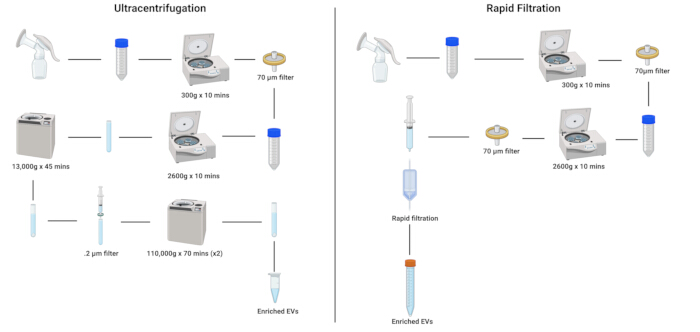
Figure 1: Schematic delineating the differences between ultracentrifugation and rapid filtration methods of EVs isolation from human breast milk. Please click here to view a larger version of this figure.
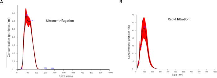
Figure 2: Nanosight tracking analysis of EVs isolated from human breast milk using ultracentrifugation and rapid filtration method. (A) NTA of human breast milk EVs (diluted 1:1000 in 1x PBS) isolated using ultracentrifugation. The data shown is a single representation of 3-4 independent trials. (B) NTA of human breast milk EVs isolated using rapid filtration. The data shown is a single representation of 3-4 independent trials. Please click here to view a larger version of this figure.
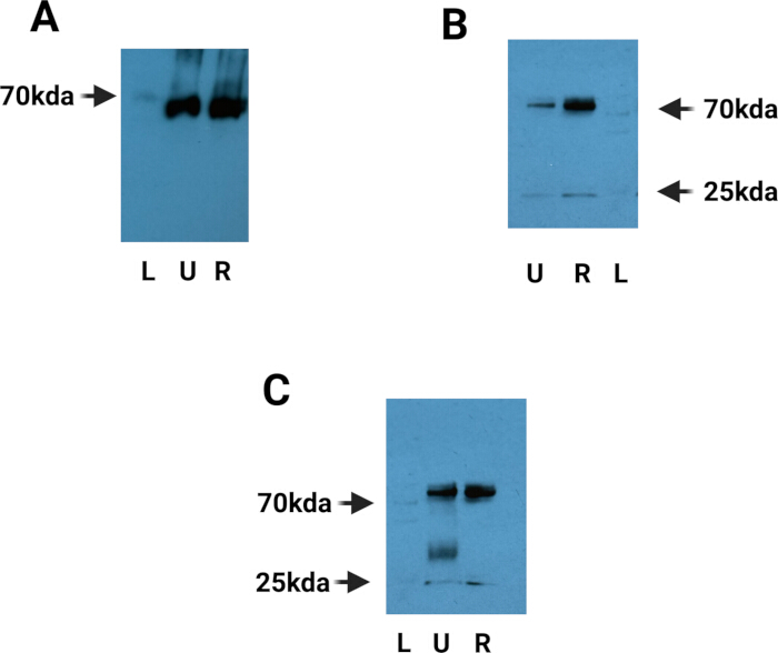
Figure 3: Western blot analysis of EVs isolated using ultracentrifugation and rapid filtration. (A) One-second exposure of 10 µg of heat shock protein 70 (Hsp70). "L" denotes ladder, "U" denotes ultracentrifugation, and "R" denotes rapid filtration. Samples obtained by rapid filtration were further concentrated by centrifugation at 110,000 × g for 70 min, performed twice. The image shown represents one of four independent trials. (B) Twenty-second exposure of 10 µg of CD9. "L" denotes ladder, "U" denotes ultracentrifugation, and "R" denotes rapid filtration. Rapid filtration samples were concentrated by centrifugation at 110,000 × g for 70 min, performed twice. The image shown represents one of four independent trials. (C) Twenty-second exposure of 10 µg of CD63. "L" denotes ladder, "U" denotes ultracentrifugation, and "R" denotes rapid filtration. Rapid filtration samples were concentrated by centrifugation at 110,000 × g for 70 min, performed twice. The image shown represents one of four independent trials. Please click here to view a larger version of this figure.
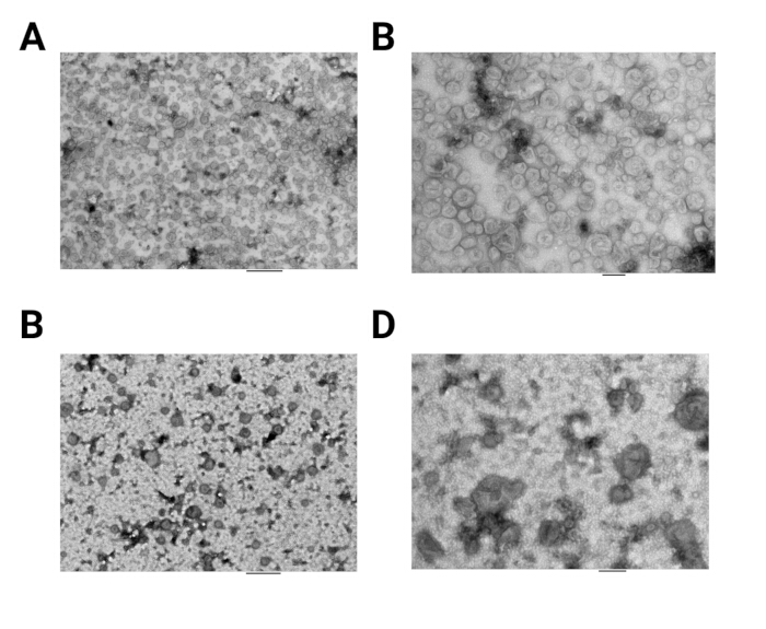
Figure 4: Transmission electron microscopy analysis of EVs isolated using ultracentrifugation and rapid filtration. (A) Electron micrograph of EVs isolated using ultracentrifugation at 8,000× magnification. Scale bar = 800 nm. The image shown represents one of three independent trials. (B) Electron micrograph of EVs isolated using ultracentrifugation at 20,000× magnification. Scale bar = 200 nm. The image shown represents one of three independent trials. (C) Electron micrograph of EVs isolated using rapid filtration at 8,000× magnification. Scale bar = 800 nm. EVs were concentrated by centrifugation at 110,000 × g for 70 min, performed twice. The image shown represents one of three independent trials. (D) Electron micrograph of EVs isolated using rapid filtration at 20,000× magnification. Scale bar = 200 nm. EVs were concentrated by centrifugation at 110,000 × g for 70 min, performed twice. The image shown represents one of three independent trials. Please click here to view a larger version of this figure.
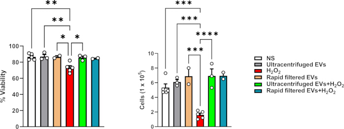
Figure 5: Functional analysis of EVs isolated using ultracentrifugation and rapid filtration. (A) Cell viability of IEC-6 cells cultured in EV-free complete RPMI-1640 medium. Cells were treated overnight with 400 µM H2O2 and 10 µg/mL of EVs simultaneously. The data shown represents one of two independent trials. (B) Cell counts of IEC-6 cells cultured in EV-free complete RPMI-1640 medium. Cells were treated overnight with 400 µM H2O2 and 10 µg/mL of EVs simultaneously. The data shown represents one of two independent trials. Please click here to view a larger version of this figure.
Supplementary Figure 1: Western blot characterization of Hsp70 in EVs isolated using ultracentrifugation and rapid filtration. One-second exposure of 10 µg of heat shock protein 70 (Hsp70). "L" denotes ladder, "U" denotes ultracentrifugation, and "R" denotes rapid filtration. Rapid filtration samples were concentrated by centrifugation at 110,000 × g for 70 min, performed twice. Each sample represents a single biological replicate. Please click here to download this File.
Supplementary Figure 2: Western blot characterization of CD9 in EVs isolated using ultracentrifugation and rapid filtration. Five-minute exposure of 10 µg of CD9. "L" denotes ladder, "U" denotes ultracentrifugation, and "R" denotes rapid filtration. Rapid filtration samples were concentrated by centrifugation at 110,000 × g for 70 min, performed twice. Each sample represents a single biological replicate. Please click here to download this File.
Supplementary Figure 3: Western blot characterization of CD63 in EVs isolated using ultracentrifugation and rapid filtration. Ten-minute exposure of 10 µg of CD63. "L" denotes ladder, "U" denotes ultracentrifugation, and "R" denotes rapid filtration. Rapid filtration samples were concentrated by centrifugation at 110,000 × g for 70 min, performed twice. Each sample represents a single biological replicate. Please click here to download this File.
Discussion
EVs are garnering attention as literature suggests that they may regulate various inflammatory mechanisms and may offer protective benefits2,7. However, a limitation of the practical use of EVs in clinical settings is the time needed to isolate EVs and extract a sufficient yield8,15. Here, a step-by-step protocol for rapid and efficient isolation of EVs from HBM using an FDA-approved filtration system has been outlined. The utility of this method cuts traditional EV isolation methods in half without sacrificing yield. Hence, this protocol may allow for practical therapeutic application of human breast milk-derived EVs in a clinical setting, should EV therapy be approved in the foreseeable future.
Existing EV isolation protocols have demonstrated consistent and effective yield of EVs from their source fluid. However, traditional means of isolation, whether it be the gold standard, differential ultracentrifugation, or the use of commercial kits, are not without their limitations. Isolation of EVs via differential ultracentrifugation, while effective, is time-consuming with protocols involving combined spin times of a minimum ~3 h2,8,15. Isolation time combined with protocols to properly quantify EV numbers would make their use under time constraints in a clinical setting challenging. Moreover, commercial kits, while effective, are costly. In addition, the number of samples that can be processed per kit is low, given the high costs of these kits8,15.
This protocol offers a solution to these limitations that saves time and money without sacrificing EV yield or purity. For this protocol to be effective, there are key steps that must be followed when carrying out the protocol. Dilution and initial centrifugation of the human breast milk are necessary for the removal of extracellular debris and milk fat from the milk sample. Removing this debris is critical for preventing clogging of the filter extension system and thus prevents having to use multiple filters for one sample. Additionally, flushing the filter extension system with PBS is critical as it primes the filter in preparation for the sample. Priming helps ensure that the sample is not lost during the priming process. It should be noted that, depending on the content of the breast milk sample, more than one filter extension set may be needed, regardless of diluting and centrifuging the sample prior to filtration. In the event this occurs, more than one filter may be used; however, the new filter extension set needs to be primed prior to filtration of the sample.
This protocol is effective and consistent in isolating EVs from human breast milk, however, there are a couple of limitations to consider. The centrifuge steps (~20 min total time between both spins) prior to filtration of the sample are necessary. Attempts to filter the diluted sample without centrifugation will lead to repeated clogging of the filter extension system, loss of sample, and add additional time to the EV isolation protocol. Moreover, the sample was not pure, as evidenced by NTA and TEM analysis. Attempts to use samples that have not been centrifuged prior to filtration may introduce unwanted contaminants into the sample, which may produce off-target effects depending on future downstream application of isolated EVs. Additionally, this protocol has only been implemented with respect to the isolation of EVs from human breast milk. Hence, the efficacy of this protocol in isolation of EVs from other fluids has not been investigated. Given the aqueous nature of all bodily fluids, it is not anticipated that there will be any differences in EV purity and yield if this protocol is used to isolate EVs from other sources.
It should also be noted that this protocol is efficient in isolating small extracellular vesicles (sEV). According to NTA, HBM-derived EVs were ~100 nm in size, suggesting that larger EVs were not present in the sample. The reason is that samples were filtered using a 0.22 µm filter, which is primarily used for the isolation of exosomes and other smaller EVs, a primary focus of the lab at the time of this study. In theory, utilization of a filter with larger pore sizes may allow for isolation of larger EVs. However, these questions are beyond the scope of this present study.
Human breast milk-derived EVs represent a natural therapeutic source that is easily accessible for clinical use. One limitation to bringing EV work to the clinical setting is isolating EVs in a practical method that any facility could potentially use. This protocol is designed to overcome that limitation and can easily be applied in the hospital setting and implemented using an existing hospital-grade, FDA-approved filtration system, commercially available, or can be purchased for much less than the standard ultracentrifuge. This method is cost-effective and produces an EV-rich and pure sample from a filtration system that is already used in the clinical setting.
Disclosures
The author(s) declared no potential conflicts of interest with respect to the research, authorship, and/or publication of this article.
Acknowledgements
Images and figures were generated using biorender.com. Research reported in this publication was supported by the UAB High Resolution Imaging Facility. This work was supported by NIH/NICHD 5R21HD104481.
Materials
| Name | Company | Catalog Number | Comments |
|---|---|---|---|
| (H+L) HRP-conjugate anti-mouse | Bio-Rad | 1721011 | |
| (H+L) HRP-conjugate anti-rabbit | Bio-Rad | 1706515 | |
| 0.20 syringe filter | Fisherbrand | 09-719C | |
| 0.70 um cell strainer | Corning | 431751 | |
| 1.5M Tris HCL | Made in-house | ||
| 15 mL tubes | Falcon | 352096 | |
| 16-inch filter extension set | B-BRAUN | FE2012F | |
| 1M Tris HCL | Teknova | T1068 | |
| 1x PBS | Gibco | 10010-031 | |
| 50 mL tubes | Falcon | 353070 | |
| 5x Reducing Sample Buffer | Thermo Scientific | 39000 | |
| Acylamide | Fisher Scientifc | BP1410-1 | |
| Amphotericin B | Gibco | 15290-018 | |
| AMT NanoSprint43 Mark-II camera | AMT | ||
| Anti-Anti | Gibco | 15240-062 | |
| Anti-CD63 | Novusbio | NBP2-32830 | |
| Anti-CD9 | Invitrogen | 10626D | |
| APS | Thermo Scientific | 17874 | |
| BCA Protein Assay Kit | Pierce | 23227 | |
| Beckman tubes | Beckman coulter | 344059 | |
| BrightStar ECL Chemiluminescent | Alkali Scientific | XR92 | |
| Exosome Depleted FBS | Fisher Scientific | NC0464480 | |
| Fast Blocking Buffer | Pierce | 37575 | |
| Fetal Bovine Serum | Life Technologies | 10082147 | |
| Hsp70 | Invitrogen | MA3-006 | |
| Human insulin | Sigma-Aldrich | I9278-5ML | |
| Hydrogen Peroxide | Fisher Scientific | H325-500 | |
| IEC-6 | ATCC | CRL-1592 | |
| JEOL 1400 FLASH TEM | JEOL USA Inc | ||
| Lane marker reducing sample buffer | Thermo Scientific | 39000 | |
| Milk powder | Bioworld | 30620074-1 | |
| Nanosight NS 300 | Malvern Instruments Ltd. | ||
| Nitrocellulose membrane | Life technologies | 88018 | |
| PELCO easiGlow discharge apparatus | Ted Pella | ||
| Protease inhibitor mini tablets | Pierce | A32955 | |
| RIPA buffer | Pierce | 89901 | |
| RPMI 1640 | Fisher Scientifc | 11875119 | |
| SDS | Fisher Scientifc | BP2436-200 | |
| T-25 flask | Thermo Scientific | 156367 | |
| TBS | Made in-house | ||
| TEMED | Bio-Rad | 161-0801 | |
| Trypsin EDTA | Gibso | 25200-056 | |
| Tween 20 | Thermo Scientific | J20605-AP |
References
- Galley, J. D., Besner, G. E. The therapeutic potential of breast milk-derived extracellular vesicles. Nutrients. 12 (3), 745(2020).
- Martin, C., et al. Human breast milk-derived exosomes attenuate cell death in intestinal epithelial cells. Innate Immun. 24 (5), 278-284 (2018).
- Hock, A., et al. Breast milk-derived exosomes promote intestinal epithelial cell growth. J Pediatr Surg. 52 (5), 755-759 (2017).
- Alexander, M., et al. Exosome-delivered microRNAs modulate the inflammatory response to endotoxin. Nat Commun. 6, 7321(2015).
- Lasser, C., et al. Human saliva, plasma, and breast milk exosomes contain RNA: Uptake by macrophages. J Transl Med. 9, 9(2011).
- Chan, C. C., et al. Type I interferon sensing unlocks dormant adipocyte inflammatory potential. Nat Commun. 11 (1), 2745(2020).
- Miyake, H., et al. Human breast milk exosomes attenuate intestinal damage. Pediatr Surg Int. 36 (2), 155-163 (2020).
- Gao, J., et al. Recent developments in isolating methods for exosomes. Front Bioeng Biotechnol. 10, 1100892(2022).
- Patel, G. K., et al. Comparative analysis of exosome isolation methods using culture supernatant for optimum yield, purity and downstream applications. Sci Rep. 9 (1), 5335(2019).
- Gandhi, J., Naik, M. N., Mishra, D. K., Joseph, J. Proteomic profiling of Aspergillus flavus endophthalmitis-derived extracellular vesicles in an in vivo murine model. Med Mycol. 60 (9), myac064(2022).
- Palinski, W., et al. Lysosome purinergic receptor p2x4 regulates neoangiogenesis induced by microvesicles from sarcoma patients. Cell Death Dis. 12 (9), 797(2021).
- Tantengco, O. aG., Radnaa, E., Shahin, H., Kechichian, T., Menon, R. Cross talk: Trafficking and functional impact of maternal exosomes at the feto-maternal interface under normal and pathologic statesdagger. Biol Reprod. 105 (6), 15621576(2021).
- Caruso Bavisotto, C., et al. Exosomal hsp60: A potentially useful biomarker for diagnosis, assessing prognosis, and monitoring response to treatment. Expert Rev Mol Diagn. 17 (9), 815-822 (2017).
- Feng, X., et al. Latest trend of milk-derived exosomes: Cargos, functions, and applications. Front Nutr. 8, 747294(2021).
- Li, P., Kaslan, M., Lee, S. H., Yao, J., Gao, Z. Progress in exosome isolation techniques. Theranostics. 7 (3), 789-804 (2017).
Reprints and Permissions
