Cell lines are useful in studies on intracellular organisms that cannot be cultured outside their host cells. Although Wolbachia-infected lines have enabled many discoveries, the heterogeneity within these lines can lead to variable experimental outcomes. We present a protocol for generating single-cell clones and continuous cultures from Wolbachia-infected JW18 cells.
Method Article
Generation of Monoclonal Cultures from Wolbachia-infected Drosophila melanogaster JW18 Cell Line
In This Article
Summary
Abstract
Cell lines are widely used models in biological research. They are particularly useful in studies of intracellular bacteria that cannot be efficiently propagated outside host organisms. Wolbachia, an intracellular symbiont of many invertebrates, induces strong reproductive and antiviral effects in its insect hosts. Wolbachia-infected insect cells have been widely used to study Wolbachia phenotypes. However, these cell lines often consist of mixed populations of cells with potentially varying phenotypes and responses to experimental treatments. In particular, cell lines with Wolbachia have been reported to exhibit variable growth rates and variable Wolbachia infection prevalence from one passage to the next. To remedy this, we generated monoclonal cell lines from the Wolbachia-infected Drosophila melanogaster-derived JW18 cell line. These clonal lines were established at different timepoints and show different Wolbachia infection statuses. This variability suggests that any treatment applied to a parental JW18 population could lead to the selection of sub-populations as opposed to influencing the physiology of the entire culture. Here, we present a protocol for generating single-cell clones and continuous clonal cultures from Wolbachia-infected insect cells, enabling more controlled and reproducible experiments.
Introduction
Drosophila melanogaster cells are widely used in genetic studies, disease research, and as a platform for high-throughput drug screening1. They are also helpful in studying one of the most prevalent intracellular microbes on the planet: Wolbachia. Wolbachia is a gram-negative obligate intracellular bacterium found in 40-70% of insect species2,3,4, where it spreads maternally and induces reproductive manipulations5,6,7,8. Wolbachia can also provide infected hosts with antiviral protection9,10,11,12,13,14,15 which together with reproductive manipulations is being exploited to control arbovirus transmission from mosquitoes to humans16,17. In various systems, Wolbachia has been shown to control apoptosis18, supply essential nutrients19, and alter gene and protein expression20,21,22,23,24,25.
Cell lines emerged as a model to study Wolbachia because of their simple biology, convenience, and ability to propagate in large quantities. Cell lines have proven useful in pre-adapting Wolbachia to symbiosis with novel hosts26 and allowing Wolbachia to accumulate mutations27,28. They were used to screen for anti-Wolbachia compounds, which can be helpful in the treatment of filarial diseases29 and studying Wolbachia-host translation interaction30. Although cell lines are indispensable in studies on Wolbachia symbioses29,30,31, the mechanisms underlying most Wolbachia phenotypes remain unknown. This suggests that the cell culture model system can still be improved.
Wolbachia-infected insect cell lines are derived from insect embryos and larvae. Aa23, derived from Aedes albopictus embryos, was the first stable Wolbachia-infected line32, subsequently used for many studies on Wolbachia symbiosis33,34,35. The JW18 cell line was one of the first naturally Wolbachia-infected lines generated from D. melanogaster29,36. This line, derived from 1 h to 15 h embryos of wMel-infected flies, has also served as a source of Wolbachia for other Drosophila cell lines: 1182-4H and S2R+37. As embryos have a high proportion of pluripotent cells, by adapting to grow in plastic, they can acquire new characteristics, sometimes different for individual cell lineages. This may explain the heterogeneity of cell morphologies and Wolbachia infection frequencies observed in these cells at any one time and over generations29.
The cellular diversity within the JW18 culture raises concerns, as studies on mosquito cell lines, such as Aag2, have shown that monoclonal and parental cell lines may respond differently to treatments due to variations in cell characteristics38. Consequently, administering treatments to a mixed cell population could yield misleading results. To study the response of a homogenous cell population to a treatment (not selection for individual cell types), we have generated monoclonal cultures from the parental JW18 cell line. There have been no previous attempts made to generate monoclonal cultures from these cells. The protocol involves isolating single cells from the parental JW18 cell line, followed by their maintenance and transfer to flasks to establish continuous clonal cultures. We checked the Wolbachia infection status of individual lines, showing that they differ in this key characteristic. Homogeneous cell cultures are critical in ensuring the accuracy and reproducibility of future experiments.
Protocol
NOTE: All work with live cells and their media must be performed in a biological safety cabinet to avoid contamination.
1. Media preparation
- Prepare fresh Culture Medium by supplementing Shield and Sang Insect medium with 10% fetal bovine serum (FBS).
- Spent and conditioned media
- Grow JW18 cells in a 25 cm2 flask containing fresh Shield and Sang Insect medium supplemented with 10% FBS at 25 °C. Incubate until the cells reach confluency (~1 week).
- Filter the 1 week-old spent cell culture medium used to grow the cells, to remove Wolbachia, cell debris, and other contaminants: use a 5 µm sterile filter and then a 0.2 µm sterile filter fitted to a 50 mL syringe.
- Prepare conditioned medium by supplementing fresh medium with 20% of spent medium. Prepare enough conditioned medium for one 96-well plate (~30 mL); using serological pipette, mix 6 mL of the filtered spent medium with 24 mL of fresh Shield and Sang Insect medium supplemented with 10% FBS.
2. Preparation of the 96-well plate
- Preparation of JW18 cell suspension
- Scrape the cells from a confluent, 1-week-old flask using a cell scraper and resuspend them using a serological pipette.
- Count the cells using a Neubauer chamber and dilute them with conditioned medium to achieve a final concentration of 2 x 10³ cells/mL.
- Adding conditioned medium to the plate
- Transfer the prepared conditioned medium to a reagent reservoir.
- Using a multichannel pipette, add 100 µL of conditioned medium to each well of the 96-well plate, except well A1.
- Serial dilution of the cell suspension
- Add 200 µL of the cell suspension to well A1.
- Using a single-channel pipette, transfer 100 µL from well A1 to well B1 and mix gently (avoid bubbles) as in Figure 1A.
- Repeat the serial dilution down column 1, discarding the final 100 µL from the last well.
- Dilution across the plate
- Add 100 µL of conditioned medium to each well in the first column, to a total volume of 200 µL. Mix gently.
- Using the multichannel pipette, transfer 100 µL from the first column to the second. Mix gently.
- Repeat this process for all columns (1-12) across the plate (Figure 1A).
- After resuspension, discard 100 µL from the last column. At this point, each well contains 100 µL of cell suspension.
- Add an additional 100 µL of culture medium to all wells, bringing the final volume in each well to 200 µL.
3. Identification of single cells
- Cover the plate, seal it with Parafilm, and label it appropriately.
- Observe the plate under a microscope, checking each well carefully for single cells (Figure 1B). Be sure to scan the wells and the edges thoroughly to confirm the presence of only one cell.
NOTE: This is one of the most critical steps in the protocol. - Record the well number of single cells on a 96-well plate map (on paper).
NOTE: Single cells are more likely to be found in wells toward the right and bottom of the plate. - Once single cells are identified and marked, store the plate in an incubator at 25 °C. Monitor the cells every 2 days.
4. Maintenance and transfer of cells
- Cell monitoring
NOTE: JW18 cells typically take 3 days to start dividing from a single cell, and clonal populations may grow at different rates (Figure 2).- Monitor the wells every 2-3 days for bacterial contamination and to ensure the medium has not dried out. If the wells appear to be drying out, top them up with fresh medium as required.
- Cell expansion
- Once cells reach confluence in the 96-well plate, transfer them to a 24-well plate (Figure 1D).
- Subsequently, transfer the cells to a 6-well plate and finally to a flask (Figure 1E, F).
NOTE: This stepwise expansion increases the likelihood of establishing a continuous clonal cell line.
- Cell freezing
- Once a clonal culture is established, freeze a portion of the cells for future use.
- Scrape the cells from the flask using a cell scraper and transfer them to a 15 mL centrifuge tube. Centrifuge at 250 x g for 5 min at 4 °C.
- Prepare a 2x freezing medium (Shield and Sang Insect Medium supplemented with 20% FBS and 10% DMSO).
- After centrifugation, discard part of the supernatant, leaving half of the final desired volume. Add an equal volume of the 2x freezing medium to the remaining supernatant and gently resuspend the pellet using a pipette.
- Aliquot 1 mL of the suspension into each 1.5 mL cryovial and place the vials on ice. Store the vials at −80 °C overnight, then transfer them to a liquid nitrogen tank the next day for long-term storage.
- Once a clonal culture is established, freeze a portion of the cells for future use.
5. PCR to confirm Wolbachia status
- Extract DNA from the monoclonal culture samples using the phenol-chloroform method39.
- Place 1 mL of confluent scraped and resuspended cells in medium in a 1.5 mL microcentrifuge tube and centrifuge the cells at 290 x g for 5 min at 4 °C. Discard the supernatant (cell culture medium) and place the tube on ice.
- Add 250 µL of Solution A, which consists of 0.1 M Tris HCl (pH 9.0), 0.1 M EDTA, and 1% SDS. Resuspend the cells and incubate at 70 °C for 30 min in a heating block.
- After incubation, add 35 µL of 8 M potassium acetate, shake the tube gently (avoid vortexing), and incubate on ice for another 30 min.
- Spin at 13,000 rpm for 15 min at 4 °C, transfer the supernatant to a new tube, and add an equal volume of phenol-chloroform. Shake well and spin again, repeating this step once.
- Transfer the supernatant to a new tube, then add 150 µL of isopropanol, shake, and spin at 10,000 rpm for 5 min. Remove the supernatant carefully, wash the pellet with 1 mL of 70% ethanol, and spin at 13,000 rpm.
- After drying the pellet for 10 min at room temperature, resuspend it in 100 µL of TE buffer. Quantify the DNA and store at -20 °C.
- Perform a conventional PCR with the DNA extracted from the monoclonal culture samples to detect Wolbachia using DreamTaq Green PCR master mix. Use wsp 81F and wsp 691R primers40 for Wolbachia and Drosophila rpl32 primers41 for DNA quality control.
- Add 7.5 µL of master mix, 0.5 µL of each primer at 10 µM, 1 µL of DNA diluted 1:50, and 5.5 µL of molecular grade water to get 15 µL of the reaction mixture.
- Use the following PCR program: 94 °C for 4 min, 30 cycles x (94 °C for 40 sec, 55 °C for 40 sec, 72 °C for 30 sec), 72 °C for 10 min. Post PCR, save samples at 4 °C.
- Carry out Agarose gel electrophoresis in a 2% agarose gel in TAE buffer at 90 V with the obtained PCR products to visualize the bands. Add a DNA ladder alongside to identify the bands for wsp (632 bp) and rpl32 (194 bp).
6. FISH staining to visualize Wolbachia infection in cell lines
- Perform fluorescent in situ hybridization (FISH) as described in Stellaris FISH protocol for adherent cells to stain Wolbachia with Quasar 670-tagged 16s rRNA FISH probe set. Stain the nucleic acids with Hoechst 33342.
NOTE: This probe set was designed based on wMel 16S rDNA sequence and matches the probe set published in Schneider et al.42 (see Supplemental Table S1).- Prepare reagents (Fixation buffer, Hybridization buffer, Wash buffer A and B, reconstituted custom probe set) following the manufacturer's instructions43.
- Grow adherent cells on 70% ethanol-sterilized 18 mm round cover glass in 24-well culture plate. Once the cells reach the desired confluency, aspirate the growth medium and wash with 1 mL of 1x PBS. Add 1 mL of fixation buffer and incubate at room temperature for 10 min. Wash the cells twice with 1 mL of 1x PBS.
- Permeabilize by immersing the coverslip with cells in 1 mL of 70% ethanol and incubate overnight at 4 °C; store the samples in ethanol at this temperature for up to 1 week before hybridization. Aspirate the ethanol and add 1 mL of Wash Buffer A; incubate at room temperature for 2-5 min.
- Prepare a humidified chamber by placing a water-saturated paper towel and a layer of Parafilm in a 150 mm culture dish. Pipette 100 µL of hybridization buffer containing 1 µL of 12.5 µM probe stock onto the Parafilm. Gently invert the cover glass, placing cells side down onto the drop of hybridization buffer. Cover and seal the chamber with Parafilm, then incubate in the dark at 37 °C for at least 4 h (up to 16 h if needed).
- After hybridization, carefully transfer the cover glass cells side up to a fresh well containing 1 mL of Wash Buffer A and incubate at 37 °C for 30 min. Aspirate the buffer and add 0.5 µL of 20 mM Hoechst stock to 700 µL of Wash Buffer A; incubate at 37 °C for 30 min in the dark.
- Remove the Hoechst solution and add 1 mL of Wash Buffer B; incubate at room temperature for 2-5 min. Finally, place a small drop (10 µL) of mounting medium on a microscope slide and mount the cover glass cells side down onto the drop. Gently wick away excess mounting medium and seal the perimeter of the cover glass with clear nail polish.
- Store the slides at 4 °C and proceed to imaging as soon as possible.
- Process the images using ImageJ44. Split channels and change the color palette as required.
Results
We visualized the process of establishing monoclonal cultures with light microscopy from the day of preparing the single-cell suspension and plating (day 0) to establishing of continuous cell lines (day 45 or 77, Figure 2). The first signs of cell division were observed on day 3 (4 days post plating, as plating was performed on day 0) when there were already four cells per well (Figure 2). On day 14, the clones grew either as a monolayer (JW18-B9, C7, and E5) or a sphere (JW18-G4), which was disrupted during passage. On day 28, the differences in the growth between cell lines were most obvious. JW18-C7 culture was confluent on day 45, while the remaining clonal cultures were only established at day 77. We have also visualized the four different cultures 30 passages post establishment to capture differences in cell morphologies (Figure 3).
To determine Wolbachia infection status of the monoclonal cultures, we conducted PCR (Figure 4) and FISH (Figure 5). We observed that all but one clone (JW18-C7) produced a band for wsp gene, indicative of a Wolbachia infection. FISH corroborated the PCR results: clones JW18-B9, JW18-E5, and JW18-G4 retained Wolbachia infection, while the clone JW18-C7 did not have Wolbachia. Overall, from a mixed JW18 cell population, we were able to establish four clonal cultures, three of which retained the original Wolbachia infection. The raw data can be accessed at Figshare: https://figshare.com/s/15fde47ca7338f31dda9.
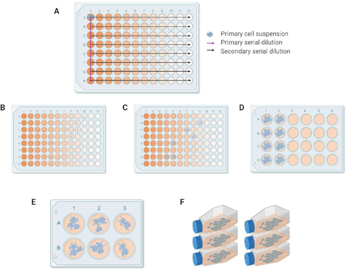
Figure 1: Workflow for monoclonal culture generation. (A) Primary and secondary dilutions in the 96-well plate. (B) Single cells post serial dilution. (C-E) Progression in multiplication of cells and transfer from 96-well to 24- and 6-well plate, respectively. (F) Continuous cultures in 25 cm2 cell culture flasks. Please click here to view a larger version of this figure.
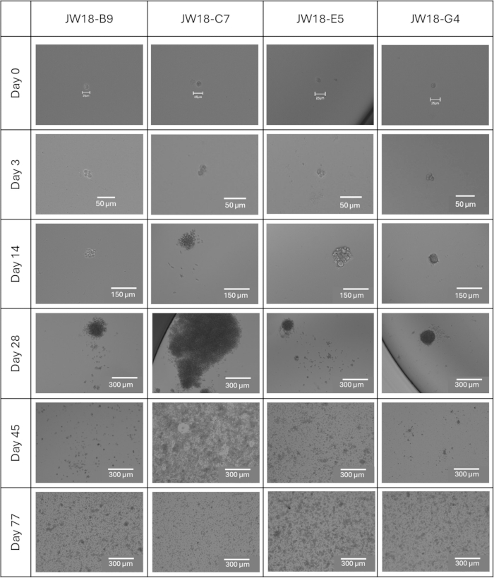
Figure 2: Progression of JW18 clonal lines from single cell to multicellular culture. Brightfield images of clonal lines from day 0 (day of plating) to day 77, when all lines were successfully established in a 25 cm2 cell culture flask. Scale bars = 25 µm (day 0), 50 µm (day 3), 150 µm (day 14), 300 µm (days 28, 45, 77). Please click here to view a larger version of this figure.
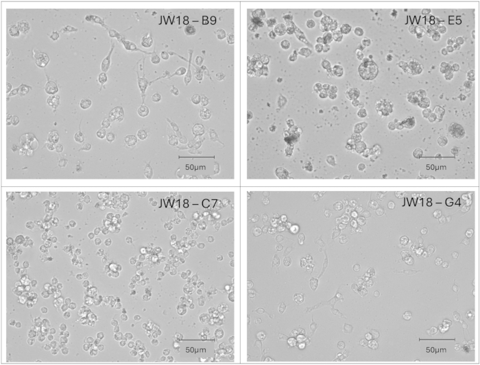
Figure 3: Morphology of individual JW18 clonal lines. Brightfield images of clonal lines showing cell morphologies at passage 30 post establishment. Scale bars = 50 µm. Please click here to view a larger version of this figure.
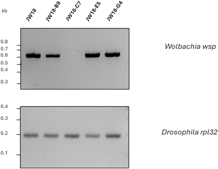
Figure 4: Wolbachia status of clonal cultures examined by PCR. PCR for Wolbachia in JW18 parental cell line and clonal lines JW18-B9, JW18-C7, JW18-E5, and JW18-G4 at passage 11 post establishment. Wolbachiawsp band confirms the presence of Wolbachia, while Drosophilarpl32 band is a DNA quality control PCR. Please click here to view a larger version of this figure.
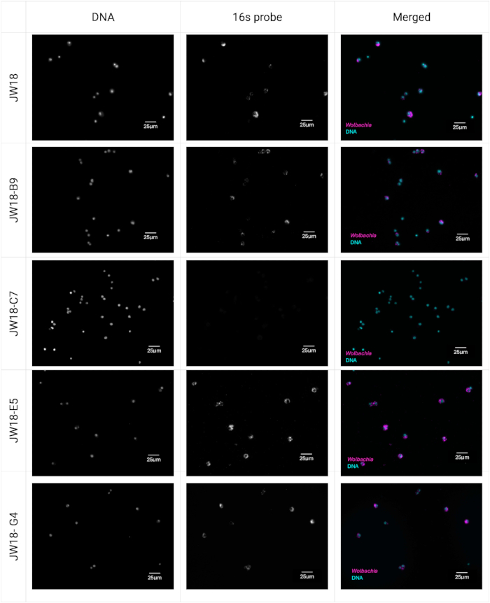
Figure 5: Wolbachia status of clonal cultures examined by FISH. JW18 parental cell line and clones JW18-B9, JW18-C7, JW18-E5, JW18-G4 visualized by FISH staining at passage 13 post establishment. Wolbachia is stained with Quasar 670-tagged 16s rRNA Stellaris FISH probe set in magenta, and nucleic acids are stained with Hoechst 33342 in cyan. Scale bars = 25 µm. Please click here to view a larger version of this figure.
Supplemental Table S1: Oligonucleotide probes designed against Wolbachia wMel 16S rDNA and their comparison to the probes published by Schneider et al.42. Please click here to download this table.
Discussion
Here we outline a standardized method for generating single-cell clones from the JW18 D.melanogaster cell line, which can be applied to other Drosophila and insect cell lines. The protocol involves serial dilution of a prepared cell suspension across a 96-well plate to isolate individual cells. These cells are then incubated under optimal conditions to establish continuous cultures. The protocol also includes the addition of 20% conditioned media to promote cell survival and single-cell division45,46.
A critical step in the protocol is verifying the presence of a single cell in each well following serial dilution. This step requires meticulous attention, as it is essential for the success of the entire protocol. To facilitate future confirmation of the single-cell origin, capturing images of the selected wells from various angles is recommended.
Another key step involves transferring cells between culture dishes. To ensure the successful establishment of a continuous culture, cells should only be transferred once they have reached confluence. If cell growth stagnates for more than two weeks, refreshing the media can promote further growth. Premature transfer of cells before confluence may result in cell death.
While this protocol is more time-consuming and labor-intensive than other commonly used methods, such as Fluorescence-Activated Cell Sorting (FACS)38,46, it offers several advantages. These include cost-effectiveness, minimal risk of cell damage, no requirement for fluorescent markers, expensive equipment, and optimization time. Overall, this protocol is well-suited for generating homogeneous insect cell populations from continuous cell lines.
Properties of the monoclonal cultures
Our results show that the monoclonal cultures differ in terms of speed of their establishment and Wolbachia infection status (Figure 2, Figure 4, and Figure 5). Wolbachia-free clone JW18-C7 was the fastest one to reach confluence in our single-cell isolation protocol. Wolbachia could either have always been absent from this cell lineage, or it could have been lost due to the inability to keep up with the division rate of the host cell. It could also have been lost due to the initial low cell density in our cloning protocol. However, as this did not happen for the other clonal cell lines, we consider this unlikely. The experimental infection of JW18-C7 with Wolbachia (alongside other, tetracycline-treated clones) could answer whether this line is permissive to the symbiont.
The cellular heterogeneity is not unique to the JW18 cell line and has been reported before for other Wolbachia-infected cell lines32,47,48,49,50,51,52. A previous attempt at its amelioration involved a transfer of Wolbachia from D. simulans eggs or Aa23 cells to a C7-10 A. albopictus cell line53. C7-10 has been cloned many years prior to Wolbachia transfers54,55, and the transfers do not constitute the natural infection we intend to study.
The difference in Wolbachia infection status and time of monoclonal culture establishment (indicative of differences in cell division rate) for individual JW18 clones can have profound implications for previous studies linking Wolbachia titer and insect cell growth rate in response to specific treatments. Altered cell numbers, symbiont densities, or changes in gene expression upon treatment might result from selection for cell types with specific properties rather than changes in the state of all cells in a mixed population. Administering treatments to a mixed cell population with the variability in key characteristics may hence yield biased, unreproducible results.
Disclosures
The authors have no conflicts of interest to disclose.
Acknowledgements
We thank William Sullivan (University of California, Santa Cruz, USA), Laura Serbus (Florida International University, USA) and Luis Teixeira (Universidade Católica Portuguesa, Lisbon, Portugal) for the JW18 cell line and useful suggestions on how to work with it. We are grateful to Nidhi Krishna Shrivastava (Jagiellonian University, Cracow, Poland) for useful suggestions on the protocol. We thank Yuliya Chykunova (Jagiellonian University, Cracow, Poland) for proofreading the manuscript.
Figure 1 was created with BioRender (Chrostek, E. (2024) https://BioRender.com/o69b306).
This work is supported by ERC Starting Grant (Grant agreement ID: 101040311) awarded to EC and doctoral fellowship from the Doctoral School of Exact and Natural Sciences (Jagiellonian University) awarded to NF. Views and opinions expressed are, however, those of the authors only and do not necessarily reflect those of the European Union or the European Research Council Executive Agency. Neither the European Union nor the granting authority can be held responsible for them.
Materials
| Name | Company | Catalog Number | Comments |
|---|---|---|---|
| 0.22 µm syringe fliter | TPP | 99722 | Sterile |
| 10x Phosphate Buffered Saline (PBS) | ThermoFisher Scientific | 70011044 | Sterile |
| 12 or 18 mm round coverglass | Any brand | ||
| 15 mL Falcon tubes | Genoplast | 601052 | Sterile |
| 24-well plate | CytoOne | CC7682-7524 | Tissue culture treated |
| 5 µm syringe filter | Whatman | 10462000 | Sterile |
| 6-well plate | CytoOne | CC7682-7506 | Tissue culture treated |
| 96-well plate | CytoOne | CC7682-7596 | Tissue culture treated |
| Agarose | ThermoFisher Scientific | J66501.30 | |
| Cell culture flask, 25 cm2 | Greiner Bio-one | 391-3103 | Sterile |
| Cell scrapers | Greiner Bio-one | 391-3010 | Sterile |
| Clear nail polish | Any brand | ||
| CryoPure vials | Sarstedt | 72.379 | Sterile |
| DMSO, cell culture grade | Merck | D2650-100ML | Sterile |
| DreamTaq PCR Master Mix | ThermoFisher Scientific | K1081 | |
| EDTA | Merck | 324503-100GM | Sterile |
| Electrophoresis Power Supply | Merck | PS 251-2 | |
| Eppendorf Concentrator Plus | Merck | EP5305000509 | Vaccum concentrator of any brand can be used instead. |
| Ethanol | Merck | 1009861000 | |
| EVOS M5000 Imaging System | ThermoFisher Scientific | AMF5000 | Inverted microscope of any brand can be used to monitor clone progression. Fluorescent microscope is required to visualise FISH. |
| Fetal Bovine Serum | Merck | F9665 | Heat-inactivated |
| Formaldehyde | ThermoFisher Scientific | 047377.9L | |
| Formamide | Merck | F7503-100ML | |
| Gel electrophoresis tanks | Cleaver Scientific | MSMIDI | |
| Gel Imager | BioRad | 12009077 | GelDoc Go Gel Imaging System |
| GeneRuler 100 bp DNA Ladder | ThermoFisher Scientific | SM0243 | |
| Heating Block | ThermoFisher Scientific | 88870005 | |
| Herasafe 2025 Class II Biological Safety Cabinet | ThermoFisher Scientific | 51033314 | Biological safety cabinet of any brand can be used as long as it maintains sterile environment inside. |
| Hoechst 33342 Solution (20 mM) 5 mL | ThermoFisher Scientific | 62249 | Store in the dark. |
| Hydrochloric acid | Merck | 258148-2.5L | |
| Integra Biosciences Corp PIPETBOY acu 2 Pipet Aid | Fisher Scientific | NC0085686 | Serological pipette controler of any brand can be used. |
| Isopropanol | Merck | I9516-1L | |
| Laboratory freezer | Liebherr | LGUex 1500 MediLine | Any freezer capable of maintaining constant temperature is suitable. |
| Liquid nitrogen tank | ThermoFisher Scientific | CY50985 | |
| Microscope slides | Merck | CLS294875X25-72EA | |
| Molecular grade water | |||
| Multichannel pipette | Eppendorf | 3125000036 | |
| Nanodrop | Peqlab | ND-1000 | |
| Neubauer Hemocytometer | Merck | BR717810-1EA | |
| Parafilm M Sealing Film | Merck | HS234526B-1EA | |
| Peltier-cooled incubator | Memmert | IPP55plus | Any incubator capable of maintaining constant 25?C is suitable. No CO2 control is required for JW18 cell line or its clones. |
| Phenol/Chloroform/Isoamyl alcohol (25:24:1) | ThermoFisher Scientific | 327115000 | |
| Pipette set | Eppendorf | EP2231300008 | |
| Potassium acetate | Merck | P1190-500G | Sterile |
| Quasar 670-tagged 16s rRNA FISH probe set | Biosearch Technologies | Custom made, sequences of the probes can be found in Table 1. | |
| Refrigerated MicroCentrifuge | Labnet | C2500-R | |
| rpl32 primer forward 5'-CCGCTTCAAGGGACAGTATC-3' | Eurofins genomics | Custom made based on the reference cited in the paper | |
| rpl32 primer reverse 5'-CAATCTCCTTGCGCTTCTTG-3' | Eurofins genomics | Custom made based on the reference cited in the paper | |
| SDS | ThermoFisher Scientific | AM9823 | Sterile |
| Serological pipettes | Corning | 357771 | |
| Shields and Sang M3 Insect Medium | Merck | S8398 | Sterile |
| Stellaris RNA FISH Hybridization Buffer | Biosearch Technologies | SMF-HB1-10 | Sterile |
| Stellaris RNA FISH Wash Buffer A | Biosearch Technologies | SMF-WA1-60 | Sterile |
| Stellaris RNA FISH Wash Buffer B | Biosearch Technologies | SMF-WB1-20 | Sterile |
| Sterile Reagent Reservoirs | ThermoFisher Scientific | 8094 | Sterile |
| TAE buffer | Merck | 1061741000 | 1x |
| TE buffer | Merck | 93283 | Sterile |
| Thermal cycler | Syngen Biotech | Thermoblock 96 | Thermal cycler of any brand can be used instead. |
| Tris base | Merck | 10708976001 | Sterile |
| Ultra low temperature freezer | PHC corporation | MDF-DU502VH-PE | Any laboratory freezer capable of maintaining -80 ?C can be used. |
| Vectashield Mounting Medium | Vector Laboratories | H-1000-10 | |
| wsp primer forward 5'-TGGTCCAATAAGTGATGAAGAAAC-3' | Eurofins genomics | Custom made based on the reference cited in the paper | |
| wsp primer reverse 5'-AAAAATTAAACGCTACTCCA-3' | Eurofins genomics | Custom made based on the reference cited in the paper |
References
- Luhur, A., Klueg, K. M., Zelhof, A. C. Generating and working with Drosophila cell cultures: current challenges and opportunities. Wiley Interdiscip Rev Dev Biol. 8 (3), e339(2019).
- Hilgenboecker, K., Hammerstein, P., Schlattmann, P., Telschow, A., Werren, J. H. How many species are infected with Wolbachia? A statistical analysis of current data. FEMS Microbiol Lett. 281 (2), 215-220 (2008).
- Zug, R., Hammerstein, P. Still a host of hosts for Wolbachia: analysis of recent data suggests that 40% of terrestrial arthropod species are infected. PLoS One. 7 (6), e38544(2012).
- Weinert, L. A., Araujo-Jnr, E. V., Ahmed, M. Z., Welch, J. J. The incidence of bacterial endosymbionts in terrestrial arthropods. Proc R Soc B. 282 (1807), 20150249(2015).
- Hoffmann, A. A., Turelli, M. Unidirectional incompatibility in Drosophila simulans: inheritance, geographic variation and fitness effects. Genetics. 119 (2), 435-444 (1988).
- O'Neill, S. L., Giordano, R., Colbert, A. M., Karr, T. L., Robertson, H. M. 16S rRNA phylogenetic analysis of the bacterial endosymbionts associated with cytoplasmic incompatibility in insects. Proc Natl Acad Sci U S A. 89 (7), 2699-2702 (1992).
- Stouthamer, R., Breeuwer, J. A. J., Luck, R. F., Werren, J. H. Molecular identification of microorganisms associated with parthenogenesis. Nature. 361 (6407), 66-68 (1993).
- Rousset, F., Bouchon, D., Pintureau, B., Juchault, P., Solignac, M. Wolbachia endosymbionts responsible for various alterations of sexuality in arthropods. Proc R Soc Lond B Biol Sci. 250 (1328), 91-98 (1992).
- Hedges, L. M., Brownlie, J. C., O'Neill, S. L., Johnson, K. N. Wolbachia and virus protection in insects. Science. 322 (5902), 702(2008).
- Teixeira, L., Ferreira, Á, Ashburner, M. The bacterial symbiont Wolbachia induces resistance to RNA viral infections in Drosophila melanogaster. PLoS Biol. 6 (12), e1000002(2008).
- Glaser, R. L., Meola, M. A. The native Wolbachia endosymbionts of Drosophila melanogaster and Culex quinquefasciatus increase host resistance to West Nile virus infection. PLoS One. 5 (8), e11977(2010).
- van den Hurk, A. F., et al. Impact of Wolbachia on infection with chikungunya and yellow fever viruses in the mosquito vector Aedes aegypti. PLoS Negl Trop Dis. 6 (11), e1892(2012).
- Aliota, M. T., et al. The wMel strain of Wolbachia reduces transmission of chikungunya virus in Aedes aegypti. PLoS Negl Trop Dis. 10 (4), e0004677(2016).
- Kean, J., et al. Fighting arbovirus transmission: natural and engineered control of vector competence in Aedes mosquitoes. Insects. 6 (1), 236-278 (2015).
- Joubert, D. A., et al. Establishment of a Wolbachia superinfection in Aedes aegypti mosquitoes as a potential approach for future resistance management. PLoS Pathog. 12 (2), e1005434(2016).
- Ross, P. A. Designing effective Wolbachia release programs for mosquito and arbovirus control. Acta Trop. 222, 106045(2021).
- Flores, H. A., O'Neill, S. L. Controlling vector-borne diseases by releasing modified mosquitoes. Nat Rev Microbiol. 16 (8), 508-518 (2018).
- Pannebakker, B. A., Loppin, B., Elemans, C. P. H., Humblot, L., Vavre, F. Parasitic inhibition of cell death facilitates symbiosis. Proc Natl Acad Sci U S A. 104 (1), 213-215 (2007).
- Hosokawa, T., Koga, R., Kikuchi, Y., Meng, X. -Y., Fukatsu, T. Wolbachia as a bacteriocyte-associated nutritional mutualist. Proc Natl Acad Sci U S A. 107 (2), 769-774 (2010).
- Zheng, Y., et al. Differentially expressed profiles in the larval testes of Wolbachia-infected and uninfected Drosophila. BMC Genomics. 12 (1), 595(2011).
- Xi, Z., Gavotte, L., Xie, Y., Dobson, S. L. Genome-wide analysis of the interaction between the endosymbiotic bacterium Wolbachia and its Drosophila host. BMC Genomics. 9 (1), 1(2008).
- Lindsey, A. R. I., Bhattacharya, T., Hardy, R. W., Newton, I. L. G. Wolbachia and virus alter the host transcriptome at the interface of nucleotide metabolism pathways. mBio. 12 (1), e03472-e03520 (2021).
- Detcharoen, M., Schilling, M. P., Arthofer, W., Schlick-Steiner, B. C., Steiner, F. M. Differential gene expression in Drosophila melanogaster and D. nigrosparsa infected with the same Wolbachia strain. Sci Rep. 11 (1), 11336(2021).
- Gruntenko, N. E., et al. Drosophila melanogaster transcriptome response to different Wolbachia strains. Int J Mol Sci. 24 (24), 17411(2023).
- Frantz, S. I., Small, C. M., Cresko, W. A., Singh, N. D. Ovarian transcriptional response to Wolbachia infection in D. melanogaster in the context of between-genotype variation in gene expression. G3 (Bethesda). 13 (5), jkad047(2023).
- McMeniman, C. J., et al. Host adaptation of a Wolbachia strain after long-term serial passage in mosquito cell lines. Appl Environ Microbiol. 74 (22), 6963-6969 (2008).
- Woolfit, M., et al. Genomic evolution of the pathogenic Wolbachia strain, wMelPop. Genome Biol Evol. 5 (11), 2189-2204 (2013).
- Martinez, J., Sinkins, S. P. Rapid evolution of Wolbachia genomes in mosquito cell culture. bioRxiv. , (2023).
- Serbus, L. R., et al. A cell-based screen reveals that the albendazole metabolite, albendazole sulfone, targets Wolbachia. PLoS Pathog. 8 (9), e1002922(2012).
- Grobler, Y., et al. Whole genome screen reveals a novel relationship between Wolbachia levels and Drosophila host translation. PLoS Pathog. 14 (11), e1007445(2018).
- Rainey, S. M., et al. Wolbachia blocks viral genome replication early in infection without a transcriptional response by the endosymbiont or host small RNA pathways. PLoS Pathog. 12 (4), e1005536(2016).
- O'Neill, S. L., et al. In vitro cultivation of Wolbachia pipientis in an Aedes albopictus cell line. Insect Mol Biol. 6 (1), 33-39 (1997).
- Fenollar, F., Maurin, M., Raoult, D. Wolbachia pipientis growth kinetics and susceptibilities to 13 antibiotics determined by immunofluorescence staining and real-time PCR. Antimicrob Agents Chemother. 47 (5), 1665-1671 (2003).
- Fallon, A. M., Witthuhn, B. A. Proteasome activity in a naïve mosquito cell line infected with Wolbachia pipientis wAlbB. In Vitro Cell Dev Biol Anim. 45 (8), 460-466 (2009).
- Molloy, J. C., Sommer, U., Viant, M. R., Sinkins, S. P. Wolbachia modulates lipid metabolism in Aedes albopictus mosquito cells. Appl Environ Microbiol. 82 (10), 3109-3120 (2016).
- Andrianov, B. V., Goriacheva, I. I., Alexandrov, I. D., Gorelova, T. V. Establishment of a new continuous cell line of Drosophila melanogaster strain infected by the intracellular endosymbiotic bacterium Wolbachia pipientis under natural conditions. Russ J Genet. 46 (1), 9-12 (2010).
- Fattouh, N., Cazevieille, C., Landmann, F. Wolbachia endosymbionts subvert the endoplasmic reticulum to acquire host membranes without triggering ER stress. PLoS Negl Trop Dis. 13 (3), e0007218(2019).
- Fredericks, A. C., et al. Aedes aegypti (Aag2)-derived clonal mosquito cell lines reveal the effects of pre-existing persistent infection with the insect-specific bunyavirus Phasi Charoen-like virus on arbovirus replication. PLoS Negl Trop Dis. 13 (11), e0007346(2019).
- Kirby, K. S. A new method for the isolation of ribonucleic acids from mammalian tissues. Biochem J. 64 (3), 405-408 (1956).
- Zhou, W., Rousset, F., O'Neill, S. Phylogeny and PCR–based classification of Wolbachia strains using wsp gene sequences. Proc R Soc Lond B Biol Sci. 265 (1395), 509-515 (1998).
- Chrostek, E., et al. Wolbachia variants induce differential protection to viruses in Drosophila melanogaster: a phenotypic and phylogenomic analysis. PLoS Genet. 9 (12), e1003896(2013).
- Schneider, D. I., Parker, A. G., Abd-alla, A. M., Miller, W. J. High-sensitivity detection of cryptic Wolbachia in the African tsetse fly (Glossina spp). BMC Microbiol. 18 (S1), 140(2018).
- Stellaris RNAFISH protocol for adherent cells. , LGC Biosearch Technologies. https://biosearchassets.biosearchtech.com/assetsv6/bti_stellaris_protocol_adherent_cell.pdf (2021).
- Schneider, C. A., Rasband, W. S., Eliceiri, K. W. NIH Image to ImageJ: 25 years of image analysis. Nat Methods. 9 (7), 671-675 (2012).
- Freshney, I. Cloning and selection. Culture of Animal Cells: a Manual of Basic Technique. , 207(2010).
- Housden, B. E., et al. Identification of potential drug targets for tuberous sclerosis complex by synthetic screens combining CRISPR-based knockouts with RNAi. Sci Signal. 8 (393), (2015).
- Fenollar, F., et al. Culture and phenotypic characterization of a Wolbachia pipientis isolate. J Clin Microbiol. 41 (12), 5434-5441 (2003).
- Dobson, S. L., Marsland, E. J., Veneti, Z., Bourtzis, K., O'Neill, S. L. Characterization of Wolbachia host cell range via the in vitro establishment of infections. Appl Environ Microbiol. 68 (2), 656-660 (2002).
- Frentiu, F. D., Robinson, J., Young, P. R., McGraw, E. A., O'Neill, S. L. Wolbachia-mediated resistance to dengue virus infection and death at the cellular level. PLoS One. 5 (10), e13398(2010).
- Voronin, D. A., Bochernikov, A. M., Baricheva, E. M., Zakharov, I. K., Kiseleva, E. V. Influence of Drosophila melanogaster genotype on biological effects of endosymbiont Wolbachia (stamm wMelPop). Tsitologiia. 51 (4), 335-345 (2009).
- Venard, C. M. -P., Crain, P. R., Dobson, S. L. SYTO11 staining vs FISH staining: a comparison of two methods to stain Wolbachia pipientis in cell cultures. Lett Appl Microbiol. 52 (2), 168-176 (2011).
- Casper-Lindley, C., et al. Rapid fluorescence-based screening for Wolbachia endosymbionts in Drosophila germ line and somatic tissues. Appl Environ Microbiol. 77 (14), 4788-4794 (2011).
- Khoo, C. C. H., Venard, C. M. P., Fu, Y., Mercer, D. R., Dobson, S. L. Infection, growth and maintenance of Wolbachia pipientis in clonal and non-clonal Aedes albopictus cell cultures. Bull Entomol Res. 103 (3), 251-260 (2013).
- Singh, K. R. P. Cell cultures derived from larvae of Aedes albopictus (Skuse) and Aedes aegypti (L.). Curr Sci. 36 (19), 506-508 (1967).
- Nouri, N., Fallon, A. M. Pleiotropic changes in cycloheximide-resistant insect cell clones. In Vitro Cell Dev Biol. 23 (3), 175-180 (1987).
Reprints and Permissions
