This study aims to present the feasibility of macro- and microdissection of the mesenteric lymphatics in the root of the mesentery with accompanying morphometry of the lymphatic clearances.
Method Article
The Role of Anatomical Dissection in Defining Colic and Small Bowel Artery Lymphovascular Bundles in the D3 Volume of Small and Large Bowel Mesentery
In This Article
Summary
Abstract
The D3 lymphadenectomy with extended mesenteric/mesocolic excision is becoming a standard in the surgery of small and large bowel carcinoma. The aim of this study is to present the feasibility of macro- and microdissection of the mesenteric lymphatics in the root of the mesentery with accompanying morphometry of the lymphatic clearances. The study was carried out on three embalmed cadavers from the body donor program. After the removal of the anterolateral abdominal wall, the greater omentum was retracted cranially, giving access to the mesentery, right, and transverse mesocolon. After marking the pertinent landmarks (ileocolic fold, middle colic fold, duodenojejunal angle), the visceral peritoneum was carefully incised along the approximate larger perimeter of the D3 volume and removed. The subperitoneal fatty and connective tissue was removed by gentle centrifugal scraping, revealing the underlying deep lymphatic and blood vessel network. We used narrow spatulas, micro-dissection scissors, small tweezers, curved forceps, and 5x magnifying lens with a fluorescent ring. The centrally positioned superior mesenteric vessels were pivotal in identifying their branches, affluents, and the accompanying lymphatic network. The lymph vessels were identified by continuity with the collector vessels and their link to the lymph nodes. Finally, the lymphatic clearances, i.e., distances between the mentioned arteries and their neighboring vessels, were measured by a digital caliper. In conclusion, the dissection of the mesenteric lymphatics gives a synoptic view of the lymph vessels network and provides valuable information for D3 surgery of small and large bowel tumors.
Introduction
Surgery of colorectal cancers and tumors of the small bowel is nowadays oriented towards D3 lymphadenectomy, i.e., removal of as much lymphatic tissue as possible around the central vascular axis of superior (or inferior) mesenteric vessels1,2. The knowledge of the macro-anatomy of the mesenteric lymphatics is clearly a prerequisite for such challenging interventions. For that reason, the D3 volume of the mesentery has been defined as a rectangle with the following limits: 2 cm cranial to the origin of the middle colic artery, 2 cm caudal to the origin of the ileocecal artery, 3 cm to the right of the lateral border of the superior mesenteric vein, and along the left-hand side of the superior mesenteric artery3. As for the third dimension, the D3 volume includes tissue anterior and posterior to the superior mesenteric vessels, respecting the embryonic planes4.
The lymphatic contents of this delicate area have been addressed in historical literature5, but also in contemporary studies, aimed at the visualization of the lymph affluents using different methodologies3,6,7. Some sources follow a didactic and scholarly approach to the matter of the lymphatic system5,8,9, while the others2,3,6,7,10 take a more direct view, but only in regard to a certain portion of the mesentery and its affiliated entities. What seems to be missing in the literature is a synoptic view of the lymph nodes and vessels of the small and the large bowel, realized in a stratigraphic manner. With dissection being the cornerstone of anatomy and anatomy being one of the bases of surgery, it stands to reason that the lymphatics of the D3 volume by minute anatomical dissection should be thoroughly presented for educational and training purposes. The aim of this study, therefore, is to recreate a detailed step-by-step anatomical approach to the mesenteric blood and lymph vessels, comparable to a certain extent, to the operative setting of the D3 lymphadenectomy.
Protocol
The protocol of the study follows the guidelines of the body donor program of the Unit of Anatomy, Faculty of Medicine, University of Geneva, Switzerland. The body donors officially signed a statement agreeing to using the whole body or body parts for teaching or research purposes. The legislative auspices of the body donation program are based on the Federal Act on Research Involving Human Beings (Human Research Act, HRA), on the Guidelines of the Swiss Academy of Medical Sciences, and the principles of the Swiss Society of Anatomy, Histology, and Embryology (SGAHE/SSAHE).
1. Materials required
- Use a Jores-embalmed (1,875 mL of 40% formaldehyde, 750 mL of chloralhydrate, 750 g of Carlsbad salt, and 12,375 mL of distilled water; stored at 5 °C prior to dissection) cadaver without a medical history of abdominal surgery, trauma, or pathology.
- Check for surgical or trauma scars on the abdominal wall.
- For video registration purposes, use a head-on camera, possibly wireless.
2. First step of the procedure
- Make a wide, deep scalpel incision of the anterolateral wall of the abdomen, cutting through all the layers, including the parietal peritoneum, but avoiding the intraperitoneal organs. Ensure that the incision line begins from the midpoint of the inguinal fold, crosses posteriorly toward the midaxillary line, and then moves upward until the costal arch.
- Ensure that this incision follows the tip of the xiphoid process and continues symmetrically on the other side. Divide the falciform and round ligaments of the liver (Figure 1), then recline the whole abdominal wall flap caudally.
3. Preparation of the dissection field
- Stand on the right-hand side of the cadaver.
- Have the assistant, standing on the left side, expose the mesentery of the cadaver by manually pulling the greater omentum, transverse colon, and transverse mesocolon cranially.
- Subsequently, pull the small bowel loops caudally and to the left. Pull the cecum to the right and present the ileocecal fold.
4. Dissecting peritoneum
- Delineate the wider area of the D3 volume according to the limits given above, but with approximately 3 cm margins around it for all sides of the rectangle. Using a scalpel, incise very shallowly (1-2 mm) the visceral peritoneum overlying it (Figure 2). Take care not to cut too deep to preserve the subperitoneal structures.
- Carefully detach the peritoneal leaf from the underlying fat and connective tissue using forceps, scissors, and/or tweezers (Figure 3). Pay particular attention to the area of the transverse mesocolon where the double serosal leaflet is thin.
5. Subperitoneal dissection - starting point
- Pull the cecum again caudally, which will tighten and enhance the ileocecal vessels.
- Start dissection of the lymphovascular bundles accompanying these vessels using the fine curved dissection forceps, tweezers, dissection needles, small spatulas, and micro-surgical sharp-sharp scissors.
- Ensure that the dissection parallels the course of the lymph vessels (Figure 4). Use mostly scraping of fat lobules instead of sharp dissection. Identifying the lymph vessels can be difficult in the context of the connective tissue strands; therefore, use a twofold differentiation: the lymph vessels are longer, more elastic, and they commence/terminate in the lymph nodes.
6. Clearing lymph nodes
- Find the lymph nodes of variable size along the ileocecal vessels and proximally toward the superior mesenteric axis.
- Each node is to be cleared by scraping the fat and connective tissue from it radially, in a centrifugal direction (Figure 5). This will present its afferent and efferent lymph vessels, which are to be followed in both directions.
7. Dissecting and separating blood vessels
- Dissect and mutually separate the underlying blood vessels (e.g., ileocecal artery and vein) within their vaginae vasorum without disturbing the overlying lymphatic network (Figure 6). To achieve this, use gaps in the vascular network, with minimal tension produced on lymph vessels. In appropriate sections of the dissection field, use a pair of scissors of the sharp-sharp type, gently forcing the blades apart parallel to the direction of the ileocecal blood vessels.
- Spray the dissection area regularly with a phenol solution to avoid drying of fine structures. Aspirate the excess fluid from the peritoneal crevices and fossae.
- Once the ileocolic blood and lymph vessels are liberated from the surrounding connective tissue and fat, measure the lymphatic clearance, i.e., distances between the artery and the accompanying lymph vessels on both sides, superior and inferior to it, using a finely calibrated caliper. Measure at the level of crossing with the right-hand side of the superior mesenteric vein and 1 cm distal to it.
8. Dissecting superior mesenteric vessels
- By following the ileocolic vessels proximally, identify their origins on the superior mesenteric vessels. Prior to their dissection, continue following the lymph vessels using a finely curved forceps and a probe, and present their network at the root of the D3 volume.
- Here, identify the collector lymphatic channel that courses longitudinally along the left-hand side of the superior mesenteric artery (Figure 7). Preserve this vessel and centrifugally from it, dissect its affluents on both sides.
- Now proceed to dissect the superior mesenteric artery and vein, keeping the superficial lymph vessels and nodes intact (Figure 8). The paravascular nerve sheath of the superior mesenteric artery requires effort in this dissection. Remove as much of this nerve tissue using the sharp-sharp scissors.
- Observe the syntopy of the ileocolic artery to the superior mesenteric vein, i.e., whether it forms a posterior or an anterior crossing. Ensure the dissection, in the same manner (liberating the blood vessels but preserving the surrounding lymphatic vessels), proceeds caudally, along the superior mesenteric artery, including levels of origin of 2-3 ileal arteries and cranially above the level of the middle colic artery origin.
9. Dissecting small bowel vessels
- Switch places with the assistant and stand on the left-hand side of the dissected specimen. Once the origins of the jejunal and ileal arteries have been established, continue the dissection in a centrifugal direction from the superior mesenteric axis. Loop the arteries with surgical thread for identification purposes and to separate them from the accompanying lymph vessels.
- Pay attention in cases of jejunal vein(s) crossing the superior mesenteric artery anteriorly. If so, use curved forceps or a probe, gently lift the vein, and continue dissection below it. Perform a careful approach and minute dissection, as the jejunal and ileal arteries are more numerous than the colic arteries, and their accompanying lymph vessels form a denser network. Perform this assisted by a large 5x magnifying lens with a fluorescent ring lamp.
- Perform the same measurement of the lymphatic clearance as for the colic vessels (Figure 9). In the region of the pancreatic notch, identify the highest, i.e., superior jejunal artery. Check if this vessel has a common trunk with the inferior pancreaticoduodenal artery.
10. Dissecting middle colic artery
- Follow the vessels from the origin of the middle colic artery and the accompanying lymph plexuses upward into the transverse mesocolon.
- Using two forceps and/or a sharp/sharp scissors, present the middle colic artery's primary and secondary bifurcation, then clearly separate the transverse mesocolic and the superior jejunal lymphatic network.
NOTE: The distal branch(es) of the middle colic artery should arch over the corridor of the superior mesenteric vessels as they leave the area of the pancreatic notch. - Again, measure the maximal aberration of the neighboring lymph vessels in relation to the middle colic artery (Figure 10).
Results
A total of three Jores-embalmed bodies (all 3 women, aged 59, 70, and 84 years., marked as A, B, C) from the body donor program of the Unit of Anatomy, Faculty of Medicine, University of Geneva, Switzerland, were included in the study. In all three cases, the minute dissection obtained a comprehensive and synoptic view of the lymph vessels and nodes in the D3 volume and their relations to the superior mesenteric vascular axis and their branches and affluents. Concerning the colic branches, the right colic artery was not present in any of the cases. The number of the jejunal arteries was A = 4, B = 5, C = 4. The lower limit of the dissected area included up to three ileal arteries and their neighbouring lymphatics. The lymphatic network included the lymphovascular bundles adjacent to intestinal arteries, the anastomotic branches, the independent branches which course between the bundles, and, finally, the constant collector lymph channel, coursing along the superior mesenteric artery left hand border, thus receiving drainage from the large bowel (right) and the small bowel (left). The mean lymphatic clearances, i.e., distances between a particular artery and its adjacent lymph vessels within a lymphovascular bundle, are given in Table 1. The data underwent statistical analyses with the aid of Statistica 64-bit v. 14.0.0.15 (TIBCO Software Inc. 2020). The small sample was qualified using a two-variable Mann-Whitney U test. There were no statistical differences between the three groups in regard of the jejunal artery clearance (p > 0.05). The same result (p > 0.05) was obtained when comparing ileal clearances between the three cases. On the other hand, when all the jejunoileal clearances (taken together) were compared to the colic (ileocolic and middle colic) clearances, the sample size permitted the use of the Student's t test. The colic clearances were significantly larger than those of the small intestine (t-value 14.35, df = 26, p<0.05). In two out of the three cases, we observed a jejunal vein crossing the superior mesenteric artery and jejunal arteries anteriorly. The cranially oriented dissection revealed no cases of the common trunk for the superior jejunal artery and the inferior pancreaticoduodenal artery, and a clear separation of the superior mesenteric artery lymphatic sheath and the lymph vessels following the middle colic artery left branch(es).
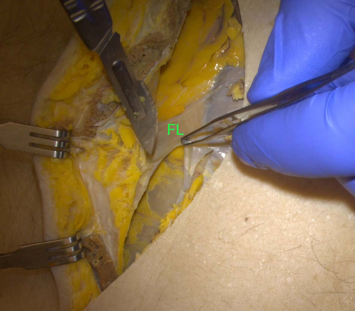
Figure 1: Ligaments. Dividing the falciform (FL) and round ligament of the liver Please click here to view a larger version of this figure.

Figure 2: Peritoneum - incision. Incising the peritoneum over the D3 volume (arrows). On the right side of the image, transverse colon (TC) Please click here to view a larger version of this figure.
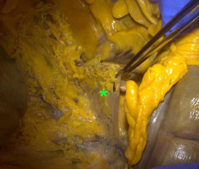
Figure 3: Peritoneum - elevation. Forceps lifting the incised peritoneum (*) and denudating the D3 volume Please click here to view a larger version of this figure.
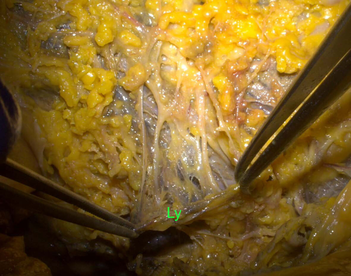
Figure 4: Tissue clearance. Left forceps holding a lymph vessel (Ly), the right one clearing the surrounding fat and connective tissue Please click here to view a larger version of this figure.
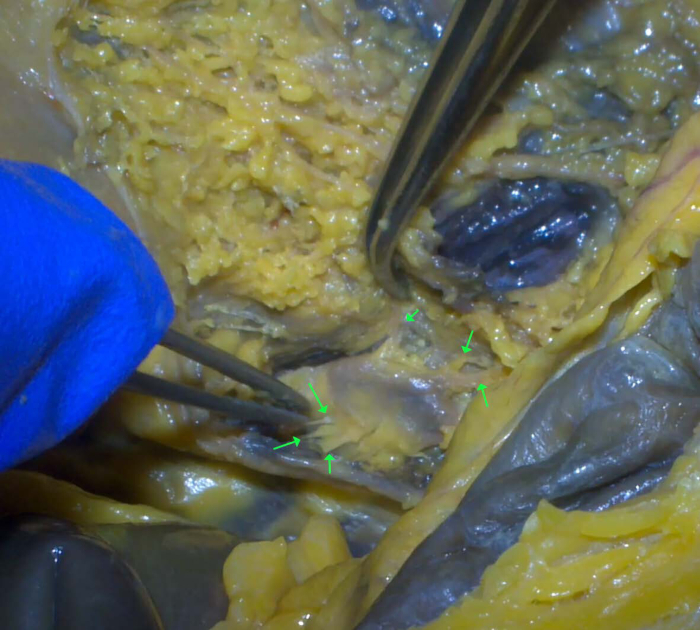
Figure 5: Lymph node. Dissection of the lymph node and its afferent and efferent vessels (arrows) Please click here to view a larger version of this figure.
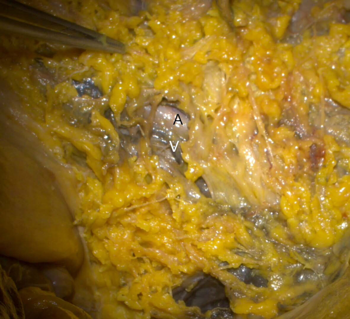
Figure 6: Ileocolic vessels. Ileocolic artery (A) and vein (V), with overlying lymph vessels Please click here to view a larger version of this figure.
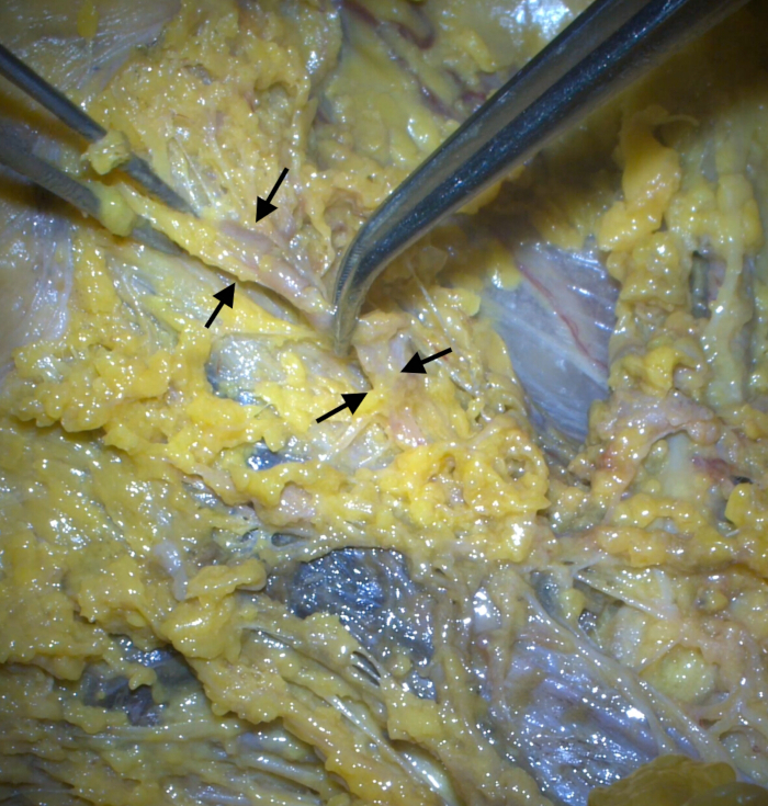
Figure 7: Collector channel. The two forceps holding the collector lymph vessel (arrows) lying along the superior mesenteric artery (white) on the left side. Please click here to view a larger version of this figure.
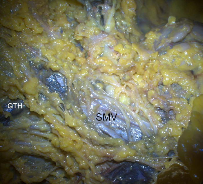
Figure 8: Synoptic view. The superior mesenteric vein (blue, SMV) and the superior mesenteric artery (white), with numerous lymph vessels crossing it anteriorly. GTH: gastrocolic trunk of Henle. Please click here to view a larger version of this figure.
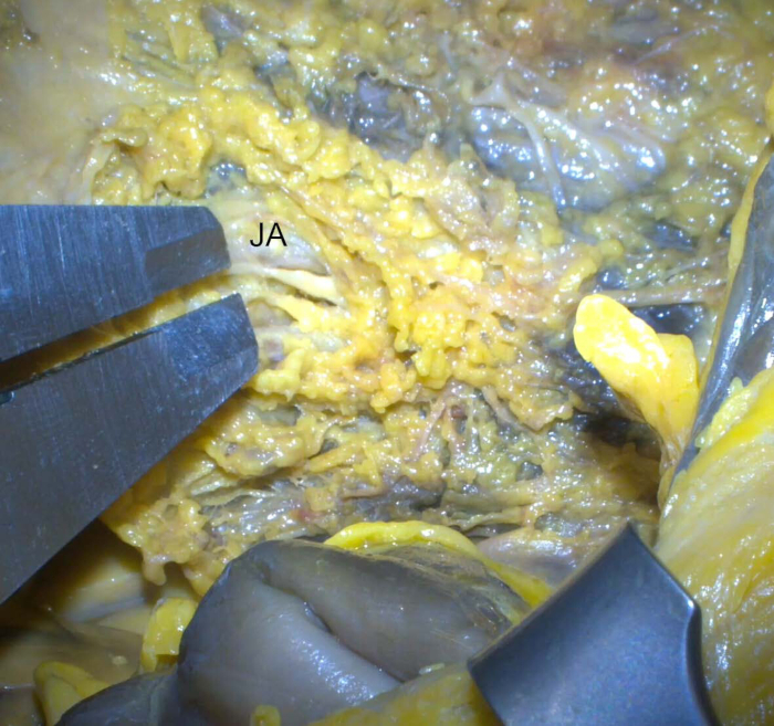
Figure 9: Lymphatic clearance. Measuring the lymphatic clearance of a jejunal artery (JA, at the upper arm of the caliper) Please click here to view a larger version of this figure.

Figure 10: Middle colic artery. Middle colic artery (MCA) arising from the superior mesenteric artery (SMA) and bifurcating in the upper part of the image. Please click here to view a larger version of this figure.
| A | B | C | |
| Mean lymphatic clearances for: | |||
| Ileocolic artery | 4.9 mm | 5.3 mm | 3.7 mm |
| Middle colic | 5.4 mm | 6.6 mm | 7.1 mm |
| Jejunal arteries | 0.9 ± 0.14 mm | 1.2 ± 0.22 mm | 1.8 ± 0.17 mm |
| Ileal arteries | 1.0 ± 0.10 mm | 0.7 ± 0.10 mm | 0.5 ± 0.08 mm |
Table 1: Mean lymphatic clearances of the colic and jejunoileal arteries
| Reference | Study type | Comment |
| Rouvière, 1981 (9) | Anatomical | Detailed study of lymphatic vessels and nodes, albeit no morphometry |
| Conley et al. 2010 (12) | Anatomical | Dissection of small bowel mesenteries with morphometry of the jejunal and ileal arteries |
| Thakur et al. 2011 (13) | Anatomical | Manual dissection of mesorectal lymph nodes |
| Delrivière et al., 2000 (14) | Anatomical | Cadaveric small bowel retrieved, and reduced in size. Mesenteric transillumination, angiography, disssection of blood vessels. Lymphatics of ileal mesentery preserved |
| Açar et al., 2014 (15) | Anatomical | CME-like stepwise dissection of vascular structures, fascias and autonomic nerves |
| Stelzner 2016 (16) | Anatomical | CME-like stepwise dissection of vascular structures, diaphanoscopy, lymphatics presented by histology |
| Nesgaard et al, 2018 (10) | Anatomical | Lymphatic clearances for right colectomy, morphometry |
| Ueki et al., 2019 (17) | Surgical | D3 lymph node dissection for transverse colon cancer, clearing the lympho-adipose tissue above the SMV and SMA and around the MCA |
| Petz et al., 2021 (18) | Surgical | Indocyanine Green (ICG) endoscopic submucosal injection to intraoperatively identify tumour lymphatic basin by near-infrared (NIR) fluorescence |
| Luo et al., 2022 (19) | Sugical | Traction of MCA and ICA pedicles, defining the peritoneum and lymphoadipose tisue overlying the SMA/SMV |
| Tei et al. 2023 (20) | Surgical | Changing positions during surgery for obtaining optimal field of view on mesenteric vessels |
| Efetov et al., 2024 (21) | Surgical | Posterior, i.e. retroperitoneal interfacial approach to superior mesenteric vessels |
| Gao, et al., 2024 (22) | Surgical | Comparison of D3 boundaries in CME right colectomy; the SMV as a medial boundary has significat oncological advantages |
| Shah et al., 2025 (23) | Surgical | Step-by-step CME right colectomy with emphasis on bowel positioning, identification of vascular anatomy, caudal to cranial mesocolic dissection and vascular ligation |
| Vasic et al., 2025 (24) | Anatomical | Mesenteric 3D reconstruction from preoperative CTs, and anatomical dissection. Key points: SMA bi/trifurcation, ileal clearances |
Table 2: List of published dissection studies.
Discussion
The high complexity of the lymphatic system, in comparison to the blood vessels, has already been established8. It has come in the limelight of intestinal surgery with the introduction of total mesenteric excision with D3 lymphadenectomy1. The classical anatomical dissection has been introduced as a method of examining the mesenteric nerve plexuses11, and this study tool has proven to be applicable in mesenteric lymphatics.
The advantage of this study is its precise, step-by-step presentation of the anatomical approach to the D3 volume lymphatic vessels. The salient points have been underlined, from defining the limits of the volume to the precise technique of identification and dissection of minute structures. The difficulties and possible pitfalls are also presented - this dissection requires subtle manipulation, particularly when separating the lymphatics from the underlying blood vessels, or when dissecting the paravascular nerve sheaths through the gaps of the lymph network.
The morphometric portion of the study has a definite clinical significance. The measured distances between the blood vessels and their adjacent lymphovascular bundles give guidelines for lymphatic clearances in surgery. The results, although based on a small sample, present a smaller clearance in jejunoileal than in colic branches, which is in concordance with the smaller mutual spaces between the small bowel arteries, in comparison to the colic ones. Furthermore, the values obtained are in accord with the ones already published2,10.
We have made a literature search on PubMed with the following keywords: dissection, mesentery, lymph, and anatomy, using the Boolean operator AND. This search revealed 196 results, which underwent further filtering, excluding case reports and articles without presentation of dissection techniques. The search results are presented in Table 29,10,12,13,14,15,16,17,18,19,20,21,22,23,24. Most papers deal with the dissection of blood vessels12,14,15,20,21, but also with fascias and autonomic nerves15, and lymph vessels and nodes9,13,16,18. As for the latter, some papers use an underdefined general term of lymphoadipose tissue around the principal blood vessels in the mesentery, without giving details of the lymphovascular network17,19. Two references give a true synoptic view of the particular lymph vessels within the D3 volume and in its vicinity, one with the adjoining morphometry10, and the other without9. Considering the integrated view (anatomical and morphometric) of the mesenteric lymphatics, our study is comparatively superior to the ones analyzed.
Three latest references come in line with this dissection methodology protocol. The first22 addresses the definition of the D3 volume boundaries, stressing the fact that the superior mesenteric vein as a medial limit carries significant oncological advantages, including a higher number of harvested lymph nodes, both total and positive ones. The second study23 proposes a standardized procedure for complete mesocolic excision during right colectomy for cancer, with detailed step-by-step descriptions. Although there are slight differences from our method (e.g., initial identification of superior mesenteric vessels by traction of the transverse mesocolon), the remaining steps are in accordance with the methodological approach presented here: identification of ileocolic vessels, caudal to cranial mesocolic dissection, incision of mesentery, entering the perivascular plane, etc. The third one24 combines segmentation and 3D reconstruction of preoperative CTs with detailed anatomical dissection. It includes morphometry of the first three ileal arteries, the same as in our study.
One can clearly see that the available literature on complete mesocolic/mesenteric excision vastly covers the field of right colectomy. The data on D3 lymphadenectomy in cases of small bowel cancer is sparse2,24. Today, the mesentery of the whole abdominal intestinal tube is considered as one organ, therefore, its lymphatics should be studied as a whole. In this sense, our study offers this combined, intuitive, and manifold methodological approach to the mesentery lymphatics, including morphometry as a valuable addition.
The dissection technique presented in this paper and results derived from it are the basis for the operative technique applied in our two clinical trials: a) Surgery With Extended (D3) Mesenterectomy for Small Bowel Tumors (https://clinicaltrials.gov/study/NCT05670574), and b) Safe D3 Right Hemicolectomy for Cancer Through Multidetector Computed Tomography (MDCT) Angio (https://clinicaltrials.gov/study/NCT01351714). These are ongoing trials; currently, they have included 107 and 623 patients, respectively. The preliminary and intermediary results have proven that survival (overall and disease-free) is achievable in most patients with metastasis in the D3 volume of the right colon after radical surgery25. The overall survival of 1, 3, and 5 years was 100%, 87.5%, and 72.9%, respectively, while the disease-free survival at 1, 3, and 5 years was 86.5%, 78.4%, and 73.1%, respectively. The short-term outcomes present an acceptable level of complications, notably 9.5% vascular injuries, otherwise no intraoperative complications. The latter is related to the preoperative anatomy CT reconstruction ("road mapping"), which significantly decreases the operating time and lowers the incidence of vascular events26.
Concerning the complexity of the vessels relevant to D3-lymph node dissection, the study of Efeteov et al.27 presented a simple, yet useful classification. Three interposition criteria were used: the superior mesenteric artery and vein (crossing or parallel), the ileocolic artery and superior mesenteric vein (anterior or posterior crossing), and the jejunal vein in relation to the superior mesenteric artery (anterior or posterior crossing). Concluding, the authors underline the value of the personalized approach to each patient with regard to the variant vascular anatomy. We could only add that in a number of cases, there exist two or more jejunal veins crossing the SMA both anteriorly and posteriorly.
There are two limitations of this study. First, it was carried out on embalmed human material, which resembles live tissue to a certain extent, but without blood circulation. The second limitation is the number of cases included (3); however, this was intended as a pilot study, close to a proof-of-principle.
In conclusion, the anatomical dissection of mesenteric lymphatics gives valuable data for high-quality surgery of tumors in the large and small bowels. It can also serve as an educational tool for surgical training.
Disclosures
The authors have no conflicts of interest to disclose.
Acknowledgements
The authors wish to express their deepest gratitude to the donors of the Body Donation Program of the Anatomy Unit, Faculty of Medicine, University of Geneva. This research received no funding.
Materials
| Name | Company | Catalog Number | Comments |
|---|---|---|---|
| Camera 7.5 mm | CinVivo, Redwood City, CA | Serial No. 0131 | Model CVCAM0028CT |
| Hub | CinVivo, Redwood City, CA | Serial No. 402917004000069 | Model CV4002SHC |
| Sugical head camera system Titan - 4k | CinVivo, Redwood City, CA | Serial No 0069 |
References
- Weber, K., Hohenberger, W. Right hemicolectomy with central vascular ligation in colon cancer. Surg Endosc. 26 (1), 282(2012).
- Vasic, T., Stimec, B. V., Stimec, M., Kjaestad, E., Ignjatovic, D. Jejunal lymphatic and vascular anatomy defines surgical principles for treatment of jejunal tumors. Dis Colon Rectum. 68 (5), 553-561 (2025).
- Stimec, B. V., Ignjatovic, D. Visible lymph affluents in the D3 volume: An MDCTA pictorial essay. Diagnostics (Basel). 12 (10), 2441(2022).
- Chemtob, A., Ignjatovic, D., Stimec, B. V. Retrocolic fascia - An anatomical and multidetector computed tomographic angiography (MDCTA) morphometric analysis in patients with right colon cancer. Diagnostics (Basel). 14 (17), 1952(2025).
- Servelle, M., Nogués, C. Anatomy of the Lymphatics of the Small Intestine. The Chyliferous Vessels. , Expansion Scientifique Française. Paris, France. (1981).
- Sun, X., Shen, W., Chen, X., Wen, T., Duan, Y., Wang, R. Primary intestinal lymphangiectasia: Multiple detector computed tomography findings after direct lymphangiography. J Med Imaging Radiat Oncol. 61 (5), 607-613 (2017).
- Watanabe, J., Ota, M., Suwa, Y., Ishibe, A., Masui, H., Nagahori, K. Real-time indocyanine green fluorescence imaging-guided complete mesocolic excision in laparoscopic flexural colon cancer surgery. Dis Colon Rectum. 59 (7), 701-705 (2016).
- Skandalakis, J. E., Skandalakis, L. J., Skandalakis, P. N. Anatomy of the lymphatics. Surg Oncol Clin N Am. 16 (1), 1-16 (2007).
- Rouvière, H. Anatomie des Lymphatiques de l'Homme. , Masson. Paris. (1981).
- Nesgaard, J. M., et al. Defining minimal clearances for adequate lymphatic resection relevant to right colectomy for cancer: a post-mortem study. Surg Endosc. 32 (9), 3806-3812 (2018).
- Luzon, J. A., et al. Reconstructing topography and extent of injury to the superior mesenteric artery plexus in right colectomy with extended D3 mesenterectomy: a composite multimodal 3-dimensional analysis. Surg Endosc. 36 (10), 7607-7618 (2022).
- Conley, D., Hurst, P. R., Stringer, M. D. An investigation of human jejunal and ileal arteries. Anat Sci Int. 85 (1), 23-30 (2010).
- Thakur, S., Somashekar, U., Chandrakar, S. K., Sharma, D. Anatomic study of distribution, numbers, and size of lymph nodes in mesorectum in Indians: A autopsy study. Int J Surg Pathol. 19 (3), 315-320 (2011).
- Delrivière, L., et al. Size reduction of small bowels from adult cadaveric donors to alleviate the scarcity of pediatric size-matched organs: an anatomical and feasibility study. Transplantation. 69 (7), 1392-1396 (2000).
- Açar, H. I., Cömert, A., Avşar, A., Çelik, S., Kuzu, M. A. Dynamic article: surgical anatomical planes for complete mesocolic excision and applied vascular anatomy of the right colon. Dis Colon Rectum. 57 (10), 1169-1175 (2014).
- Stelzner, S., et al. Anatomy of the transverse colon revisited with respect to complete mesocolic excision and possible pathways of aberrant lymphatic tumor spread. Int J Colorectal Dis. 31 (2), 377-384 (2016).
- Ueki, T., et al. Vascular anatomy of the transverse mesocolon and bidirectional laparoscopic D3 lymph node dissection for patients with advanced transverse colon cancer. Surg Endosc. 33 (7), 2257-2266 (2019).
- Petz, W., et al. Fluorescence-guided D3 lymphadenectomy in robotic right colectomy with complete mesocolic excision. Int J Med Robot. 17 (3), e2217(2021).
- Luo, W., et al. Laparoscopic complete mesocolic excision with central vascular ligation (CME + CVL) for right-sided colon cancer: A new "Superior Mesenteric Artery First" approach. Ann Surg Oncol. 29 (8), 5066-5073 (2022).
- Tei, M., Sueda, T., Yoshikawa, Y., Hasegawa, J. Laparoscopic Right hemicolectomy with complete mesocolic excision and central vascular ligation using a right colon rotation technique (Flip-Flap Method). Am Surg. 89 (5), 1793-1797 (2023).
- Efetov, S. K., Semchenko, B. S., Rychkova, A. K., Panova, P. D. A new technique of primary retroperitoneal approach for minimally invasive surgical treatment of cecal colon cancer with D3 lymph node dissection. Tech Coloproctol. 28 (1), 144(2024).
- Gao, F., et al. Comparison of effect of different medial boundaries in laparoscopic right hemicolectomy: a meta-analysis. Zhonghua Wei Chang Wai Ke Za Zhi. 27 (12), 1276-1283 (2024).
- Shah, S. Simplified and reproducible laparoscopic complete mesocolic excision with D3 right hemicolectomy. Colorectal Dis. 27 (1), 17242(2025).
- Vasic, T., Stimec, M., Stimec, B. V., Ignjatovic, D. Lymphatic and vascular anatomy define surgical principles for bowel-sparing radical treatment of ileal tumors. (forthcoming) Surg Endosc. 39 (4), 2711-2720 (2025).
- Banipal, G. S., et al. Are metastatic central lymph nodes (D3 volume) in right-sided colon cancer a sign of systemic disease? A sub-group analysis of an ongoing multicenter trial. Ann Surg. 279 (4), 648-656 (2024).
- Willard, C. D., et al. Preoperative anatomical road mapping reduces variability of operating time, estimated blood loss, and lymph node yield in right colectomy with extended D3 mesenterectomy for cancer. Int J Colorectal Dis. 34 (1), 151-160 (2019).
- Efetov, S. K., Zubayraeva, A. A., Rychkova, A. K. Personalized evaluation of D3-lymph node dissection complexity for right colorectal cancer considering anatomy of superior mesenteric vessels. Khirurgiia (Mosk). 10, 29-37 (2024).
Reprints and Permissions
