This protocol describes a step-by-step procedure for harvesting a vascularized and reinnervated abdominal wall allograft, detailing its anatomic landmarks, technical requirements, and future potential clinical applications.
A subscription to JoVE is required to view this content. Sign in or start your free trial.
Method Article
Procurement for a Vascularized and Reinnervated Abdominal Wall Allotransplantation
In This Article
Summary
Abstract
Extensive and complex abdominal wall defects, particularly those associated with intestinal or visceral organ damage, pose significant medical and surgical challenges. An ideal reconstruction must restore anatomy, function, sensation, and body image. Currently, no conventional reconstruction method fulfills all these criteria in such complex scenarios. However, vascularized composite allotransplantation (VCA) offers a unique solution, providing satisfactory anatomical and functional outcomes -- albeit at the cost of lifelong immunosuppression.
Since the first reported human case in 2003, approximately 40 abdominal wall transplants have been performed worldwide, all in conjunction with intestinal or multivisceral transplantation. While various technical adaptations have been described, the procedure has proven to be both safe and effective for patients with complex abdominal wall defects. To date, no reinnervated abdominal wall allograft has been attempted in humans. However, reinnervation appears to be the next frontier, with the potential to enhance functional outcomes and reduce complications.
This protocol outlines a standardized procedure for harvesting and preparing a vascularized abdominal wall composite allograft, designed to ensure optimal results and minimize tissue damage. The graft, vascularized via the deep inferior epigastric vessels, is harvested with generous margins to enable tension-free reconstruction. We also detail the specific steps required to collect an innervated specimen. At the end of the procedure, the two deep inferior epigastric arteries are cannulated, and the graft is perfused with preservation solution for transport. Ultimately, this protocol aims to standardize abdominal wall graft procurement. It is intended as a valuable resource for both translational research and clinical applications, particularly as interest in abdominal wall transplantation continues to grow.
Introduction
Successful closure of the abdominal wall following intestinal transplantation (ITx) or multivisceral transplantation (MVTx) presents a significant challenge for reconstructive surgeons due to various factors -- such as the limited volume of the recipient's abdominal cavity, morphological discrepancies between donor and recipient, postoperative edema, a scarred abdomen, and the presence of multiple enterocutaneous fistulas1,2,3,4,5,6.
Over the past few decades, several techniques for abdominal closure have been proposed to address or at least mitigate the complications associated with complex abdominal defects. These include the use of synthetic meshes, skin expansion, negative pressure therapy, and pedicled or free flaps2,6,7,8,9,10,11,12,13.
However, when conventional methods are not feasible, two alternative strategies may be considered: selecting a donor with smaller organs, though this can increase waiting times and the risk of mortality for patients on transplant lists (Fishbein et al. reported an ideal donor-to-recipient weight ratio between 0.76 and 1.1)14 or proposing an associated abdominal wall transplant.
This concept was first introduced by Levi et al. in 200315 and later improved with a microvascular version by Cipriani et al.16. It is a feasible and safe procedure with numerous advantages, particularly given that immunosuppression is already required for intestinal or multivisceral transplants. It is, however, crucial to screen for donor contraindications, such as a history of abdominal hernias or bulges, prior intra-abdominal surgeries, or obesity.
A major benefit of abdominal wall allotransplantation is the ability to monitor immunosuppression and rejection through the skin component of the graft, which serves as both an immune modulator and a visible sentinel of host immune activity. This offers a valuable tool for early detection of intestinal graft rejection and, more importantly, helps avoid unnecessary over-immunosuppression in cases of graft dysfunction unrelated to immune rejection17,18.
Despite its promise, abdominal wall allotransplantation remains a rare surgical intervention, with only about forty cases reported worldwide19,20. These cases are described mainly through case series and reviews5,15,16,19,20,21,22,23,24,25,26,27,28,29,30. Furthermore, literature on the procurement of reinnervated abdominal wall grafts is extremely limited; to date, no such procedures have been performed in living human subjects31. However, reinnervation is expected to significantly enhance functional outcomes27. This article aims to provide a step-by-step protocol for the procurement of an abdominal wall allograft-including the technical specifics of its reinnervated variant.
Access restricted. Please log in or start a trial to view this content.
Protocol
The Anatomy Laboratory of the Faculty of Medicine in Nice, France, generously provided the specimens and materials used in this study. The research was approved by the French National Ethics Committee (approval number 83.2024) and conducted in accordance with the principles of the Declaration of Helsinki.
The abdominal wall allograft, roughly pentagonal in shape, extends from the xiphoid process to the pubic symphysis. It includes, in a single block, the skin and subcutaneous tissue, both rectus abdominis muscles, portions of the internal, external, and transversus abdominis muscles, the deep muscular fascia, the transversalis fascia, and the parietal peritoneal fascia. The graft is vascularized by the bilateral deep inferior epigastric vessels, which are transected proximally at their origin from the external iliac vessels.
1. Preoperative drawing (Figure 1)
- Position the patient supine on the surgical table, with the arms alongside the body. Protect the eyes and ensure appropriate padding of pressure points.
- Using a dermographic pencil, draw the superior boundary by marking a line starting at the xiphoid process of the sternum. Extend the line laterally, following the inferior margin of the rib cage on both sides, until it reaches a vertical line located 2-3 cm lateral to the anterior superior iliac spine.
- Define the lateral boundary using the vertical line positioned 2-3 cm lateral to the anterior superior iliac spine. From this point, draw a line downward to meet the femoral artery pulse, approximately 7-10 cm below the inguinal ligament.
- Draw the inferior boundary by extending a line from the femoral artery pulse toward the pubic spine on each side, forming a triangle whose apex lies below the inguinal ligaments. Connect both sides of the drawing along the midline, ending at the pubic symphysis.
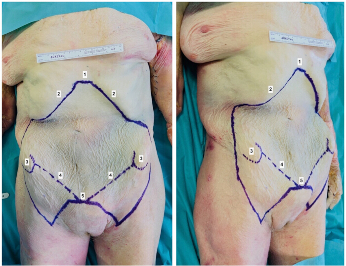
Figure 1: Preoperative drawing. The continuous blue line represents the surgical skin incision of the abdominal wall allotransplantation. (1) xiphoid process, (2) lower costal margin, (3) anterosuperior iliac spine, (4) inguinal ligament, (5) pubic symphysis. Please click here to view a larger version of this figure.
2. Upper incision (Figure 2)
- Perform a bilateral subcostal skin incision along the previously marked line using a cold scalpel blade.
- Continue the incision through the subcutaneous tissue using an electrocautery device until reaching the muscular fascia.
- Incise the fascia overlying the rectus abdominis muscles to expose the muscle bodies.
- Transect the costal insertions of the rectus abdominis muscles with the electrocautery.
- Identify the superior epigastric vessels, ligate them, and section them.
- Behind each mobilized rectus muscle, incise successively the deep muscular aponeurosis, the transversalis fascia, and the parietal peritoneal fascia.
- Laterally, dissect the external oblique muscles and identify the semilunar line.
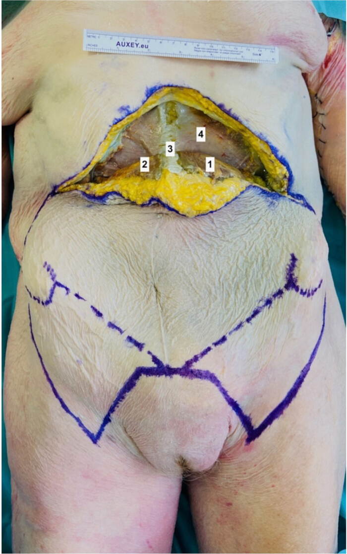
Figure 2: Upper Incision. The upper detachment of the abdominal allograft allows visualization of the abdominal wall muscles. (1) left rectus abdominis muscle, (2) right rectus abdominis muscle, (3) white line, (4) deep muscular aponeurosis. Please click here to view a larger version of this figure.
3. Lateral incisions (Figure 3)
- Perform a skin incision with a cold blade following the preestablished lateral design.
- Continue the incision through the subcutaneous tissue using an electrocautery device until reaching the aponeurotic layer of the external oblique muscles.
- Identify the semilunar line, then make a vertical incision in the aponeurosis of the external oblique muscle, approximately 2-3 cm lateral to the semilunar line.
- Incise the external oblique muscle, followed by the internal oblique muscle, maintaining a 2 to 3 cm distance lateral to the semilunar line to avoid damaging the thoracolumbar nerves.
- Identify the cleavage plane between the internal oblique and transverse abdominis muscles.
- Locate the thoracolumbar nerves running between the internal oblique and transverse abdominis muscles, and carefully dissect them laterally as far as possible.
- Once the thoracolumbar nerves are protected, incise the transverse abdominis muscle, followed by the transversalis fascia, and finally the parietal peritoneal fascia.
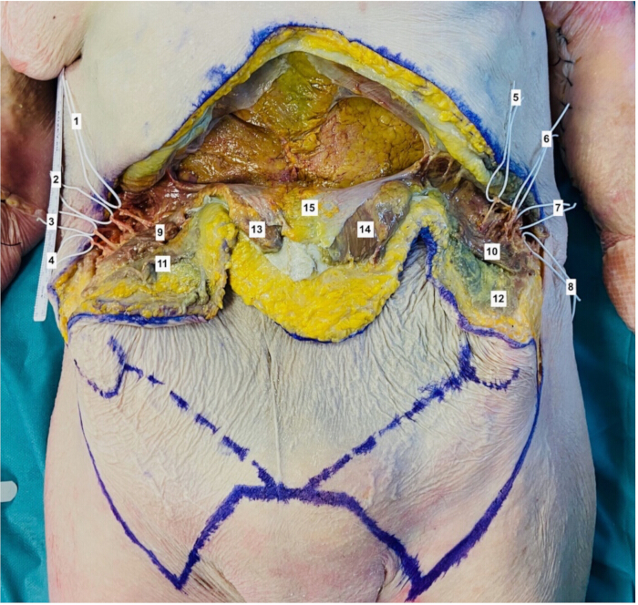
Figure 3: Lateral Incision and identification of thoracolumbar nerves. The lateral dissection shows the thoracolumbar nerves running between the internal oblique and transverse abdominis muscles. The allograft is raised from cranial to caudal. (1) right thoracolumbar nerve n°1, (2) right thoracolumbar nerve n°2, (3) right thoracolumbar nerve n°3, (4) right thoracolumbar nerve n°4, (5) left thoracolumbar nerve n°1, (6) left thoracolumbar nerve n°2, (7) left thoracolumbar nerve n°3, (8) left thoracolumbar nerve n°4, (9) right internal oblique muscle, (10) left internal oblique muscle, (11) right external oblique muscle, (12) left external oblique muscle, (13) right rectus abdominis muscle, (14) left rectus abdominis muscle, (15) parietal peritoneal fascia. Please click here to view a larger version of this figure.
4. Release of the allograft from cranial to caudal
- Begin the release of the allograft from cranial to caudal, leaving it attached to the donor body by its lower margins.
- Identify the deep inferior epigastric pedicles located on the deep surface of the rectus abdominis muscles.
5. Lower incision and pedicle dissection (Figure 4, Figure 5, and Figure 6)
- Make a skin incision along the lower preestablished design using a cold blade.
- Continue the subcutaneous dissection with an electric scalpel until reaching the crural muscular aponeurotic plane of the thigh.
- Identify the great saphenous vein; ligate and cut it. Locate the lateral femoral cutaneous nerve and transect it.
- Incise the crural fascia, which must be included in the graft specimen.
- Elevate the two triangular fascio-cutaneous flaps located beneath the inguinal ligaments until the femoral vessels are exposed.
- On both sides, carefully dissect the femoral artery and vein along with their collateral branches (superficial inferior epigastric and superficial circumflex iliac vessels). Continue the dissection toward the external iliac artery and vein, identifying and preserving the deep inferior epigastric and deep circumflex iliac vessels. Loop the deep inferior epigastric vessels with a silicone loop for protection. In heart-beating donors, ensure arterial pulsatility is maintained throughout the procedure, and confirm that the skin edges of the allograft bleed as an indicator of perfusion.
- Continue the dissection above the inguinal ligament by removing the muscular fascia over the iliac muscle to avoid damaging the deep circumflex iliac vessels.
- Using an electric scalpel, detach the rectus abdominis muscles from their pubic insertions, taking care to preserve the integrity of the deep inferior epigastric vessels.
- Ligate and transect the contents of the inguinal canal: the spermatic cord and ilioinguinal nerve in men, or the round ligament of the uterus and ilioinguinal nerve in women.
- Complete the dissection by ligating and cutting the deep circumflex iliac, superficial circumflex iliac, and superficial inferior epigastric vessels at their origins on the external iliac and femoral vessels. Ensure that the two deep inferior epigastric vascular pedicles remain attached to both the abdominal wall allograft and the donor's body.
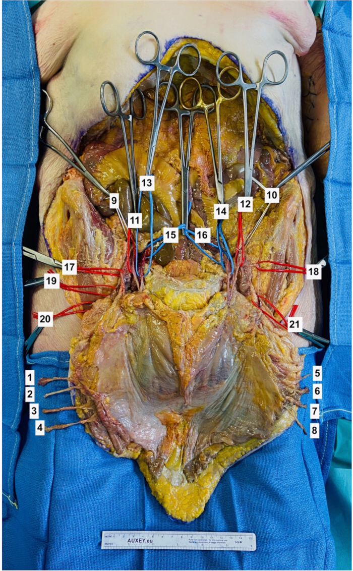
Figure 4: Allograft vascularization. This anterior view exposes all the vessels (arteries and veins) and nerves that need to be identified. In the background, in the abdominal cavity, we can see the abdominal organs. (1) right thoracolumbar nerve n°1, (2) right thoracolumbar nerve n°2, (3) right thoracolumbar nerve n°3, (4) right thoracolumbar nerve n°4, (5) left thoracolumbar nerve n°1, (6) left thoracolumbar nerve n°2, (7) left thoracolumbar nerve n°3, (8) left thoracolumbar nerve n°4, (9) right external iliac artery, (10) left external iliac artery, (11) right deep epigastric inferior artery, (12) left deep epigastric inferior artery, (13) right deep epigastric inferior vein, (14) left deep epigastric inferior vein, (15) right external iliac vein, (16) left external iliac vein, (17) right deep circumflex iliac artery, (18) left deep circumflex iliac artery, (19) right superficial circumflex iliac artery, (20) right superficial epigastric inferior artery, (21) common trunk of left superficial epigastric inferior artery and left superficial circumflex iliac artery. Please click here to view a larger version of this figure.
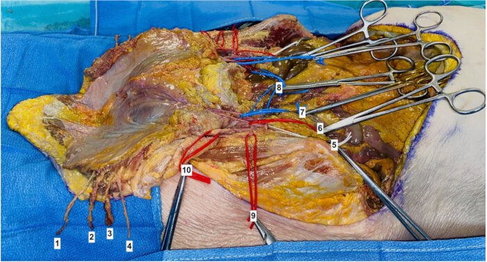
Figure 5: Allograft vascularization (left side). On this left side view, we can see four thoracolumbar nerves. Moreover, in that case, there is a common trunk between the left superficial epigastric inferior artery and the left superficial circumflex iliac artery. (1) left thoracolumbar nerve n°1, (2) left thoracolumbar nerve n°2, (3) left thoracolumbar nerve n°3, (4) left thoracolumbar nerve n°4, (5) left external iliac artery, (6) left deep epigastric inferior artery, (7) left deep epigastric inferior vein, (8) left external iliac vein, (9) left deep circumflex iliac artery, (10) common trunk of left superficial epigastric inferior artery and left superficial circumflex iliac artery. Please click here to view a larger version of this figure.
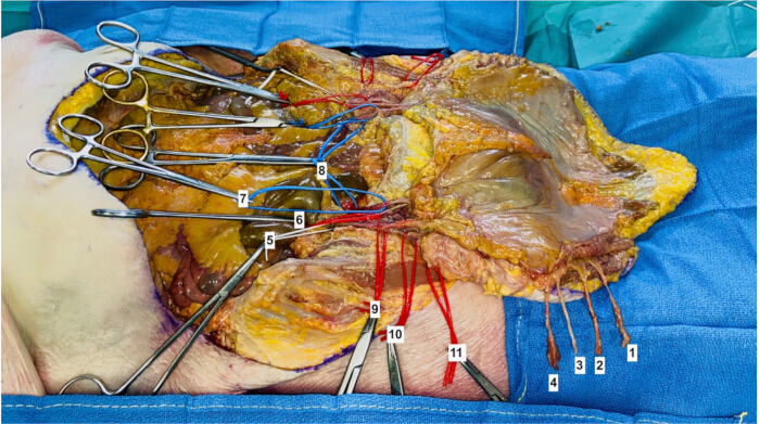
Figure 6: Allograft vascularization (right side). On this right side view, we can see four thoracolumbar nerves. Here, the right superficial epigastric inferior artery and the right superficial circumflex iliac artery begin separately from the femoral artery. (1) right thoracolumbar nerve n°1, (2) right thoracolumbar nerve n°2, (3) right thoracolumbar nerve n°3, (4) right thoracolumbar nerve n°4, (5) right external iliac artery, (6) right deep epigastric inferior artery, (7) right deep epigastric inferior vein, (8) right external iliac vein, (9) right deep circumflex iliac artery, (10) right superficial circumflex iliac artery, (11) right superficial epigastric inferior artery. Please click here to view a larger version of this figure.
6. Graft weaning and conditioning (Figure 7)
- Keep the abdominal wall allograft pedicled on the deep inferior epigastric vessels while the abdominal viscera are being removed. During this time, place the graft in a sterile basin filled with cold saline solution.
- After the complete removal of the abdominal viscera, identify and mark the deep inferior epigastric vessels on both sides-typically one artery and two veins per side. Carefully dissect and ligate the vessels at their origins from the external iliac vessels. Transect the vessels to release the graft.
- Catheterize each deep inferior epigastric artery using a vascular cannula equipped with a stopcock. Secure the cannula to the arterial lumen with a ligature.
- Attach an irrigation set to the cannulas and flush the graft with 1-3 L of University of Wisconsin (UW) preservation solution cooled to 4 °C. Maintain a flow rate of approximately 100 mL/min under low pressure (<100 mmHg) until the effluent runs clear. This step ensures optimal cooling and preservation of the graft prior to transplantation.
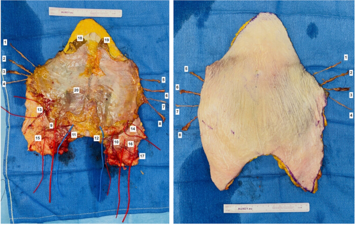
Figure 7: Free abdominal allograft. The left image shows the deep side of the free allograft, and the right image shows the superficial side of the free allograft. This abdominal wall allotransplantation is now ready to receive the preservation solution and to be transported. (1) left thoracolumbar nerve n°1, (2) left thoracolumbar nerve n°2 left, (3) left thoracolumbar nerve n°3, (4) left thoracolumbar nerve n°4 left, (5) right thoracolumbar nerve n°1, (6) right thoracolumbar nerve n°2, (7) right thoracolumbar nerve n°3, (8) right thoracolumbar nerve n°4, (9) left deep epigastric inferior artery, (10) right deep epigastric inferior artery, (11) left deep epigastric inferior vein, (12) right deep epigastric inferior vein, (13) left deep circumflex iliac artery, (14) right deep circumflex iliac artery, (15) common trunk of left superficial epigastric inferior artery and left superficial circumflex iliac artery, (16) right superficial circumflex iliac artery, (17) right superficial epigastric inferior artery, (18) left rectus abdominis muscle, (19) right rectus abdominis muscle, (20) arcuate line. Please click here to view a larger version of this figure.
Access restricted. Please log in or start a trial to view this content.
Results
The donor body dissected for this study was a female subject measuring 1.69 m in height and presenting a standard body morphology. The harvested abdominal wall allograft measured 40 cm in height and 21 cm in width, with a relatively uniform thickness of 43 mm.
On each side, four thoracolumbar nerves of good caliber (approximately 1 mm in diameter) were successfully isolated. These nerves were dissected laterally within the anatomical plane between the internal oblique and transverse abdominis ...
Access restricted. Please log in or start a trial to view this content.
Discussion
According to current literature, abdominal wall allotransplantation has proven to be an effective reconstructive option in complex organ transplantation settings. The current indications for this surgical technique remain limited to cases where conventional methods of abdominal wall reconstruction are not feasible during intra-abdominal organ transplantation. These include situations such as a multi-scarred and fibrotic abdomen resulting from repeated abdominal surgeries, the presence of multiple enterocutaneous fistulas...
Access restricted. Please log in or start a trial to view this content.
Disclosures
The authors have no conflicts of interest to declare.
Acknowledgements
The authors wish to express their sincere gratitude to the individuals who generously donated their bodies to science, thereby enabling anatomical research. The insights gained from such studies have the potential to significantly advance scientific knowledge and improve patient care. As such, these donors and their families deserve our deepest appreciation.
Access restricted. Please log in or start a trial to view this content.
Materials
| Name | Company | Catalog Number | Comments |
|---|---|---|---|
| 11.5” Medium Premium Surgiclip II Auto Suture vessel clip applier | Covidien | ||
| 2-0 silk suture | |||
| Adson Forceps | MPM | 106-2112A | |
| Bipolar Coagulation Forceps | Olsen | 20-1320I | |
| Custodiol HTK Solution for limb perfusion | Essential Pharmaceuticals Inc. | off-label use | |
| Cysto/ Bladder Irrigation Set | Baxter Healthcare Corp. | ||
| Disposable Scalpel #15 | Sklar | ||
| DLP 3 mm vessel cannula blunt tip | Medtronic Inc | ||
| Fine needle cautery | Cormedica | ||
| Forceps Dilators | WPI | 15910 | |
| IV stopcock | |||
| Micro scissors | WPI | 504492 | |
| Monopolar Diathermy | Valleylab | ||
| Oscillating saw | GPC Medical | ||
| Saline solution 0,9% | GenDepot | S0600-101 | |
| Sterile Esmarch bandage | |||
| Sterile surgical marking pen | Cardinal health | 212PR | |
| Strabismus scissors | Surtex | 102-4109 | |
| Sutures Ethilon 4.0 | Ethicon | 1667G | |
| Syringe 10 mL | Agilent | 9301-6474 | |
| Three sterile procurement plastic bags, and three sterile zip ties | |||
| Tissue Forceps | MPM | 106-0511 | |
| Vessel loop | Deroyal | 30-711 |
References
- Alexandrides, I. J., Liu, P., Marshall, D. M., Nery, J. R., Tzakis, A. G., Thaller, S. R. Abdominal wall closure after intestinal transplantation. Plast Reconstr Surg. 106 (4), 805-812 (2000).
- Di Benedetto, F., et al. Use of prosthetic mesh in difficult abdominal wall closure after small bowel transplantation in adults. Transplant Proc. 37 (5), 2272-2274 (2005).
- Carlsen, B. T., Farmer, D. G., Busuttil, R. W., Miller, T. A., Rudkin, G. H. Incidence and management of abdominal wall defects after intestinal and multivisceral transplantation. Plast Reconstr Surg. 119 (4), 1247-1255 (2007).
- Zanfi, C., et al. Incidence and management of abdominal closure-related complications in adult intestinal transplantation. Transplantation. 85 (11), 1607-1609 (2008).
- Berli, J. U., et al. Current concepts and systematic review of vascularized composite allotransplantation of the abdominal wall. Clin Transpl. 27 (6), 781-789 (2013).
- Fortunato, A. C., et al. Technique for closing the abdominal wall in intestinal and multivisceral transplantation. Ann Transplant. 27, e934595(2022).
- Kimata, Y., et al. Anterolateral thigh flap for abdominal wall reconstruction. Plast Reconstr Surg. 103 (4), 1191-1197 (1999).
- Ng, R. W., Chan, J. Y., Mok, V., Li, G. K. Clinical use of a pedicled anterolateral thigh flap. J Plast Reconstr Aesthet Surg. 61 (2), 158-164 (2008).
- Gondolesi, G. E., Aguirre, N. F. Techniques for abdominal wall reconstruction in intestinal transplantation. Curr Opin Organ Transplant. 22 (2), 135-141 (2017).
- Alleyne, B., Ozturk, C. N., Rampazzo, A., Johnson, J., Gurunluoglu, R. Combined submuscular tissue expansion and anterior component separation technique for abdominal wall reconstruction: long-term outcome analysis. J Plast Reconstr Aesthet Surg. 70 (6), 752-758 (2017).
- Wooten, K. E., Ozturk, C. N., Ozturk, C., Laub, P., Aronoff, N., Gurunluoglu, R. Role of tissue expansion in abdominal wall reconstruction: a systematic evidence-based review. J Plast Reconstr Aesthet Surg. 70 (6), 741-751 (2017).
- Patel, N. G., Ratanshi, I., Buchel, E. W. The best of abdominal wall reconstruction. Plast Reconstr Surg. 141 (1), 113e-136e (2018).
- Aliotta, R. E., Gatherwright, J., Krpata, D., Rosenblatt, S., Rosen, M., Gurunluoglu, R. Complex abdominal wall reconstruction, harnessing the power of a specialized multidisciplinary team to improve pain and quality of life. Hernia. 23 (2), 205-215 (2019).
- Fishbein, T. M., Bodian, C. A., Miller, C. M. National sharing of cadaveric isolated intestinal allografts for human transplantation: a feasibility study. Transplantation. 69 (5), 859-863 (2000).
- Levi, D. M., et al. Transplantation of the abdominal wall. Lancet. 361 (9376), 2173-2176 (2003).
- Cipriani, R., et al. Abdominal wall transplantation with microsurgical technique. Am J Transplant. 7 (5), 1304-1307 (2007).
- Gerlach, U. A., et al. Abdominal wall transplantation: skin as a sentinel marker for rejection. Am J Transplant. 16 (6), 1892-1900 (2016).
- Barnes, J., Issa, F., Vrakas, G., Friend, P., Giele, H. The abdominal wall transplant as a sentinel skin graft. Curr Opin Organ Transplant. 21 (5), 536-540 (2016).
- Honeyman, C., et al. Abdominal wall transplantation: indications and outcomes. Curr Transplant Rep. 7, 279-290 (2020).
- Reed, L. T., Echternacht, S. R., Shanmugarajah, K., Hernandez, R., Langstein, H. N., Leckenby, J. I. Twenty years of abdominal wall allotransplantation: a systematic review of the short- and long-term outcomes. Plast Reconstr Surg. 150 (5), 1062e-1070e (2022).
- Selvaggi, G., et al. Expanded use of transplantation techniques: abdominal wall transplantation and intestinal autotransplantation. Transplant Proc. 36 (5), 1561-1563 (2004).
- Selvaggi, G., Levi, D. M., Cipriani, R., Sgarzani, R., Pinna, A. D., Tzakis, A. G. Abdominal wall transplantation: surgical and immunologic aspects. Transplant Proc. 41 (2), 521-522 (2009).
- Allin, B. S. R., et al. A single center experience of abdominal wall graft rejection after combined intestinal and abdominal wall transplantation. Am J Transplant. 13 (8), 2211-2215 (2013).
- Giele, H., et al. Remote revascularization of abdominal wall transplants using the forearm. Am J Transplant. 14 (6), 1410-1416 (2014).
- Avashia, Y. J., Mackert, G. A., May, B., Erdmann, D., Ravindra, K. V. Abdominal wall transplantation. Curr Transplant Rep. 2, 269-275 (2015).
- Giele, H., Vaidya, A., Reddy, S., Vrakas, G., Friend, P. Current state of abdominal wall transplantation. Curr Opin Organ Transplant. 21 (2), 159-164 (2016).
- Park, S. H., Eun, S. H. Abdominal wall transplant surgery. Exp Clin Transplant. 16 (6), 745-750 (2018).
- Molitor, M., Oliverius, M., Sukop, A. Abdominal wall transplantation. Biomed Pap Med Fac Univ Palacky Olomouc Czech Repub. 162 (3), 184-189 (2018).
- Erdmann, D., et al. Small bowel and abdominal wall transplantation: a novel technique for synchronous revascularization. Am J Transplant. 19 (7), 2122-2126 (2019).
- Bustos, V. P., et al. Abdominal wall vascularized composite allotransplantation: a scoping review. J Reconstr Microsurg. 38 (6), 481-490 (2022).
- Lupon, E., et al. Allotransplantations de tissus composites en France : mise au point [Vascularized composite allografts in France: an update]. Ann Chir Plast Esthet. 70 (2), 140-147 (2025).
- Atia, A., Hollins, H., Hernandez, J. A., Erdmann, D. Abdominal wall transplantation. Reconstructive Transplantation. , Springer Nature. Switzerland. 301-309 (2023).
- Nasir, S., Bozkurt, M., Klimczak, A., Siemionow, M. Large antigenic skin load in total abdominal wall transplants permits chimerism induction. Ann Plast Surg. 61 (5), 572-579 (2008).
- Jin, J., et al. Use of abdominal wall allotransplantation as an alternative for the management of end stage abdominal wall failure in a porcine model. J Surg Res. 162 (2), 314-320 (2010).
- Yang, J., Erdmann, D., Chang, J. C., et al. A model of sequential heart and composite tissue allotransplant in rats. Plast Reconstr Surg. 126 (1), 80-86 (2010).
- Quigley, M. A., Fletcher, D. R., Zhang, W., Nguyen, V. T. Development of a reliable model of total abdominal wall transplantation. Plast Reconstr Surg. 132 (4), 988-994 (2013).
- Lao, W. W., Wang, Y. L., Ramirez, A. E., Cheng, H. Y., Wei, F. C. A new rat model for orthotopic abdominal wall allotransplantation. Plast Reconstr Surg Glob Open. 2 (4), e136(2014).
- Broyles, J. M., et al. Reconstruction of large abdominal wall defects using neurotized vascular composite allografts. Plast Reconstr Surg. 136 (4), 728-737 (2015).
- Cipriani, R., Negosanti, L., Pinto, V., Sgarzani, R., Gelati, C., Contedini, F. Abdominal wall transplantation and technique. Abdominal Solid Organ Transplantation: Immunology, Indications, Techniques and Early Complications. Pinna, A. D., Ercolani, G. , Springer International. 379-389 (2015).
- Light, D., et al. Total abdominal wall transplantation: an anatomical study and classification system. Plast Reconstr Surg. 139 (6), 1466-1473 (2017).
- Huger, W. E. Jr The anatomic rationale for abdominal lipectomy. Am Surg. 45 (9), 612-617 (1979).
- Hollenbeck, S. T., et al. The extended abdominal wall flap for transplantation. Transplant Proc. 43 (5), 1701-1705 (2011).
- Broyles, J. M., et al. Functional abdominal wall reconstruction using an innervated abdominal wall vascularized composite tissue allograft: a cadaveric study and review of the literature. J Reconstr Microsurg. 31 (1), 39-44 (2015).
- Singh, D. P., et al. Novel technique for innervated abdominal wall vascularized composite allotransplantation: a separation of components approach. Eplasty. 14, e34(2014).
- Duchateau, J., Declety, A., Lejour, M. Innervation of the rectus abdominis muscle: implications for rectus flaps. Plast Reconstr Surg. 82 (2), 223-228 (1988).
- Hammond, D. C., Larson, D. L., Severinac, R. N., Marcias, M. Rectus abdominis muscle innervation: implications for TRAM flap elevation. Plast Reconstr Surg. 96 (1), 105-110 (1995).
- Mori, H., Akita, A., Hata, Y. Anatomical study of innervated transverse rectus abdominis musculocutaneous and deep inferior epigastric perforator flaps. Surg Radiol Anat. 29 (2), 149-154 (2007).
- Rozen, W. M., Tran, T. M. N., Ashton, M. W., Barrington, M. J., Ivanusic, J. J., Taylor, G. I. Refining the course of the thoracolumbar nerves: a new understanding of the innervation of the anterior abdominal wall. Clin Anat. 21 (4), 325-333 (2008).
- Gurunluoglu, R., Rosen, M. J. Recipient vessels for microsurgical flaps to the abdomen: a systematic review. Microsurgery. 37 (6), 707-716 (2017).
- Atia, A., et al. Surgical techniques for revascularization in abdominal wall transplantation. J Reconstr Microsurg. 36 (7), 522-527 (2020).
- Atia, A., et al. Synchronous abdominal wall and small-bowel transplantation: a 1-year follow-up. Plast Reconstr Surg Glob Open. 8 (7), e2995(2020).
- Severance, G., Walsh, L. Rehabilitation after bilateral hand transplantation in the quadrimembral patient: review and recommendations. Tech Hand Up Extrem Surg. 17 (4), 215-220 (2013).
- Lupon, E., et al. Engineering vascularized composite allografts using natural scaffolds: a systematic review. Tissue Eng Part B Rev. 28 (3), 677-693 (2022).
- Sasaki, H., et al. Selective Bcl-2 inhibition promotes hematopoietic chimerism and allograft tolerance without myelosuppression in nonhuman primates. Sci Transl Med. 15 (690), eadd5318(2023).
Access restricted. Please log in or start a trial to view this content.
Reprints and Permissions
