This protocol enables the isolation of sufficient primary chondrocytes within 6-8 h, facilitating further in-depth investigations of cartilage biology and cartilage disease mechanisms.
A subscription to JoVE is required to view this content. Sign in or start your free trial.
Method Article
In vitro Isolation and Culturing of Mouse Primary Chondrocytes for Cartilage Biology
In This Article
Summary
Abstract
Articular cartilage destruction leads to altered chondrocyte activity, which is a major causative factor in a variety of cartilage diseases, including osteoarthritis (OA) and rheumatoid arthritis (RA). As the most abundant cell type in cartilage, an in-depth study of the biological properties of chondrocytes is essential for the development of effective disease treatment strategies. Isolation and culturing of primary chondrocytes provide important experimental materials for the investigation of cartilage disease and contribute to unraveling the mechanisms of cartilage injury and repair. This protocol provides a detailed description of the in vitro isolation and culturing methods for primary chondrocytes derived from mice. By using the optimized collagenase II protocol, chondrocyte isolation can be completed within 8 h for neonatal mouse knee joint cartilage specimens. The cell yield is approximately 1-2 × 10³ cells/mg of cartilage tissue, varying with tissue freshness and initial cell density; the isolated cells exhibit >90% viability. This protocol uses mouse primary chondrocytes as an example, showing the morphological characteristics and adhesion status of cells cultured for 1, 2, and 5 days after isolation. Reverse transcription quantitative polymerase chain reaction (RT-qPCR) and western blotting showed that interleukin-1β (IL-1β) treatment reduced Collagen Type II Alpha 1 (Col2a1) and increased Matrix Metallopeptidase 13 (Mmp13) expression, confirming that isolated chondrocytes respond to inflammatory stimuli. Compared with conventional methods, this protocol employs a higher collagenase concentration to reduce the impact of prolonged isolation time on cell viability, while avoiding potential cell damage from trypsin treatment. The obtained primary chondrocytes are pure and viable, suitable for in-depth studies on cartilage injury-related mechanisms, which have important implications for in vitro research and the clinical treatment of cartilage disease.
Introduction
Articular cartilage destruction is a major cause of various cartilage diseases, such as OA and RA1,2. Chondrocytes, the predominant cell type in cartilage, are essential for maintaining homeostasis and facilitating repair in response to injury3. Alterations in chondrocyte activity not only affect the structural integrity of cartilage but also induce inflammatory responses and progressive tissue degeneration4. Therefore, an in-depth investigation of the biological properties of chondrocytes is crucial for elucidating the etiopathogenesis of these diseases.
In recent years, the role of chondrocytes in inflammatory responses has received increasing attention. Studies have shown that inflammatory cytokines, such as IL-1β and tumor necrosis factor-α (TNF-α), significantly compromise the metabolic processes of chondrocytes, resulting in increased expression of matrix-degrading enzymes and decreased matrix synthesis5,6,7. These changes ultimately lead to the gradual degeneration of cartilage and joint dysfunction. Therefore, investigating the biological alterations of chondrocytes under pathological conditions within in vitro models has become particularly imperative. Building upon traditional two-dimensional (2D) monolayer culture, recent advances in three-dimensional (3D) chondrocyte culture and organoid technologies have been increasingly adopted in cartilage-related research. These refined systems offer a more physiologically relevant microenvironment and provide valuable insights into chondrocyte behavior in disease contexts8.
The selection of a cell source critically determines whether in vitro findings accurately recapitulate the pathophysiology of joints. Unlike immortalized chondrocyte lines or cartilage explants, primary chondrocytes retain the native genetic background, mechanosensitive calcium signaling pathways, and phenotypic stability of articular cartilage, thereby exhibiting superior chondrogenic capacity and biological behaviors that more closely reflect those of resident cartilage cells9. Systematic differences in calcium signaling sources and key ion channels and downstream pathways have been documented between primary chondrocytes and the ATDC5 cell line10. Consequently, when investigating joint physiology or pathology, primary chondrocytes should be prioritized. Therefore, optimizing the isolation and culturing of primary chondrocytes is essential for accurately modeling disease-related biology and exploring potential therapeutic drugs11,12,13,14,15. Nevertheless, existing methods compromise cell viability or phenotype integrity due to prolonged digestion16,17.
Here, we describe a protocol for the in vitro isolation and culturing of primary chondrocytes derived from mouse cartilage tissue, which rapidly yields sufficient cells. Compared with conventional low-enzyme-concentration, prolonged digestion protocols, this study has optimized the enzyme digestion conditions and tissue pretreatment steps. This optimization has significantly reduced the digestion time to 6-8 h while avoiding potential cell damage from trypsin treatment15,17. Using this method, approximately 1-2 × 10³ chondrocytes can be obtained from each mg of mouse cartilage tissue. This protocol offers a reliable guide for isolating and culturing primary chondrocytes in vitro, providing a robust model for cartilage disease studies.
Access restricted. Please log in or start a trial to view this content.
Protocol
All animal experiments were approved by the Ethics Committee of Xi'an Honghui Hospital (No. 202309009), and the experimental procedures strictly adhered to the 3R principles of replacement, reduction, and refinement for animal experimentation.
Figure 1A illustrates a schematic diagram of the preparation of reagents and equipment. The Table of Materials includes information on the reagents and equipment used in this protocol.
1. Experimental mice
NOTE: Four 3-day-old, wild-type C57BL/6J mice were used in this study.
- Maintain the cages at a constant temperature of 25 °C ± 2 °C and humidity of 50%-60%, with a 12 h/12 h light/dark cycle. Provide specific-pathogen-free (SPF)-grade sterile mouse diet (sterilized by 60Co irradiation) and sterile water ad libitum.
- Collect knee joint cartilage (~10 mg/mouse) from four 3-day-old mice post mortem to isolate primary chondrocytes.
2. Cartilage specimen collection and processing
- Mouse processing and tissue dissection
- Euthanize neonatal mice within 3 days after birth. Sterilize the body surface by wiping with 75% medical alcohol.
- Isolate mouse knee joints with sterile surgical instruments. Place them in sterile 1x PBS.
- Excise the muscles, bones, synovium, and other proliferative connective tissues surrounding the knee joint carefully with sterile surgical instruments. Retain only the cartilage tissue of the knee joint.
- Rinse the tissue specimens 3x with sterile 1x PBS. Discard the liquid.
NOTE: Figure 1B illustrates a simplified schematic diagram of cartilage tissue acquisition and preprocessing.

Figure 1: Simplified workflow of material preparation and cartilage specimen processing. (A) Schematic diagram of reagent and equipment setup, including reagents, consumables, and major instruments. (B) Step-by-step protocol for processing cartilage tissues from mouse knee joints. Abbreviations: PBS = phosphate-buffered saline. Please click here to view a larger version of this figure.
3. Isolation and culture of primary chondrocytes
- Transfer the isolated cartilage tissue into a new 6-well cell culture plate. Using a sterile No. 11 surgical blade, mince the tissue into small pieces approximately 0.25 mm3 in size. Rinse once with prechilled sterile 1x PBS. Discard the liquid.
- Add 2 mL of prewarmed (37 °C) digestion solution containing high-concentration collagenase II (25 mg/mL, dissolved in sterile Dulbecco's Modified Eagle Medium/Nutrient Mixture F-12 [DMEM/F12] basal medium) to cover the tissue block.
- Incubate the cell culture plate in a humidified 5% CO2 incubator at 37 °C for 15 min for enzymatic digestion. After incubation, discard the liquid.
- Repeat steps 3.2 and 3.3.
NOTE: High-concentration enzyme digestion is a preliminary step. It aims to predigest the tissue, partially break down its structure for better subsequent digestion, and remove matrix adhesions that may exist due to cutting from the tissue periphery. - Add 2 mL of prewarmed (37 °C) digestion solution containing low-concentration collagenase II (5 mg/mL, dissolved in sterile DMEM/F12 basal medium) to cover the tissue block.
- Incubate the cell culture plate in a humidified 5% CO2 incubator at 37 °C for 6-8 h for digestion and observe the progress of cell separation. Occasionally perform slow horizontal shaking (80-120 rpm) or gentle pipetting to enhance lysis efficiency.
NOTE: For neonatal mouse tissue, cell release is visible around 3 h, with full isolation achievable between 6 and 8 h. During the 6-8 h collagenase II treatment, observe the lysis every hour and terminate digestion when no obvious tissue chunks are visible to the naked eye and 90% of cells are free and in suspension under a microscope. During the digestion process, vacuole-like chondrocytes can be observed gradually detaching from the cartilage tissue mass under an optical microscope. This step can be repeated several times to minimize the damage to chondrocytes caused by prolonged digestion, and the digestion solution can be replaced with a fresh low-concentration collagenase II digestion solution to increase the cell yield. - Filter all digestion products through a 100 µm cell strainer into a sterile 50 mL centrifuge tube.
NOTE: Use a 100 µm cell strainer to prevent loss of chondrocyte aggregates and increase yield. Residual tissue fragments are removed during medium exchange. - Rinse the cell culture plate with sterile 1x PBS. Filter through the cell strainer again.
- Collect all filtrate from the previous step in a 15 mL centrifuge tube. Centrifuge at 300 × g for 5 min at room temperature with the brake off. Discard the supernatant after centrifugation.
- Add 2 mL of sterile 1x PBS and gently resuspend the precipitate by lightly blowing and aspirating with a pipette. Transfer to a sterile 15 mL centrifuge tube. Centrifuge at 300 × g for 5 min at room temperature with the brake off. Discard the supernatant after centrifugation.
- Resuspend the precipitate from step 4.10 with 2 mL of freshly prepared sterile DMEM/F12 complete medium, which contains 15% fetal bovine serum (FBS) and 1% P/S. Enumerate the harvested cell suspension using a hemocytometer, then plate the cells into a sterile 10 cm culture dish at 2-2.5 × 10⁵ cells/cm2. Supplement to a final volume of 10 mL with complete medium.
NOTE: Chondrocyte seeding density should be 2-2.5 × 10⁵ cells/cm2, as low or medium density seeding can lead to a phenotypic shift of articular chondrocytes towards fibroblasts. Proliferation activity and viability of the isolated primary cells can be assessed using optional MTT assays or trypan blue exclusion staining (Optional Step). - Incubate in a humidified 5% CO2 incubator at 37 °C.
- Replace with fresh DMEM/F12 complete medium (containing 10% FBS, 1% P/S) after the cells are observed to be completely adherent to the wall under an optical microscope.
- Change the medium every 2 days. Continue culturing until the confluence reaches 70%, at which point subsequent experiments can be performed.
NOTE: The serum concentration in the culture medium can be increased to 15% based on the cell condition. Figure 2 illustrates a simplified schematic diagram of cartilage tissue digestion, isolation, and culture of primary cells.
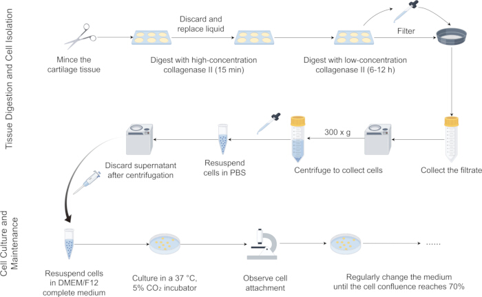
Figure 2: Simplified procedure for cartilage tissue digestion, primary cell isolation, and culture. Cartilage was minced and digested continuously with high-concentration collagenase II digestion solution for 15 min, repeated once, then digested with low-concentration collagenase II digestion solution for 6-8 h to isolate the cells. The digested tissue was then filtered, and the filtrate was collected and centrifuged. The cell pellet was washed with 1x PBS and cultured in DMEM/F12 medium until the confluence reached 70%, at which point the cells were ready for subsequent experiments. Please click here to view a larger version of this figure.
4. Functional verification of primary chondrocytes
- Metabolism and changes of chondrocytes under inflammatory conditions.
- Treat cells with 10 ng/mL IL-1β for 24 h to simulate inflammatory conditions.
- For RT-qPCR and western blotting, extract total RNA or total protein to detect the metabolic indicators of the chondrocyte extracellular matrix, such as COL2A1, MMP13.
NOTE: Figure 3 illustrates a schematic diagram of the functional validation of primary chondrocytes and the subsequent experimental procedures. The antibody information is provided in the Table of Materials. Detailed procedures for western blotting and RT-qPCR are provided in Table 1, Table 2, Table 3, Table 4, and Table 5. All methods followed standard protocols11,18.
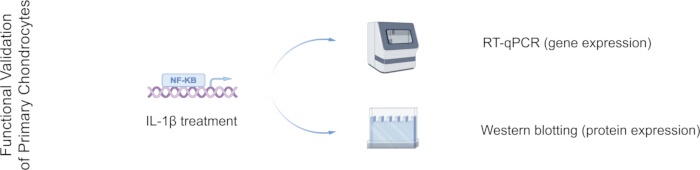
Figure 3: Schematic diagram of the primary chondrocyte functional validation program. Inflammatory conditions induced by IL-1β treatment; effects on cartilage-associated phenotypes assessed by RT-qPCR and Western blotting. Abbreviations: IL-1β = interleukin-1β; RT-qPCR = Reverse transcription quantitative polymerase chain reaction. Please click here to view a larger version of this figure.
| Western Blotting workflow conditions | |
| Step | Condition / Reagent |
| SDS-PAGE gel preparation | 8 % (w/v) Tris-HCl separating gel |
| Stacking electrophoresis | 80 V, 30–40 min |
| Resolving electrophoresis | 120 V, 1–1.5 h |
| Transfer | Semi-dry, 15 V, 30–60 min |
| Blocking | Rapid blocking buffer, 2 h at room temperature |
Table 1: Western blotting workflow conditions. Summary of western blotting conditions, including SDS-PAGE composition, electrophoresis parameters, transfer settings, and blocking procedure.
| Reverse transcription reaction mix | |
| Component | Volume (μL) |
| 5× Transcriptor Reverse Transcriptase Reaction Buffer | 4 |
| Deoxynucleotide Mix ( 10 mM each) | 2 |
| Protector RNase Inhibitor (40 U/μL) | 0.5 |
| Transcriptor Reverse Transcriptase (20 U/μL) | 0.5 |
| Nuclease-free water | to 20 |
| Total volume | 20 |
Table 2: Reverse transcription reaction mix. Reverse transcription reaction composition for cDNA synthesis.
| Primer information for RT-qPCR of mouse primary chondrocyte | ||||
| Gene name | Sequence(5’ 3’) 3’) | Annealing temperature (°C) | ||
| mGapdh | Forward | TGGCCTTCCGTGTTCCTAC | 60 | |
| Reverse | GAGTTGCTGTTGAAGTCGCA | |||
| mCol2a1 | Forward | TGACCTCAACTACATGGTCTACA | 60 | |
| Reverse | CTTCCCATTCTCGGCCTTG | |||
| mMmp13 | Forward | CTTCTTCTTGTTGAGCTGGACTC | 60 | |
| Reverse | CTGTGGAGGTCACTGTAGACT | |||
Table 3: Primer information for RT-qPCR of mouse primary chondrocyte. Sequences and annealing temperature (60 °C) of primers used to quantify Gapdh, Col2a1, and Mmp13 transcripts in mouse primary chondrocytes.
| RT-qPCR reaction mix | |
| Component | Volume (μL) |
| 2 × SYBR Green qPCR Master Mix | 10 |
| Diluted cDNA template | 8 |
| Forward primer (10 μM) | 1 |
| Reverse primer (10 μM) | 1 |
| Total volume | 20 |
Table 4: RT-qPCR reaction mix. RT-qPCR reaction mix (20 µL) with SYBR Green master mix, template, and primers.
| RT-qPCR cycling protocol | ||
| Step | Temperature (°C) | Time |
| Initial denaturation (×1) | 95 | 10 min |
| Denaturation (×40) | 95 | 5 s |
| Annealing/extension (×40) | 60 | 15 s |
| Extension (×40) | 72 | 30 s |
| Melt curve (×1) | 95 55 95 | 60 s 30 s 30 s |
Table 5: RT-qPCR cycling protocol. Denaturation at 95 °C for 5 s, annealing/extension at 60 °C for 15 s, and a final melt-curve analysis were performed for 40 cycles.
Access restricted. Please log in or start a trial to view this content.
Results
As described in Figure 1, Figure 2, and Figure 3, we performed the protocol and successfully isolated primary chondrocytes from mouse knee joints. From approximately 10 mg of neonatal mouse cartilage, we obtained about 1 × 10⁴ cells with >90% viability. On the first day of in vitro culture, primary mouse chondrocytes exhibited a rounded morphology, with most cells in suspension and not yet ful...
Access restricted. Please log in or start a trial to view this content.
Discussion
Isolation and culture of primary chondrocytes are essential for elucidating the pathophysiology of cartilage diseases22. In primary chondrocyte culture, optimizing collagenase concentration and digestion time is crucial, as it balances the degradation of the extracellular matrix while reducing potential cellular damage17. Herein, we describe a reliable and reproducible method for isolating primary chondrocytes from mouse cartilage tissues. Using this method, approximately 1...
Access restricted. Please log in or start a trial to view this content.
Disclosures
The authors declare that they have no conflicts of interest.
Acknowledgements
This work was supported by the National Natural Science Foundation of China (No. 82370909). Figure 1, Figure 2, and Figure 3 were created with Figdraw.
Access restricted. Please log in or start a trial to view this content.
Materials
| Name | Company | Catalog Number | Comments |
|---|---|---|---|
| 1× PBS Powder (2 L) | Servicebio | G0002-2L | |
| 100 mm cell culture dishes | Servicebio | CCD-100 | |
| 100 μm cell sieve | Sangon Biotech | F613463-0001 | |
| 15mL Conical Centrifuge Tube (Sterile, Enzyme-Free) | Servicebio | EP-1501-J | |
| 50mL Conical Centrifuge Tube (Sterile, Enzyme-Free) | Servicebio | EP-5001-J | |
| 6-well cell culture plate | Servicebio | CCP-6H | |
| Anti-COL2A1 rabbit polyclonal antibody | Immunoway | YT1022 | Primary Antibodies; WB: 1/500 |
| Anti-GAPDH mouse monoclonal antibody | Proteintech | 60004-1-IG | Primary Antibodies; WB: 1/2000 |
| Anti-MMP13 rabbit polyclonal antibody | Immunoway | YT2796 | Primary Antibodies; WB: 1/1000 |
| Aseptic ultra-clean bench | Thermo Fisher | 51029704 | |
| Cell culture incubator | Thermo Fisher | 51033782 | |
| Collagenase II | Solarbio | C8150 | |
| Custom primers (Gapdh, Col2a1, Mmp13) | Sangon Biotech | Designed using Primer BLAST | |
| DMEM/F12 medium | Hyclone | SH30023.01 | |
| Fetal bovine serum | ExCell | FCS500 | |
| HRP-Goat Anti-Mouse polyclonal antibody | Immunoway | RS0001 | Secondary Antibodies; WB: 1/10000 |
| HRP-Goat Anti-Rabbit polyclonal antibody | Immunoway | RS0002 | Secondary Antibodies; WB: 1/10000 |
| Inverted phase contrast microscope | OLYMPUS | CKX53 | |
| Micropipette | Thermo Fisher | ||
| Miniature benchtop centrifuge | SUNNE | SN-TDL-6 | |
| No. 11 surgical blade | Servicebio | QXJZ-11 | |
| Penicillin-Streptomycin Solution(100X) | Beyotime | C0222 | |
| Small Animal Dissection Instruments Set | Servicebio | QXTZ01 |
References
- Makris, E. A., Gomoll, A. H., Malizos, K. N., Hu, J. C., Athanasiou, K. A. Repair and tissue engineering techniques for articular cartilage. Nat Rev Rheumatol. 11 (1), 21-34 (2015).
- Bierma-Zeinstra, S., Van Middelkoop, M., Runhaar, J., Schiphof, D. Nonpharmacological and nonsurgical approaches in OA. Best Pract Res Clin Rheumatol. 34 (2), 101564(2020).
- Urlic, I., Ivkovic, A. Cell sources for cartilage repair-biological and clinical perspective. Cells. 10 (9), 2496(2021).
- Murphy, M. P., et al. Articular cartilage regeneration by activated skeletal stem cells. Nat Med. 26 (10), 1583-1592 (2020).
- Zhao, K., Ruan, J., Nie, L., Ye, X., Li, J. Effects of synovial macrophages in osteoarthritis. Front Immunol. 14, 1164137(2023).
- Wang, B. W., et al. Aucubin protects chondrocytes against IL-1β-induced apoptosis in vitro and inhibits osteoarthritis in a mouse model. Drug Des Devel Ther. 13, 3529-3538 (2019).
- Kuswanto, W., Baker, M. C. Repurposing drugs for the treatment of osteoarthritis. Osteoarthritis Cartilage. 32 (8), 886-895 (2024).
- Heywood, H. K., Thorpe, S. D., Jeropoulos, R. M., Caton, P. W., Lee, D. A. Modulation of sirtuins during monolayer chondrocyte culture influences cartilage regeneration upon transfer to a 3d culture environment. Front Bioeng Biotechnol. 10, 971932(2022).
- Piñeiro-Ramil, M., et al. Generation of human immortalized chondrocytes from osteoarthritic and healthy cartilage : A new tool for cartilage pathophysiology studies. Bone Joint Res. 12 (1), 46-57 (2023).
- Li, W., Zhou, Y., Han, L., Wang, L., Lucas Lu, X. Calcium signaling of primary chondrocytes and atdc5 chondrogenic cells under osmotic stress and mechanical stimulation. J Biomech. 145, 111388(2022).
- Zhao, Y., et al. Protein folding dependence on selenoprotein m contributes to steady cartilage extracellular matrix repressing ferroptosis via PERK/ATF4/CHAC1 axis. Osteoarthritis Cartilage. 33 (2), 261-275 (2025).
- Zhang, H., et al. Mechanical overloading promotes chondrocyte senescence and osteoarthritis development through downregulating FBXW7. Ann Rheum Dis. 81 (5), 676-686 (2022).
- Tang, H., et al. The IRF1/GBP5 axis promotes osteoarthritis progression by activating chondrocyte pyroptosis. J Orthop Translat. 44, 47-59 (2024).
- Bittner, N., et al. Primary osteoarthritis chondrocyte map of chromatin conformation reveals novel candidate effector genes. Ann Rheum Dis. 83 (8), 1048-1059 (2024).
- Ramser, A., Greene, E., Rath, N., Dridi, S. Primary growth plate chondrocyte isolation, culture, and characterization from the modern broiler. Poult Sci. 102 (1), 102254(2023).
- Mao, J., et al. Insufficiency of collagenases in establishment of primary chondrocyte culture from cartilage of elderly patients receiving total joint replacement. Cell Tissue Bank. 24 (4), 759-768 (2023).
- Lau, T. T., Peck, Y., Huang, W., Wang, D. A. Optimization of chondrocyte isolation and phenotype characterization for cartilage tissue engineering. Tissue Eng Part C Methods. 21 (2), 105-111 (2015).
- Pagani, S., et al. Rna extraction from cartilage: Issues, methods, tips. Int J Mol Sci. 24 (3), 2120(2023).
- Goldring, M. B., Otero, M., Tsuchimochi, K., Ijiri, K., Li, Y. Defining the roles of inflammatory and anabolic cytokines in cartilage metabolism. Ann Rheum Dis. 67 (Suppl 3), 75-82 (2008).
- Wang, G., et al. TGFbeta attenuates cartilage extracellular matrix degradation via enhancing FBXO6-mediated MMP14 ubiquitination. Ann Rheum Dis. 79 (8), 1111-1120 (2020).
- Pang, Y., Zhao, L., Ji, X., Guo, K., Yin, X. Analyses of transcriptomics upon IL-1β-stimulated mouse chondrocytes and the protective effect of catalpol through the NOD2/NB-κb/MAPK signaling pathway. Molecules. 28 (4), 1606(2023).
- Naranda, J., Gradisnik, L., Gorenjak, M., Vogrin, M., Maver, U. Isolation and characterization of human articular chondrocytes from surgical waste after total knee arthroplasty (TKA). PeerJ. 5, e3079(2017).
- Koyama, N., et al. Pluripotency of mesenchymal cells derived from synovial fluid in patients with temporomandibular joint disorder. Life Sci. 89 (19-20), 741-747 (2011).
- Lawrence, J. E. G., et al. Single-cell transcriptomics identifies chondrocyte differentiation dynamics in vivo and in vitro. Dev Cell. , (2025).
- Huang, C., et al. Single-cell transcriptomic analysis of chondrocytes in cartilage and pathogenesis of osteoarthritis. Genes Dis. 12 (2), 101241(2025).
- Wu, C. L., et al. Single cell transcriptomic analysis of human pluripotent stem cell chondrogenesis. Nat Commun. 12 (1), 362(2021).
- Zhang, H., Huang, J., Alahdal, M. Exosomes loaded with chondrogenic stimuli agents combined with 3d bioprinting hydrogel in the treatment of osteoarthritis and cartilage degeneration. Biomed Pharmacother. 168, 115715(2023).
- Mahapatra, C., Jin, G. Z., Kim, H. W. Alginate-hyaluronic acid-collagen composite hydrogel favorable for the culture of chondrocytes and their phenotype maintenance. Tissue Eng Regen Med. 13 (5), 538-546 (2016).
Access restricted. Please log in or start a trial to view this content.