A unique method to surgically resect oral squamous cell carcinoma tumors established in the oral cavity is shown here.
A subscription to JoVE is required to view this content. Sign in or start your free trial.
Method Article
Translationally-Relevant Tumor Resection Model for Murine Preclinical Models of Oral Squamous Cell Carcinoma
In This Article
Summary
Abstract
Currently, the first-line treatment for the most common form of head and neck squamous cell carcinoma (HNSCC), oral cavity squamous cell carcinoma (OSCC), is surgical resection, followed by risk-adapted treatments, such as chemoradiotherapy. However, with the current standard of care and even the emergence of new treatments, such as checkpoint blockade, the overall survival for advanced, recurrent disease remains high, and the prevalence of head and neck cancer is projected to continue to increase in the coming years. Thus, deeper investigation into current standard-of-care modalities, in addition to novel therapeutics, is necessary to translate potential and promising treatment options to patients with HNSCC. Preclinical models are utilized to evaluate the safety and efficacy of potential treatment interventions. Unfortunately, current preclinical models fail to fully embody the surgical procedure many patients receive, as they primarily involve non-surgical treatment modalities, like radiation, chemotherapy, or immunotherapy. Furthermore, existing surgical models are limited by additional reagents needed to support the establishment of tumors in orthotopic sites and the requirement of microsurgery for tumor removal. This warrants the generation of improved and simplified preclinical models to investigate therapeutics and gain insight into locoregional tumor recurrence and treatment resistance. This article describes a novel syngeneic clinically relevant orthotopic murine subtotal resection model that recapitulates the standard of care in OSCC. This model simplifies existing models and results in measurable recurrence following surgical resection of buccal mucosal tumors. Use of this model may lead to valuable insights into tumor recurrence and treatment resistance in patients with OSCC. The establishment of this preclinical model paves the way for further work to investigate how OSCC responds to post-resection adjuvant therapies, capturing the tumor state at the clinically relevant window at which patients generally begin such subsequent treatments.
Introduction
Malignancies originating in the larynx, pharynx, hypopharynx, nasal cavity, salivary glands, and oral cavity are categorized as head and neck cancers. Head and neck squamous cell carcinoma (HNSCC), which accounts for over 90% of these cases, develops from the squamous epithelial cells that line these anatomical sites1. More specifically, oral cavity squamous cell carcinoma (OSCC) is a malignancy confined to the oral cavity region. In 2022, head and neck cancer was the sixth most prevalent cancer globally, with an estimated 947,000 new cases recorded worldwide and 482,000 deaths annually2.
Surgical resection is generally the first-line treatment modality for head and neck cancers, including OSCC, often followed by chemoradiotherapy1,3,4. While these standard-of-care therapies are potentially curative in locally restricted or early-stage cases, there remains a high rate of locoregional and distant recurrence for patients with OSCC. In fact, fewer than two-thirds of patients with OSCC survive beyond five years and endure significant side effects5,6,7. There is increasing evidence that the traumatic stress incurred during surgery triggers the physiological processes involved in tissue damage and wound healing8,9. Moreover, studies have also reported that surgical resection contributes to immunological dysfunction that can, in turn, contribute to the potential for recurrence10.
In spite of a majority of patients undergoing surgical resection as first-line therapy for OSCC, there currently remains a need for relevant preclinical orthotopic resection models for OSCC that standardize the establishment of tumors and subtotal resection and preserve the associated immunological mechanisms for tumor recurrence in order to provide a critical context for research in this area11,12,13. Prior studies reporting animal resection models include a 4T1 breast tumor resection model that has been used to study the immunosuppressive effects of surgery14. For studies in OSCC, the use of a heterotopic MOC1-OVA tumor subtotal resection model in the murine flank has been reported15. In orthotopic sites, the use of the HPV oropharyngeal SCC cell line, mEERL95, has been used to establish submental tumors for resection and recurrence modeling, while surgically transferring draining lymph nodes to naïve mice for further study16. Relevant orthotopic studies investigating recurrence using immunodeficient mice have also been reported13.
Herein is a newly established protocol for an immunocompetent, syngeneic, orthotopic OSCC preclinical model of standard-of-care tumor resection survival surgery and recurrence, observed within one week post-resection. This procedure further simplifies existing models by performing tumor removal without microsurgery and using the murine oral cancer tumor cell line (MOC2), which does not require the addition of cell or basement membrane matrix to establish tumors.
Access restricted. Please log in or start a trial to view this content.
Protocol
The following procedure, as well as animal housing and maintenance, was conducted in the Center for Laboratory Animal Medicine and Care at The University of Texas Health Science Center at Houston (UTHealth), in coordination with the institutional Animal Welfare Committee. The associated protocol was approved by the Institutional Animal Care and Use Committee (IACUC) and is in accordance with the principles and ethical guidelines outlined in the US Public Health Service's policy on the humane care and use of laboratory animals. Please ensure observance of all policies set by local IACUC and ensure an IACUC-approved animal use protocol (or equivalent) is in place before proceeding with the following sections. The reagents and the equipment used are listed in the Table of Materials.
1. Establishing orthotopic buccal mucosal oral tumors
NOTE: For this specific protocol, MOC2 cells were used to establish tumors17. However, other cell lines or types may also be used. The quantity of tumor cells necessary to establish tumors in vivo is cell line-dependent and should be optimized prior to inoculation for surgery.
- When the tumor cell culture reaches approximately 80% confluence, harvest cells by aspirating and discarding culture media.
- To remove remaining serum, wash cells with phosphate-buffered saline (PBS) without magnesium and calcium, sufficient in volume to fully cover the vessel (e.g., flask) surface with the attached cells.
- Add 0.05% trypsin to the culture flask to sufficiently cover the bottom of the flask. Incubate cells at 37 °C with 5% CO2 for approximately 5 min for cell detachment.
NOTE: Incubation period required (2-15 min) to detach cells may vary depending on the cells (i.e., cell line) used. However, prolonged exposure to trypsin may be damaging to cells. If additional time is needed for adequate cell detachment, monitor cells periodically and closely. - After cell detachment is observed, quench with at least 2 times the volume of trypsin used with complete media (e.g., for 5 mL trypsin, quench with at least 10 mL of complete media).
- Centrifuge the cell suspension at 350 x g for 5 min at room temperature, and aspirate the supernatant.
- Resuspend in cold PBS and take an aliquot for counting. Repeat steps 1.5-1.6 for an additional wash to remove any remaining media.
- Using additional PBS, adjust to the optimized concentration of cells for inoculation (i.e., 3 x 10⁴ MOC2 cells per 30 µL volume). Keep cell suspension on ice until ready for use.
- Anesthetize the mouse using 1%-3% isoflurane, supplemented with 1.5 L/min oxygen in a small animal chamber.
NOTE: Hair removal is not necessary but recommended at this stage for help in visualizing the inoculation site and improvement of inoculation consistency. If removing at this stage, follow the instructions at steps 3.3-3.5. - Mix the cell suspension thoroughly to ensure even distribution of cells and collect in a 0.3 mL insulin syringe.
- Restrain the sedated mouse by scruffing its posterior neck with one hand so the murine anterior side faces the handler and the head faces upward for the mouth to be open and accessible.
- Carefully insert the syringe into the open mouth, at a downward angle toward the left lower mandibular vestibule. Continue to insert the needle through the lower buccal mucosa.
- Deliver 30 µL of cell suspension into this region and carefully remove the syringe, hold this position for 5 s, then slowly withdraw the syringe to avoid tracking additional cell suspension upon removal.
- Ensure the mouse is in normal physical and behavioral condition following inoculation.
- Monitor the mouse's weight and tumor growth with a balance and digital calipers, respectively. Monitor for tumor development regularly, 2 times per week or per specified protocol.
2. Material setup for surgery
NOTE: It is highly recommended that at least one other surgeon or surgical assistant be present to assist in setup and organization throughout the procedure to help ensure maintenance of sterility.
- Select a clean procedure room with a closable door and limited traffic.
- Pre-warm a new cage, containing clean, dry bedding and a nest of paper towels, by setting it partially atop an electric warmer.
- Warm a bead sterilizer to operating temperature (250-265 °C).
- Set up the isoflurane induction chamber and portable anesthesia device, with the appropriate nose cone, and connect to isoflurane, oxygen, and the relevant scavenging system.
NOTE: When using the induction chamber, anesthetize mice with the setting at approximately 3% isoflurane. When using the nose cone, anesthetize mice with 1%-2% isoflurane. - Turn on the water-heated surgical pad to 38.9 °C and secure the isoflurane source at the head of the pad.
- Sanitize the intended work surface and surgical pad with a disinfectant and cover the workspace with a clean bench pad.
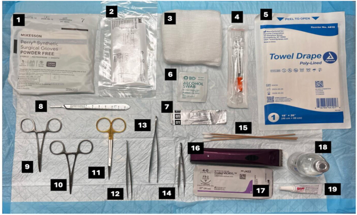
Figure 1: Selected surgical instruments and materials for tumor resection. (1) Surgical gloves, (2) drape, (3) gauze pad, (4) 0.3 mL insulin syringe, (5) towel drape, (6) isopropyl alcohol swab pad, (7) scalpel blade, (8) scalpel without blade, (9) Hartman hemostat, (10) Mosquito hemostatic forceps, (11) micro dissecting scissors, (12) micro dissecting forceps (13) Brown-Adson forceps (14) micro dissecting forceps, (15) cotton tip applicators, (16) trimmer, (17) 4-0 coated vicryl suture, (18) 0.25% Bupivacaine, and (19) eye lubricant ointment. Please click here to view a larger version of this figure.
3. Preparation of the animal for surgery
- Weigh the mouse 24 h prior to the procedure to determine appropriate anesthesia and pain relief drug dosage, per specified protocol.
- Anesthetize the mouse in the induction chamber for approximately 2 min, and then administer analgesics at least 30 min before surgery. This should be subcutaneously injected at the nape of the murine neck.
- If not done previously at the time of inoculation or if previously cleared hair has begun to regrow, add a small amount of hair removal cream using a cotton-tip applicator directly to the hair at the tumor resection site, with sufficient margins to keep hair out of the incision space through the procedure.
- Allow cream to sit for approximately 10 s and wipe hair away with dry, clean gauze.
- Remove any remaining cream from the site with gauze dampened with saline or water, then dry the area with dry, clean gauze.
NOTE: Hair removal by an electronic trimmer is an acceptable but less preferred alternative method, as the skin around the head and neck can be thin and delicate. If using a trimmer, be sure to avoid sensitive areas, such as the eyes, and keep any loose skin taut to help avoid unwanted injury to the mouse. - Administer systemic analgesic subcutaneously and local analgesic to the incision site.
- Place the anesthetized mouse on the heating pad, lying on its back with its anterior side facing upward. Its nose should be just inside the nose cone cover.
- Place a small amount (i.e., sufficient to cover the eye) of sterile lubricant eye ointment on each eye of the mouse to prevent drying of the eyes during the procedure.
- Using a clean cotton-tip applicator, swab the site of resection with 70% isopropyl alcohol followed by 10% iodine 3 times, making sure to remove the iodine after the last pass with 70% isopropyl alcohol.
- Wearing sterile gloves and appropriate personal protective equipment (robe, hair cover), prepare the sterile field of at least 18"W X 26"L, and lay out all appropriate tools and materials (sterile cotton gauze, sterile Q-tips, sterile saline, isopropyl alcohol applicators, surgical tools) (Figure 1).
- Cover the mouse with a sterile drape and cut an access hole immediately above the resection site using sterile scissors.
4. Surgical resection of established oral tumors
NOTE: Throughout the procedure, the surgeon or assistant should check for breathing and reflex of the mouse to ensure an appropriate oxygenation while the mouse is under anesthesia. Adjust isoflurane in accordance with veterinary guidance and institutional policy as needed.
- Using a new surgical scalpel, create a submandibular incision, just below the tumor and along the left jawline, taking up the length of the tumor (Figure 2A,B).
- Carefully use the Hartman hemostats to widen the opening toward the tumor. As needed, dab any blood with sterile gauze.
- Locate the tumor and use forceps to hold it in place (Figure 2B).
NOTE: An assistant may assist at this point with using forceps to maintain access to the tumor site (i.e., pull skin away). - Using surgical scissors, snip around the base of the tumor to resect all visible portions (Figure 2C). Reposition the skin over the resected tumor site and close the incision with four to six interrupted sutures, using 4-0 long-lasting vicryl sutures (Figure 2D).
NOTE: Be careful not to secure sutures too tightly to preserve skin without further hurting the mice, as well as to prevent potential injuries upon later suture removal. - To continue with additional surgeries, clean the surfaces of surgical instruments with 70% isopropyl alcohol and place them in the heated bead sterilizer for 10-15 s prior to each additional use. The same set of tools may be used to perform surgery on up to 5 mice or, as specified by institutional guidelines and practices.
NOTE: Resected tumors may be discarded or taken for processing and further analysis (i.e., fixation for histology and imaging; tissue dissociation and immune staining for flow cytometry phenotyping analysis, per the specific research study experimental plans18,19 (Figure 2E).
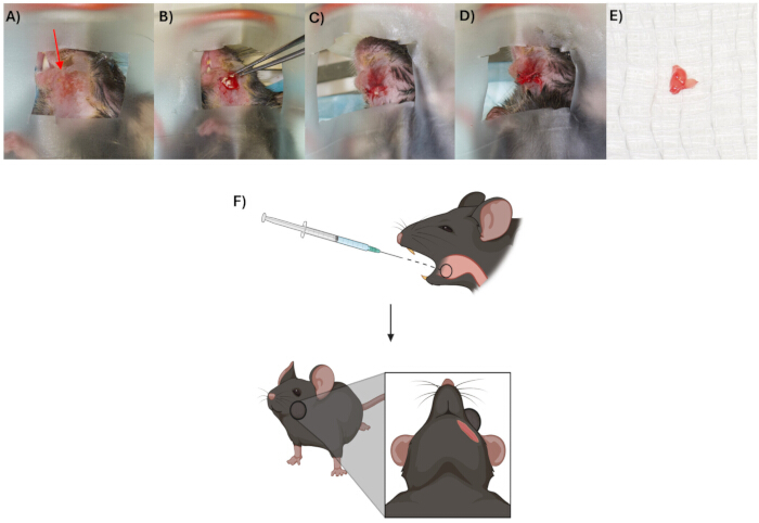
Figure 2: Oral tumors, established transorally through the lower buccal mucosa, can be resected via a submandibular approach without healing complications. (A) Established tumor (red arrow) in the oral cavity. (B) Submandibular incision exposing the tumor. (C) The resected tumor. (D) Wound closure with multiple interrupted vicryl sutures. (E) Excised tumor. (F) Summary schematic for transoral inoculation. Image created in BioRender. Please click here to view a larger version of this figure.
5. Post-resection care
- After closing the incision, turn off the anesthesia flow to 0% and continue only O2 at 1.5 L/min until the mouse begins to regain consciousness (less than 5 min). If the mouse remains motionless and unresponsive after several minutes, refer to institutional policies on veterinary intervention.
- Gently transfer the mouse to the pre-warmed cage. Continue to monitor the mouse for post-operative consciousness and mobility. It should regain full consciousness and mobility by 10 min.
- Perform daily post-operative monitoring in accordance with approved protocol and institutional policies.
- Immediately following the surgery, provide "soft" food for the mouse by setting pellets soaked in water in an accessible container (i.e., plastic weigh boat) directly into the cage. If no complications arise, the mouse can resume eating normal solid food at the next routine feeding.
- Monitor mice twice daily for the first 48 h (2 days) and once per day for a minimum of 96 h (4 days) post-operation. Subsequently, resume a normal monitoring schedule (i.e., 2 times weekly).
- At day 7 post-surgery, remove sutures under anesthesia, being careful not to cut or injure the mice.
- Continue to monitor mice and once they reach their humane endpoint, humanely euthanize animals in accordance with the approved protocol euthanasia criteria and the American Veterinary Medical Association.
NOTE: Statistical analysis of tumor growth kinetics and animal survival are relevant metrics for this methodology. For Kaplan-Meier survival curves, statistical analyses using the log-rank/Mantel-Cox test were performed via statistical analysis software. For tumor growth curves, Šídák's multiple comparisons test by way of a two-way ANOVA analysis was used to compare tumor sizes between unresected and resected groups. P-values less than 0.05 were considered significant.
Access restricted. Please log in or start a trial to view this content.
Results
Observations from MOC2 murine buccal mucosa
Using the MOC2 cell line, tumors were established in the murine buccal mucosa of the oral cavity (Figure 2A-F). Established primary tumors were resected at a tumor size of 25-36 mm2. Upon resection, mice typically regained full mobility within 5-10 min. Mice were subsequently monitored for post-operative care to confirm healing and recovery and to observe tumor recurrence. During thi...
Access restricted. Please log in or start a trial to view this content.
Discussion
Herein, this article describes a simple procedure for the establishment and subtotal resection of tumors in the buccal mucosa of the oral cavity in mice. Previous studies have similarly demonstrated the feasibility of establishing tumors through the murine tongue, buccal mucosa, or floor-of-mouth for downstream analysis of resected tumors, as well as to investigate subsequent local recurrence and regional and distant metastasis, each with its own limitations12,13...
Access restricted. Please log in or start a trial to view this content.
Acknowledgements
This work was funded by 1R21DE034543 (NIH-NIDCR). We also acknowledge the administrative, veterinary, and maintenance staff of the UTHealth Center for Laboratory Animal Medicine and Care for their support and care.
Access restricted. Please log in or start a trial to view this content.
Materials
| Name | Company | Catalog Number | Comments |
|---|---|---|---|
| 0.05% Trypsin | Corning | 25-052-Cl | 0.53 mM EDTA, 1 X [-] sodium bicarbonate |
| 0.25% Bupivacaine HCl Injection | McKesson | 1077280 | 2.5mg/kg-1mg/kg, ID/SC 20-30uL |
| 0.9% Sodium Chloride, USP | McKesson | 161736 | 5 mL/kg, SC, IV 1.5 mL |
| 23 G Needle | Becton, Dickinson and Company [McKesson] | 402 | |
| 4-0 Coated Vicryl | McKesson | 100700 | |
| 50 mL conical tubes | Thermo Scientific | 33965 | |
| Absorbent Bench Pads | VWR | 115-0684 | |
| Alcohol Swab Isopropyl Alcohol 70% | Becton, Dickinson and Company [VWR] | 326895 | |
| Autoclave Pouch Self-Sealing 5 ¼” x 10” | McKesson | 960944 | |
| Balance | Ohaus [VWR] | 30253019 | |
| Bioclave Mini | Benchmark Scientific | B4000-M | |
| Brown-Adson Forceps | ROBOZ [VWR] | RS-5231 | |
| Cotton-Tipped Applicator Sterile 6 in | McKesson | 999736 | |
| Dulbecco's Phophate Buffered Saline / Modified | Cytiva | SH30028.02 | [-] calcium, [-] magnesium |
| Ethiqa XRÒ* | Fidelis Animal Health | NDC 86084-100-30 | 3.25 mg/kg SC |
| General Purpose Drape | McKesson | 68107 | |
| Glass Bead Sterilizer | VWR | 75999-324 | |
| Glass Beads | VWR | 75999-332 | |
| Hartman Hemostats | Fine Science Tools [VWR] | 13003-10 | |
| High-Quality, Portable Anesthesia Device | Patterson Veterinary | 07-8915662 | |
| Isoflurane, USP | McKesson | 803250 | 4-5% Induction, 1-3% Maintenance |
| MedLEDÒ Chrome (Headlight) | KLS Martin | ML-MC7-HT-HK-X21 | |
| Micro Dissecting Forceps | ROBOZ [VWR] | RS-5174 | |
| Micro Dissecting Scissors | ROBOZ [VWR] | RS-5914 | |
| Monoject Insulin Syringes | Cardinal Health [McKesson] | 8881600145 | |
| Mosquito Hemostatic Forceps | ROBOZ [VWR] | RS-7100 | |
| Nair Body Cream Hair Remover | Church & Dwight Co., Inc. | ||
| Needle Holder | ROBOZ [VWR] | RS-7154 | |
| PVP Prep Solution 10% Povidone Iodine | McKesson | 911740 | |
| Scalpel | ROBOZ [VWR] | RS-9843 | |
| Scalpel Blades Carbon Steel No. 10 | McKesson | 862685 | |
| Small Animal Heated Pad | K&H Pet Products [Amazon] | 9 X 12in 20W | |
| Small Mouse Chamber | Patterson Veterinary | 07-893338 | |
| Small Nose Cone | Patterson Veterinary | 78909681 | |
| Steri-Drape | McKesson | 5713 | |
| Sterile Gauze Pads 2”x2” – 12 PLY | Dukal Corporation [Amazon] | REF: 1212 | |
| Sterile Lubricant Eye Ointment | McKesson | 730979 | |
| Surgical Gloves | McKesson | 20-2070N | |
| Syringe 1cc | McKesson | 1065984 | |
| Traceable Digital Carbon Fiber Calipers | Fischer Brand | 15-077-957 | |
| Trimmer Kit | WAHL [Amazon] | BravMini |
References
- Klein, J. D., Grandis, J. R. The molecular pathogenesis of head and neck cancer. Cancer Biol Ther. 9 (1), 1-7 (2010).
- Lee, A. M., Weaver, A. N., Acosta, P., Harris, L., Bowles, D. W. Review of current and future medical treatments in head and neck squamous cell carcinoma. Cancers (Basel). 16 (20), 3488(2024).
- Liu, Y., Zhang, N., Wen, Y., Wen, J. Head and neck cancer: Pathogenesis and targeted therapy. MedComm (Beijing). 5 (9), e702(2024).
- Qiao, X., et al. The evolving landscape of PD-1/PD-L1 pathway in head and neck cancer. Front Immunol. 11, 1721(2020).
- Bakos, O., Lawson, C., Rouleau, S., Tai, L. -H. Combining surgery and immunotherapy: turning an immunosuppressive effect into a therapeutic opportunity. J Immunother Cancer. 6 (1), 86(2018).
- Chaves, P., et al. Preclinical models in head and neck squamous cell carcinoma. Br J Cancer. 128 (10), 1819-1827 (2023).
- International Agency for Research on Cancer. Estimated number of new cases from 2022 to 2050, both sexes, age [0-85+]. Globocan. , World Health Organization. (2022).
- Bonomi, M., Patsias, A., Posner, M., Sikora, A. The role of inflammation in head and neck cancer. Adv Exp Med Biol. 816, 107-127 (2014).
- Tang, F., Tie, Y., Tu, C., Wei, X. Surgical trauma-induced immunosuppression in cancer: recent advances and the potential therapies. Clin Transl Med. 10 (1), 199-223 (2020).
- Tai, L. -H., et al. A mouse tumor model of surgical stress to explore the mechanisms of post-operative immunosuppression and evaluate novel perioperative immunotherapies. J Vis Exp. (85), e51253(2014).
- Kansal, V., Kinney, B. L. C., Schmitt, N. C. Characterization of the tumor microenvironment in the mouse oral cancer (MOC1) model after orthotopic implantation in the buccal mucosa. Head Neck. 46 (5), 1056-1062 (2024).
- Oweida, A. J., et al. Intramucosal inoculation of squamous cell carcinoma cells in mice for tumor immune profiling and treatment response assessment. J Vis Exp. (146), e59195(2019).
- Behren, A., et al. Development of an oral cancer recurrence mouse model after surgical resection. Int J Oncol. 36 (4), 849-855 (2010).
- Tai, L. -H., et al. Preventing post-operative metastatic disease by inhibiting surgery-induced dysfunction in natural killer cells. Cancer Res. 73 (1), 97-107 (2013).
- Sharon, S., et al. A platform for locoregional T-cell immunotherapy to control HNSCC recurrence following tumor resection. Oncotarget. 12 (13), 1201-1213 (2021).
- Mermod, M., et al. Mouse model of postsurgical primary tumor recurrence and regional lymph node metastasis progression in HPV-related head and neck cancer. Int J Cancer. 142 (12), 2518-2528 (2018).
- Judd, N. P., Allen, C. T., Winkler, A. E., Uppaluri, R. Comparative analysis of tumor-infiltrating lymphocytes in a syngeneic mouse model of oral cancer. Otolaryngol Head Neck Surg. 147 (3), 493-500 (2012).
- Cossarizza, A., et al. Guidelines for the use of flow cytometry and cell sorting in immunological studies (third edition). Eur J Immunol. 51 (12), 2708-3145 (2021).
- Gurina, T. S., Simms, L. Histology, staining. , StatPearls Publishing. (2025).
- Cognetti, D. M., Weber, R. S., Lai, S. Y. Head and neck cancer: An evolving treatment paradigm. Cancer. 113 (7 Suppl), 1911-1932 (2008).
- Anderson, G., et al. An updated review on head and neck cancer treatment with radiation therapy. Cancers (Basel). 13 (19), 4912(2021).
- Johnson, D. E., et al. Head and neck squamous cell carcinoma. Nat Rev Dis Primers. 6 (1), 92(2020).
- Friedman, J., et al. Neoadjuvant PD-1 immune checkpoint blockade reverses functional immunodominance among tumor antigen-specific T cells. Clin Cancer Res. 26 (3), 679-689 (2020).
- Zhou, S., et al. Neoadjuvant and adjuvant immunotherapy in the treatment of oral squamous cell carcinoma. J Biochem Mol Toxicol. 39 (3), e70199(2025).
- Shibata, H., Saito, S., Uppaluri, R. Immunotherapy for head and neck cancer: a paradigm shift from induction chemotherapy to neoadjuvant immunotherapy. Front Oncol. 11, 727433(2021).
- Saddawi-Konefka, R., et al. Lymphatic-preserving treatment sequencing with immune checkpoint inhibition unleashes cDC1-dependent antitumor immunity in HNSCC. Nat Commun. 13 (1), 4298(2022).
- Darragh, L. B., et al. Elective nodal irradiation mitigates local and systemic immunity generated by combination radiation and immunotherapy in head and neck tumors. Nat Commun. 13 (1), 7015(2022).
- Saddawi-Konefka, R., et al. Defining the role of immunotherapy in the curative treatment of locoregionally advanced head and neck cancer: Promises, challenges, and opportunities. Front Oncol. 11, 738262(2021).
Access restricted. Please log in or start a trial to view this content.
Reprints and Permissions
